. 2012;8(10):e1002984.
doi: 10.1371/journal.pgen.1002984. Epub 2012 Oct 4.

The Paramecium germline genome provides a niche for intragenic parasitic DNA: evolutionary dynamics of internal eliminated sequences

Affiliations

The Paramecium germline genome provides a niche for intragenic parasitic DNA: evolutionary dynamics of internal eliminated sequences

Olivier Arnaiz et al. PLoS Genet. 2012.

Abstract

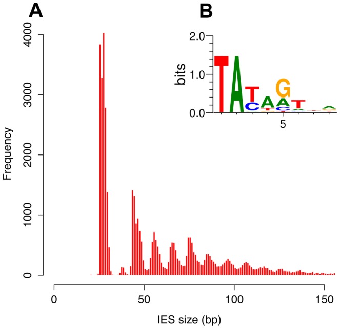
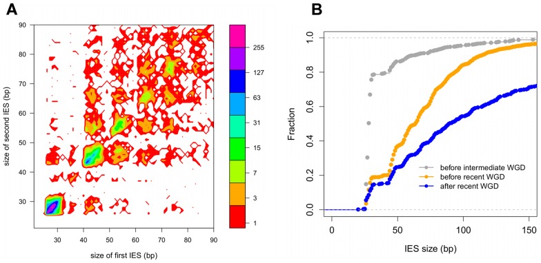
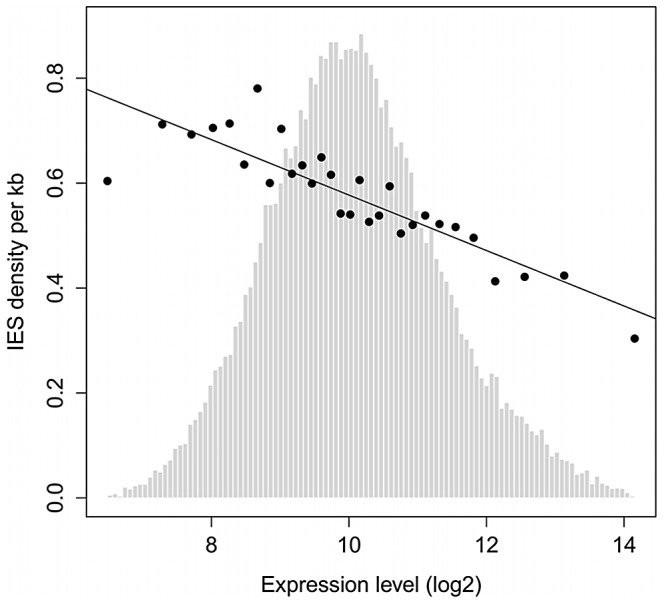
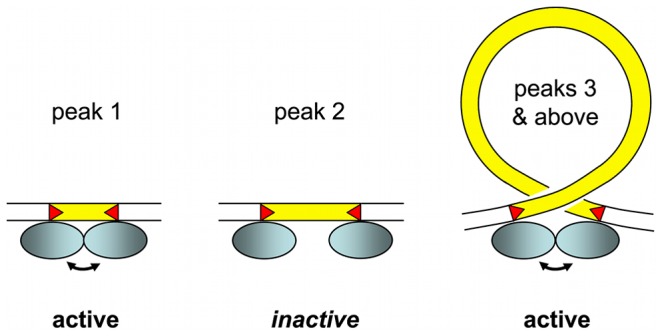
Insertions of parasitic DNA within coding sequences are usually deleterious and are generally counter-selected during evolution. Thanks to nuclear dimorphism, ciliates provide unique models to study the fate of such insertions. Their germline genome undergoes extensive rearrangements during development of a new somatic macronucleus from the germline micronucleus following sexual events. In Paramecium, these rearrangements include precise excision of unique-copy Internal Eliminated Sequences (IES) from the somatic DNA, requiring the activity of a domesticated piggyBac transposase, PiggyMac. We have sequenced Paramecium tetraurelia germline DNA, establishing a genome-wide catalogue of -45,000 IESs, in order to gain insight into their evolutionary origin and excision mechanism. We obtained direct evidence that PiggyMac is required for excision of all IESs. Homology with known P. tetraurelia Tc1/mariner transposons, described here, indicates that at least a fraction of IESs derive from these elements. Most IES insertions occurred before a recent whole-genome duplication that preceded diversification of the P. aurelia species complex, but IES invasion of the Paramecium genome appears to be an ongoing process. Once inserted, IESs decay rapidly by accumulation of deletions and point substitutions. Over 90% of the IESs are shorter than 150 bp and present a remarkable size distribution with a -10 bp periodicity, corresponding to the helical repeat of double-stranded DNA and suggesting DNA loop formation during assembly of a transpososome-like excision complex. IESs are equally frequent within and between coding sequences; however, excision is not 100% efficient and there is selective pressure against IES insertions, in particular within highly expressed genes. We discuss the possibility that ancient domestication of a piggyBac transposase favored subsequent propagation of transposons throughout the germline by allowing insertions in coding sequences, a fraction of the genome in which parasitic DNA is not usually tolerated.


Conflict of interest statement

The authors have declared that no competing interests exist.

Figures

Figure 1
Figure 1. IES excision.
Schematic representation of, from left to right, a canonical IES, a nested IES and an IES with an alternative boundary. In the case of the nested IES, the middle line represents either an intermediate in the excision pathway or an alternative final product. In the case of the alternative boundary IES, the middle line represents an alternative final product.
Figure 2
Figure 2. Anchois Tc1/mariner family transposon.
A) Alignment of the DDE domains of bacterial IS630 elements (IS630Sd, Salmonella dublin, GenBank Accession No. A43586; IS630Ss, Shigella sonnei, X05955), invertebrate and fungal Tc1 transposons (Baril, D melanogaster, Q24258; Impala, Fusarium oxysporum, AF282722; S, D melanogaster, U33463; Tc1, C elegans, X01005) and ciliate Tc1/mariner transposons (TBE1, Oxytricha fallax, L23169; Tec1 and Tec2, Euplotes crassus, L03359 and L03360; Anchois, Thon and Sardine, Paramecium tetraurelia, this article; Tennessee, Paramecium primaurelia, [34]). Asterisks mark the conserved catalytic DDE residues. B) Schematic diagram of the 3.6 Kb Anchois consensus, showing the position and orientation of the 3 ORFs. The yellow triangles represent the ∼22 nt TIRs. Asterisks mark the position of residues of the catalytic DDE triad for the ORF encoding the DDE transposase.
Figure 3
Figure 3. IES sequence properties.
A) Histogram of the sizes of the genome-wide set of IESs that are shorter than 150 bp. B) Sequence logo showing information content at each position, corrected for a G+C content of 28%, for the ends of the genome-wide set of IESs.
Figure 4
Figure 4. IES conservation in genes related by WGD.
A) Filled contour plot of the correlation between the size of IES pairs that have been conserved with respect to the recent WGD. The x axis gives the size in bp of the first IES, the y axis gives the size in bp of the second IES found in the ohnologous gene and the color of each point indicates the number of times that combination of x,y values was found in the data set. The color legend is shown to the right of the figure, the numbers represent counts of the x,y value pairs; the rainbow colors are distributed according to a log2 scale. B) Size distribution of IESs conserved in “quartets” i.e. genes that are still present in 4 copies in the genome after duplication at both the intermediate and the recent WGD events. In order to compare size distributions for different classes of IES, they are represented as experimental cumulative distribution functions. The ripples in each curve correspond to the peaks of a histogram representation as in Figure 3A. The curves are for IESs that must have originated from an ancestral IES acquired before the intermediate WGD (grey, N1111 IESs), IESs that must have originated from an ancestral IES acquired before the recent WGD (orange, N1100 IESs) and the IESs that might have been acquired since the recent WGD (blue, N1000 IESs).
Figure 5
Figure 5. TA-indels are produced by IES excision errors.
Schematic representation of the “residual” and “low frequency” TA-indels that were identified by comparing the MAC draft genome assembly (MAJOR form) with the 13× Sanger sequencing reads used to build the assembly . The TA-indels were identified by one or more reads that differed from the assembly (minor form). The residual TA-indels were assumed to be the result of occasional failure to excise an IES and the low-frequency TA-indels to result from excision of MAC-destined sequences. Comparison of the genome-wide set of IESs with the TA-indels revealed that many TA-indels result from the use of alternative IES boundaries situated inside the corresponding IES in the case of residual TA-indels and outside the IES in the case of low-frequency TA-indels. In the schema, TA dinucleotides in black boxes are bona fide IES boundaries while TA dinucleotides in blue boxes are alternative IES boundaries.
Figure 6
Figure 6. IES density is inversely proportional to gene expression level.
Genes were binned according to their median expression level across 58 microarrays representing different cellular and growth conditions as described in , . The expression levels were divided into 30 bins as in . The black points show the average IES density (per Kb) of genes in each bin. Linear regression was used to fit the points. Light gray bars show the distribution of genes according to their expression level (before binning).
Figure 7
Figure 7. IES size constraint and the assembly of an active excision complex.
Our working model is based on the assumption that oligomerization of the IES excisase (most likely the domesticated transposase PiggyMac) on DNA activates catalytic cleavage at IES ends (IESs are drawn in yellow and red triangles highlight the orientation of their ends). In the absence of any information on the stoichiometry of the complex, the excisase is represented by a shaded blue ellipse. For very short IESs from peak 1 (26–30 bp in length), the required contact between protein subunits may be established directly (double-headed arrow) and the complex is active. For IESs longer than 44 bp (peak 3 and above), we propose that looping of the intervening DNA double helix brings IES ends into close proximity and activates DNA cleavage. We have arbitrarily drawn the complex as an antiparallel arrangement of IES ends within a negatively supercoiled loop, but other conformations are possible. IESs from the “forbidden” peak 2 would be too long to allow direct contacts between protein subunits to be established, and too short to form an excision loop.

References

    1. Aury J-M, Jaillon O, Duret L, Noel B, Jubin C, et al. (2006) Global trends of whole-genome duplications revealed by the ciliate Paramecium tetraurelia. Nature 444: 171–178 doi:nature05230 - DOI - PubMed
    1. Chalker DL, Yao M-C (2011) DNA elimination in ciliates: transposon domestication and genome surveillance. Annu Rev Genet 45: 227–246 doi:10.1146/annurev-genet-110410-132432. - PubMed
    1. Coyne RS, Lhuillier-Akakpo M, Duharcourt S (2012) RNA-guided DNA rearrangements in ciliates: is the best genome defense a good offense? Biol Cell Accepted manuscript online doi:10.1111/boc.201100057. - PubMed
    1. Schoeberl UE, Mochizuki K (2011) Keeping the soma free of transposons: programmed DNA elimination in ciliates. J Biol Chem 286: 37045–37052 doi:10.1074/jbc.R111.276964. - PMC - PubMed
    1. Bétermier M (2004) Large-scale genome remodelling by the developmentally programmed elimination of germ line sequences in the ciliate Paramecium. Res Microbiol 155: 399–408. - PubMed

Publication types