. 2012;7(10):e46646.
doi: 10.1371/journal.pone.0046646. Epub 2012 Oct 5.

Generation and characterization of human heme oxygenase-1 transgenic pigs

Affiliations

Generation and characterization of human heme oxygenase-1 transgenic pigs

Hye-Jung Yeom et al. PLoS One. 2012.

Abstract

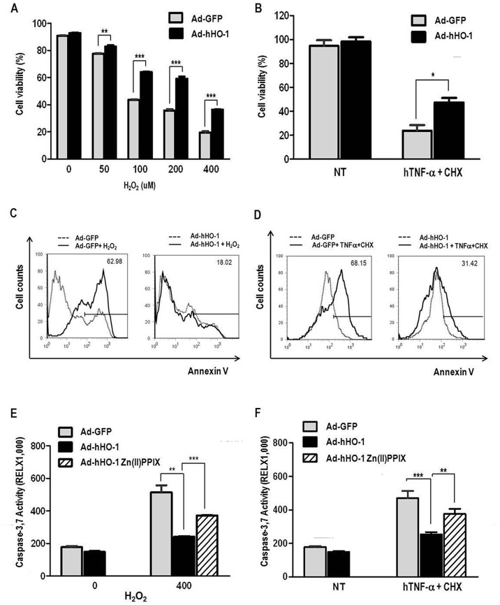
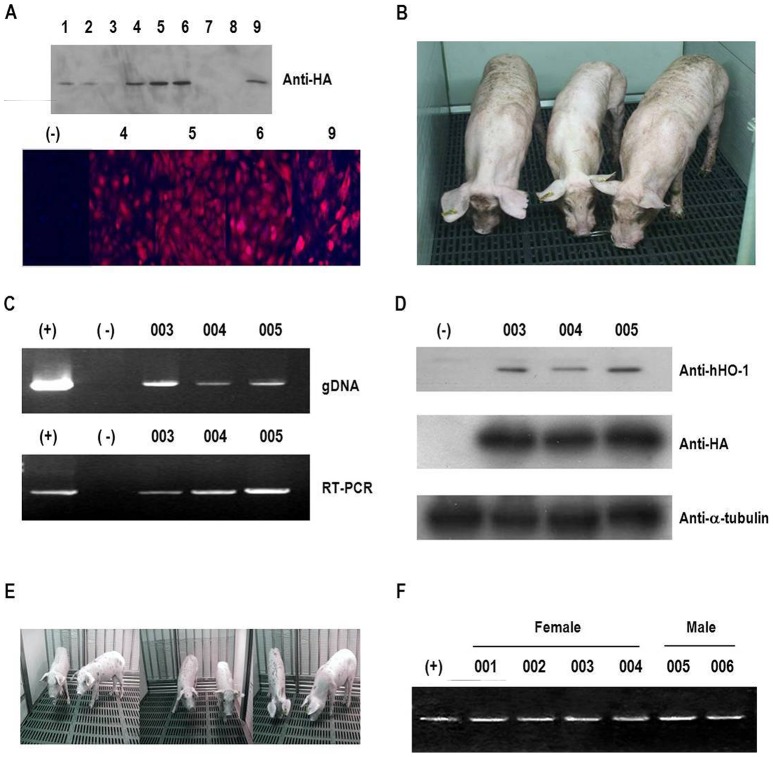
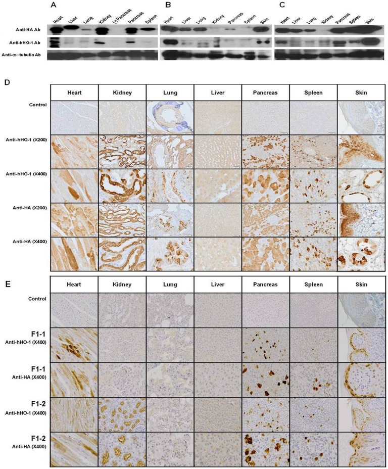
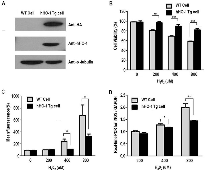
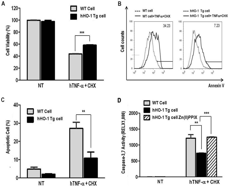
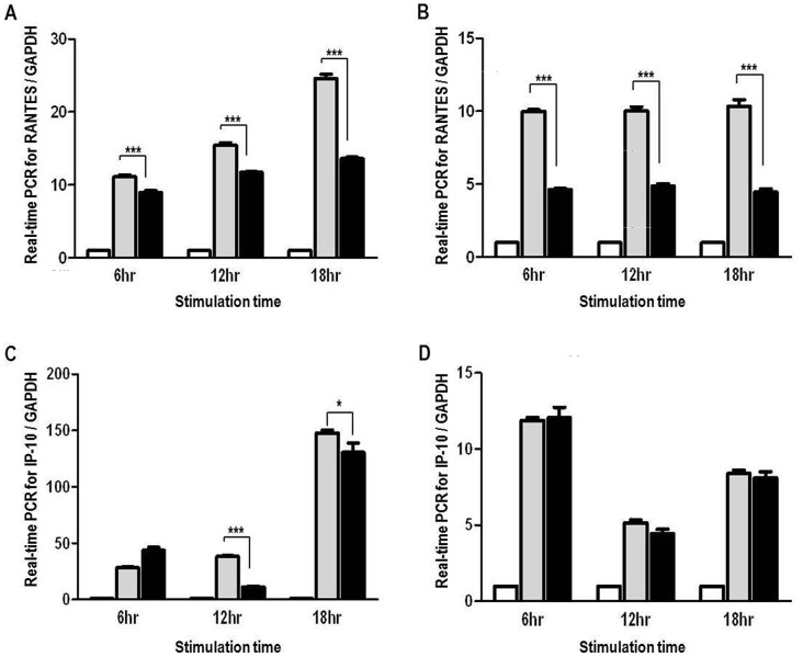
Xenotransplantation using transgenic pigs as an organ source is a promising strategy to overcome shortage of human organ for transplantation. Various genetic modifications have been tried to ameliorate xenograft rejection. In the present study we assessed effect of transgenic expression of human heme oxygenase-1 (hHO-1), an inducible protein capable of cytoprotection by scavenging reactive oxygen species and preventing apoptosis caused by cellular stress during inflammatory processes, in neonatal porcine islet-like cluster cells (NPCCs). Transduction of NPCCs with adenovirus containing hHO-1 gene significantly reduced apoptosis compared with the GFP-expressing adenovirus control after treatment with either hydrogen peroxide or hTNF-α and cycloheximide. These protective effects were diminished by co-treatment of hHO-1 antagonist, Zinc protoporphyrin IX. We also generated transgenic pigs expressing hHO-1 and analyzed expression and function of the transgene. Human HO-1 was expressed in most tissues, including the heart, kidney, lung, pancreas, spleen and skin, however, expression levels and patterns of the hHO-1 gene are not consistent in each organ. We isolate fibroblast from transgenic pigs to analyze protective effect of the hHO-1. As expected, fibroblasts derived from the hHO-1 transgenic pigs were significantly resistant to both hydrogen peroxide damage and hTNF-α and cycloheximide-mediated apoptosis when compared with wild-type fibroblasts. Furthermore, induction of RANTES in response to hTNF-α or LPS was significantly decreased in fibroblasts obtained from the hHO-1 transgenic pigs. These findings suggest that transgenic expression of hHO-1 can protect xenografts when exposed to oxidative stresses, especially from ischemia/reperfusion injury, and/or acute rejection mediated by cytokines. Accordingly, hHO-1 could be an important candidate molecule in a multi-transgenic pig strategy for xenotransplantation.


Conflict of interest statement

Competing Interests: Hyunil Kim is employed by Optifarm Solution Inc. There are no patents, products in development or marketed products to declare. This does not alter the authors' adherence to all the PLOS ONE policies on sharing data and materials.

Figures

Figure 1
Figure 1. Impact of Ad-hHO-1 on H2O2 or hTNF-α and CHX-induced cell death and apoptosis in NPCCs.
A: Ad-hHO-1 increased cell viability in H2O2-treated NPCCs. NPCCs were transduced with Ad-hHO-1 for 24 hr before the addition of H2O2 (0, 50, 100, 200, or 400 µM). After incubating the cells with H2O2 for an additional 24 hr, cell viability was measured by CCK-8. B: After treatment with hTNF-α and CHX cell viability was measured by CCK-8. An increased proportion of viable cells were detected in the Ad-hHO-1 group compared with the Ad-GFP control group. NPCCs were infected with Ad-hHO-1 for 24 hr before the addition of hTNF-α (25 ng/ml) and CHX (10 µg/ml). After treatment with hTNF-α and CHX, the NPCCs were incubated for an additional 24 hr. C: FACS analysis of apoptosis in NPCCs after treatment with 400 µM H2O2. A lower proportion of apoptotic cells were noted in Ad-hHO-1 (50 MOI) infected NPCCs relative to the Ad-GFP control. The NPCCs were treated with H2O2 (400 µM) and FACS analysis was performed using annexin V-PE. Each experiment was repeated three times (data not shown). D: FACS analysis of apoptosis in NPCCs after treatment with hTNF-α and CHX for 24 hr. A smaller proportion of apoptotic cells was detected in Ad-hHO-1 (200 MOI)-infected NPCCs than in the Ad-GFP (200 MOI) group (p<0.05). After treatment with hTNF-α and CHX, apoptosis of cultured NPCCs was measured by FACS using annexin V. The numbers indicate the proportion of apoptotic cells in the NPCCs after 24 hr of incubation with hTNF-α and CHX. E: Lower activity of caspase 3/7 in Ad-hHO-1-infected NPCCs relative to Ad-GFP-infected NPCCs after 24 hr of incubation with H2O2 (400 µM). After Zn(II)PPIX treatment, Ad-hHO-1 increased activity of caspase 3/7. Each experiment was repeated three times. (* p<0.05, ** p<0.01, *** p<0.001, NT: no treatment). F: The activities of caspase 3/7 were measured in Ad-hHO-1-infected NPCCs. The protective effect of Ad-hHO-1 in the NPCCs compared with Ad-GFP after 24 hr of incubation with hTNF-α and CHX. Each experiment was repeated three times. (** p<0.01, *** p<0.001).
Figure 2
Figure 2. Generation of hHO-1 transfected donor cell lines and hHO-1-Tg pigs.
A: Establishment of hHO-1-expressing cells using O-type white Yucatan pig fibroblasts. Expression of hHO-1 was identified by western blot analysis and immunocytochemistry. A donor cell colony with a high level of expression was chosen, and nuclear transfer was performed. B: Three hHO-1-Tg pigs were generated (F0; #003, #004, and #005). C: Expression of the hHO-1 gene was confirmed by genomic DNA PCR and RT-PCR. Genomic DNA recovered from blood was analyzed by PCR (top lane). RT-PCR analysis of whole blood isolated from the HA-hHO-1-Tg pigs is shown in the bottom lane (#003, #004, and #005). Positive control: hHO-1-transfected cells (MPN3); negative control: normal pig. D: Western blotting was performed in ear tissue from the transgenic pig using anti-hHO-1 and anti-HA antibodies. The α-tubulin was used as a control. E: Six hHO-1-Tg progenies (F1; 4 females and 2 males) pigs were generated after bred of one F0 pig with wild type female. F: Expression of the hHO-1 gene was confirmed by RT-PCR analysis of whole blood isolated from the HA-hHO-1-Tg pigs. Positive control (+): hHO-1-transfected cells (MPN3). Negative control (−): wild type pigs. All the 6 piglets were confirmed as transgenic.
Figure 3
Figure 3. Tissue distribution of hHO-1 protein in hHO-1-Tg pig tissues.
A: The tissue distribution of the hHO-1 protein was analyzed in various organs by western blot analysis in a 465-day-old male hHO-1 F0 pig. Western blot was performed on heart, kidney, lung, liver, pancreas, spleen, and skin tissue. B: The tissue distribution of the hHO-1 protein was analyzed in various organs by western blot analysis in a 74-day-old male hHO-1 F1 pig. Western blot was performed on heart, kidney, lung, liver, pancreas, spleen, and skin tissue. C: The tissue distribution of the hHO-1 protein was analyzed in various organs by western blot analysis in a 74-day-old male hHO-1 F1 pig. Western blot was performed on heart, kidney, lung, liver, pancreas, spleen, and skin tissue using anti-hHO-1 (1∶1,000) and anti-HA antibodies (1∶4,000). Twenty micrograms of protein were loaded into each lane as indicated by the α-tubulin band. D: Immunohistochemistry of heart, kidney, lung, liver, pancreas, spleen, and skin tissues of hHO-1 F0 pig were performed to analyze expression patterns of hHO-1 and HA. Paraffin sections of various organs were stained with anti-HA antibody (1∶1,000), anti-hHO-1antibody (multiple organs: 1∶100) and control (anti-hHO-1 antibody, anti-HA antibody: data not shown). E: Immunohistochemistry of heart, kidney, lung, liver, pancreas, spleen, and skin tissues of hHO-1 F1 (n = 2) were performed to analyze expression patterns of hHO-1 and HA. Paraffin sections of various organs were stained with anti-HA antibody (1∶1,000), anti-hHO-1antibody (multiple organs: 1∶100) and control (anti-hHO-1 antibody, anti-HA antibody: data not shown).
Figure 4
Figure 4. Anti-oxidant effects of fibroblasts from the hHO-1-Tg pig.
A: Western blot analysis for hHO-1 and HA in the hHO-1-Tg fibroblasts using anti-HO-1 (1∶200) and anti-HA (1∶4,000) antibodies. The α-tubulin was used as a control. B: Cell viability was higher in the hHO-1-Tg fibroblasts compared to the wild-type. After H2O2 (0, 200, 400 or 800 µM) treatment, cell viability was slightly better in the hHO-1-Tg fibroblast than in the wild-type fibroblasts. Cell viability was measured by CCK-8 after induction with H2O2 for an additional 1 hr. C: Level of ROS was significantly reduced in the hHO-1-Tg fibroblasts in comparison to the wild type fibroblasts. After H2O2 treatment, ROS production was significantly reduced in the hHO-1-Tg fibroblasts compared with the wild-type fibroblasts. Fibroblasts obtained from ear tissues were incubated with 25 µM DCFH-DA for 15 minutes at 37°C, and ROS were measured using FACS. D: Expression of the iNOS gene by real-time PCR in hHO-1-Tg fibroblasts compared with the wild-type fibroblasts. The threshold cycle (Ct) value was defined as the number of PCR cycles at which the fluorescence crossed a fixed threshold above baseline. For relative quantification, the ΔΔCt method was used to measure fold changes of cDNA. (* p<0.05, ** p<0.01, *** p<0.001).
Figure 5
Figure 5. Anti-apoptotic effects of fibroblasts from the hHO-1-Tg pig.
A: Cell viability was measured using the CCK-8. After induction with hTNF-α (25 ng/ml) and CHX (10 µg/ml) for 15 hours, the hHO-1 group had a lower percentage of viable cells. B and C: FACS analysis of hTNF-α and CHX-mediated apoptosis in fibroblasts. Apoptosis was analyzed using annexin V staining. The numbers indicate the proportion of apoptotic cells in the NPCCs after 15 hr incubation with hTNF-α and CHX. D: Activities of caspase 3/7 were measured in fibroblasts. Human HO-1 protected transgenic fibroblasts from apoptosis after 15 hr of incubation with hTNF-α and CHX. After Zn(II)PPIX treatment, Ad-hHO-1 increased activity of caspase 3/7. Each experiment was repeated three times. (* p<0.05, ** p<0.01, *** p<0.001, NT: no treatment).
Figure 6
Figure 6. Anti-inflammatory effects of fibroblasts from the hHO-1-Tg pig.
A: Expression of the RANTES gene in hHO-1-Tg fibroblasts compared with wild-type fibroblasts in response to hTNF-α stimulation (20 ng/ml) for 6, 12 or 18 hr. RANTES mRNA was measured by real-time PCR. B: Expression of the RANTES gene in hHO-1-Tg fibroblasts compared with the wild-type fibroblasts in response to LPS stimulation (1 µg/ml) for 6, 12, or 18 hr. RANTES mRNA was measured by real-time PCR. C: Expression of the IP-10 gene in hHO-1-Tg fibroblasts compared with the wild-type fibroblasts in response to hTNF-α stimulation (20 ng/ml) for 6, 12 or 18 hr. IP-10 mRNA was measured by real-time PCR. D: Expression of the IP-10 gene in hHO-1-Tg fibroblasts compared with the wild-type fibroblasts in response to LPS stimulation (1 µg/ml) for 6, 12, or 18 hr. IP-10 mRNA was measured by real-time PCR. (* p<0.05, *** p<0.001, White bars: control, gray bars: WT cell, black bars: HO-1-Tg cells).

Similar articles

Cited by

References

    1. Lai L, Kolber-Simonds D, Park KW, Cheong HT, Greenstein JL, et al. (2002) Production of alpha-1,3-galactosyltransferase knockout pigs by nuclear transfer cloning. Science 295: 1089–1092. - PubMed
    1. Miyagawa S, Yamamoto A, Matsunami K, Wang D, Takama Y, et al. (2010) Complement regulation in the GalT KO era. Xenotransplantation 17: 11–25. - PubMed
    1. Charniot JC, Bonnefont-Rousselot D, Albertini JP, Zerhouni K, Dever S, et al. (2007) Oxidative stress implication in a new ex-vivo cardiac concordant xenotransplantation model. Free radical research 41: 911–918. - PubMed
    1. Li LP, Zhang L, Peng LP, Cheng L (2010) Heme oxygenase-1 is the candidate targeting for organ transplantation. Chinese medical journal 123: 2128–2134. - PubMed
    1. Ollinger R, Pratschke J (2010) Role of heme oxygenase-1 in transplantation. Transpl Int 23: 1071–1081. - PubMed

Publication types

MeSH terms