. 2012;7(10):e46949.
doi: 10.1371/journal.pone.0046949. Epub 2012 Oct 11.

Actin recruitment to the Chlamydia inclusion is spatiotemporally regulated by a mechanism that requires host and bacterial factors

Affiliations

Actin recruitment to the Chlamydia inclusion is spatiotemporally regulated by a mechanism that requires host and bacterial factors

Elizabeth Chin et al. PLoS One. 2012.

Abstract

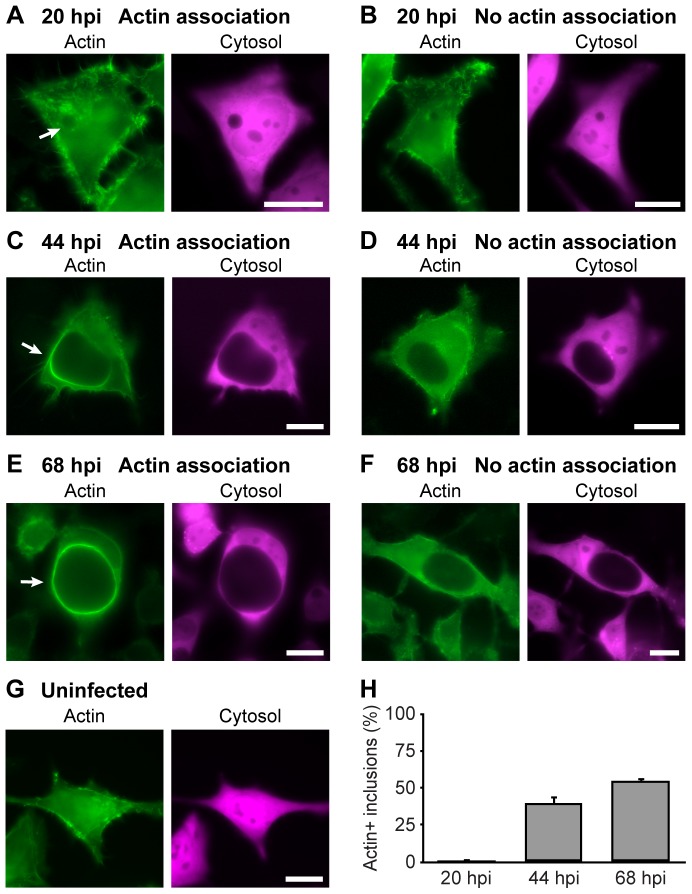
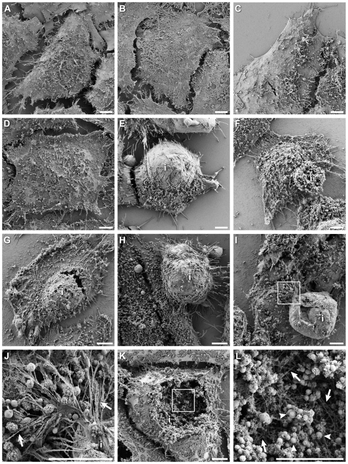
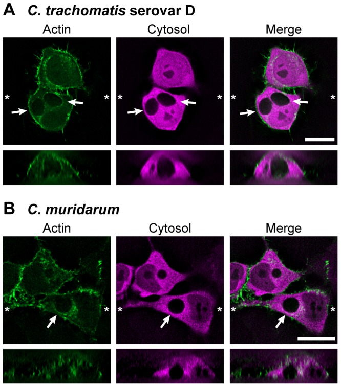
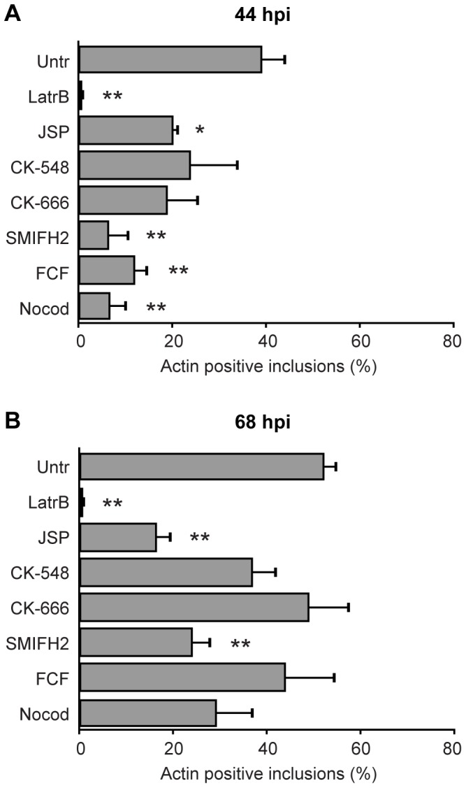
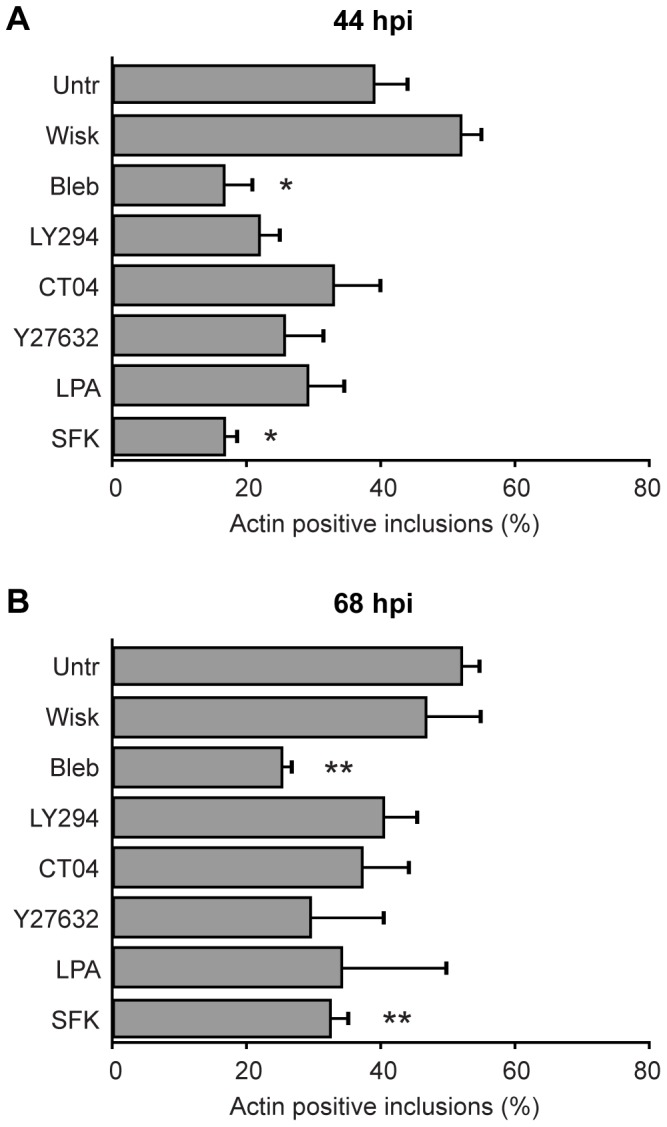
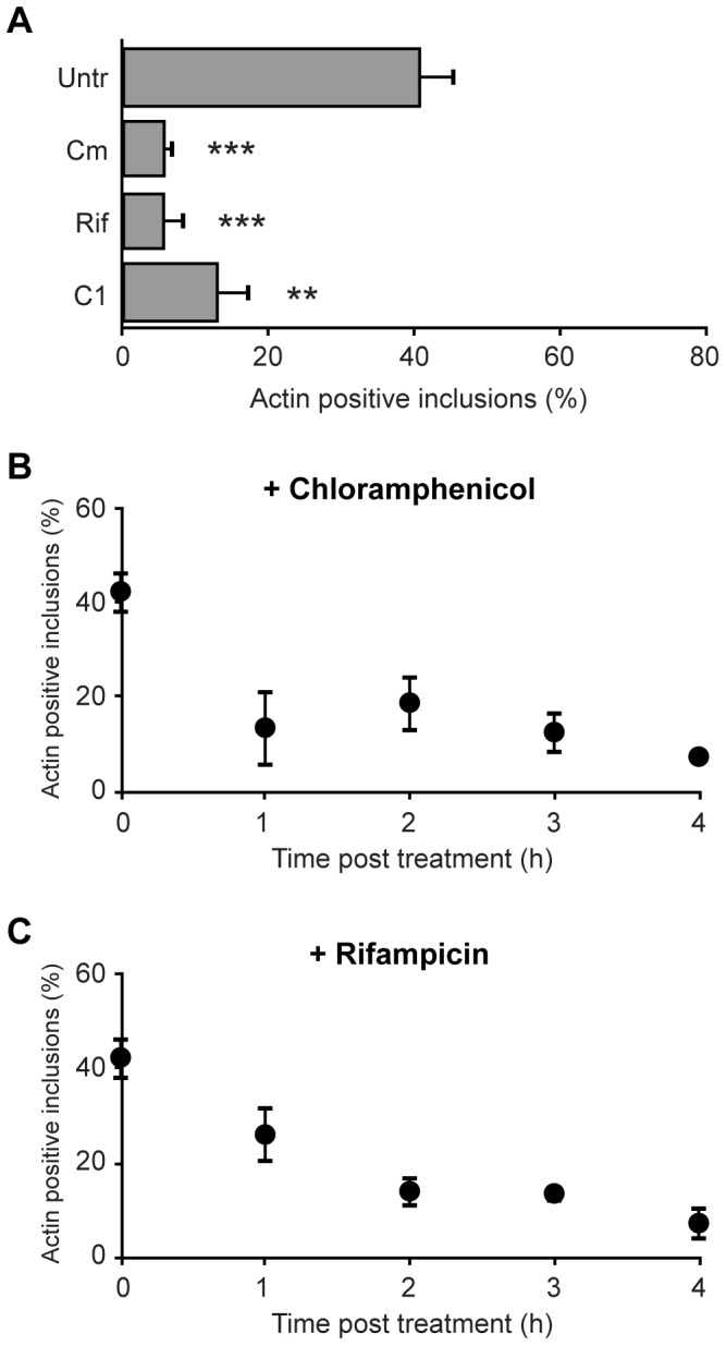
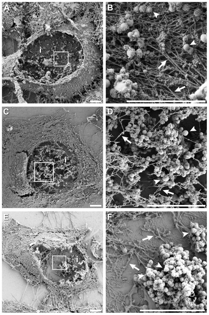
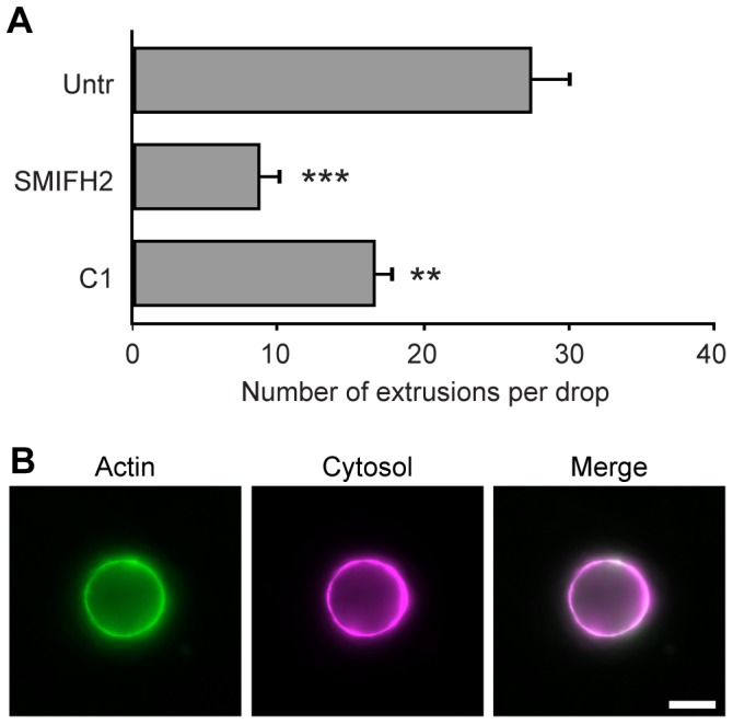
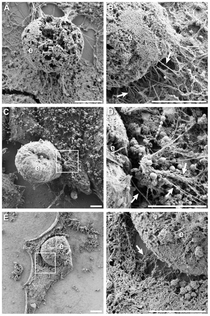
The ability to exit host cells at the end of their developmental growth is a critical step for the intracellular bacterium Chlamydia. One exit strategy, extrusion, is mediated by host signaling pathways involved with actin polymerization. Here, we show that actin is recruited to the chlamydial inclusion as a late event, occurring after 20 hours post-infection (hpi) and only within a subpopulation of cells. This event increases significantly in prevalence and extent from 20 to 68 hpi, and actin coats strongly correlated with extrusions. In contrast to what has been reported for other intracellular pathogens, actin nucleation on Chlamydia inclusions did not 'flash', but rather exhibited moderate depolymerization dynamics. By using small molecule agents to selectively disrupt host signaling pathways involved with actin nucleation, modulate actin polymerization dynamics and also to disable the synthesis and secretion of chlamydial proteins, we further show that host and bacterial proteins are required for actin coat formation. Transient disruption of either host or bacterial signaling pathways resulted in rapid loss of coats in all infected cells and a reduction in extrusion formation. Inhibition of Chlamydia type III secretion also resulted in rapid loss of actin association on inclusions, thus implicating chlamydial effector proteins(s) as being central factors for engaging with host actin nucleating factors, such as formins. In conclusion, our data illuminate the host and bacterial driven process by which a dense actin matrix is dynamically nucleated and maintained on the Chlamydia inclusion. This late stage event is not ubiquitous for all infected cells in a population, and escalates in prevalence and extent throughout the developmental cycle of Chlamydia, culminating with their exit from the host cell by extrusion. The initiation of actin recruitment by Chlamydia appears to be novel, and may serve as an upstream determinant of the extrusion mechanism.


Conflict of interest statement

Competing Interests: The authors have declared that no competing interests exist.

Figures

Figure 1
Figure 1. Assembly of actin coats on Chlamydia inclusions increases in strength and prevalence over the developmental cycle.
Cellular actin in HeLa cells was labeled with LifeAct-GFP (green) and cytosolic DsRed (magenta). Cells were infected with C. trachomatis and imaged live at 20 hpi (A,B), 44 hpi (C,D), 68 hpi (E,F) or uninfected (G). Examples of inclusions with actin association (A,C,E) and no recruitment (B,D,F) are provided. The example in (A) depicts actin clouds but not coat formation. Inclusions with actin recruitment (A,C,E) are marked with arrows. Scale bars = 20 µm. (H) Quantitation of inclusions with actin coats, at indicated times hpi. Error bars denote the SEM, >1200 inclusions were scored from a total of n≥5 experiments.
Figure 2
Figure 2. High resolution actin cytoskeleton ultrastructure in Chlamydia-infected cells by detergent extraction scanning electron microscopy.
(A) Uninfected cell processed without detergent extraction, showing membrane topology. (B) Uninfected cell with membranes removed and actin cytoskeleton preserved; cytoskeletal matrix was revealed. (C) Chlamydia infected cell at 20 hpi without detergent extraction, membranes were intact and appearance is indistinguishable from uninfected cells. (D) Chlamydia infected cell at 20 hpi with membranes extracted and actin cytoskeleton preserved; small inclusions and/or bacteria were difficult to identify at this time. (E) Chlamydia infected cell at 44 hpi with membranes intact; underlying large inclusion i is evident in this example, and plasma membrane on protruding surface was smoother with reduced microvilli density. (F) Chlamydia infected cell at 44 hpi with membranes removed and cytoskeleton preserved; in this example, little or no actin was recruited to the inclusion, which is readily visible as a spheroid structure of packed bacteria. (G) Detergent-extracted, Chlamydia infected cell at 44 hpi exhibiting significant actin polymerization on the inclusion; inclusion associated actin is continuous with actin matrix of the cell body. (H) Chlamydia infected cell at 68 hpi with membranes intact, showing large protrusion of underlying inclusion. (I) Detergent-extracted, Chlamydia infected cell at 68 hpi showing large inclusion densely wrapped with actin. Boxed region is enlarged in (J), and illustrates the fibrous actin connections between large inclusion, or possibly an extrusion, with the cytoskeletal matrix of the cell. Numerous actin bundles were observed, some are marked by arrows. (K) A ruptured Chlamydia infected cell at 68 hpi with membranes removed and actin structures preserved. Dense layers of actin on the inclusion were seen, and contents of inclusion are revealed to contain hundreds of individual bacteria. Boxed region is enlarged in (L) to highlight the network of actin filaments (arrows) effacing the inclusion surface and show individual bacteria (arrowheads). Scale bars = 5 µm.
Figure 3
Figure 3. Actin distribution in cells infected with C. trachomatis serovar D and C. muridarum.
HeLa cells transfected with LifeAct-GFP (green) and cytosolic DsRed (red) were infected with C. trachomatis serovar D (A) or C. muridarum (B) and imaged by live fluorescence microscopy at 20 hpi for C. trachomatis serovar D and 44 hpi for C. muridarum. Over 35 z-stacks were acquired at 0.5 µm intervals. Image stacks were additionally processed by deconvolution. Representative fields of cells for each time point are depicted. Asterisks (*) mark the locations where orthogonal planes in xz were taken. Arrows mark the inclusions. Scale bars = 20 µm.
Figure 4
Figure 4. Involvement of cytoskeletal signaling pathways in actin recruitment to the Chlamydia inclusion.
HeLa cells were infected with C. trachomatis and at 44 hpi (A) or 68 hpi (B) were treated for 4 h at 37°C with inhibitors targeting: actin polymerization (latrunculin B, jasplakinolide), Arp2/3 complex (CK-548, CK-666), formins (formin inhibitor SMIFH2), septins (FCF), microtubules (nocodazole), or left untreated. The numbers of inclusions with positive actin recruitment on their membranes were determined for each treatment. Error bars denote the SEM, >1000 inclusions were scored from a total of n = 4 experiments. * p<0.01, ** p<0.001.
Figure 5
Figure 5. Involvement of actin-associated and additional host signaling pathways in actin recruitment to the Chlamydia inclusion.
HeLa cells were infected with C. trachomatis and at 44 hpi (A) or 68 hpi (B) were treated for 4 h at 37°C with specific inhibitors against: N-WASP (wiskostatin), myosin II (blebbistatin), PI3K (LY294002), Rho GTPase (CT04), ROCK (Y27632), RhoA activator (LPA), Src family kinases (Src kinase inhibitor I), or left untreated. The numbers of inclusions with positive actin recruitment on their membranes were determined for each treatment. Error bars denote the SEM, >1000 inclusions were scored from a total of n = 4 experiments. * p<0.01, ** p<0.001.
Figure 6
Figure 6. Important role for Chlamydia proteins in mediating actin coat formation.
(A) HeLa cells expressing LifeAct-GFP and cytosolic DsRed were infected with C. trachomatis and at 44 hpi were treated for 4 h at 37°C with chloramphenicol, rifampicin, the type III secretion inhibitor C1, or left untreated. The numbers of inclusions with positive actin recruitment on their membranes were determined for each treatment. The effect of shorter treatments with chloramphenicol (B) or rifampicin (C) was determined by incubating infected cells with these compounds for times indicated. Error bars denote the SEM, >1000 inclusions were scored from a total of n = 3 experiments. ** p<0.001 *** p<0.0001.
Figure 7
Figure 7. Effect of chloramphenicol and rifampicin treatment on actin ultrastructure in Chlamydia infected cells.
(A) Chlamydia infected cell at 44 hpi with membranes removed and actin cytoskeleton preserved. Inclusion and cell were ruptured, thereby revealing actin structures on back face of the inclusion. (B) Enlargement of boxed region in (A), showing structural details of actin network behind the dissolved inclusion surface and individual bacteria. (C) Detergent extracted, Chlamydia infected cell treated with chloramphenicol for 4 h at 44 hpi. (D) Enlargement of boxed region in (C), illustrating a reduction in the number and density of actin filaments associated with the dissolved inclusion surface; in this example actin structures were in the foreground, where inclusion membrane was previously located. (E) Detergent extracted, Chlamydia infected cell treated with rifampicin for 4 h at 44 hpi. (F) Enlargement of boxed region in (E), showing significant reduction in number and density of actin filaments on the dissolved inclusion membrane, behind where the inclusion membrane would have been. Inclusions are marked with i, some examples of bacteria (arrowheads) and actin filaments (arrows) are also labeled. Scale bars = 5 µm.
Figure 8
Figure 8. Actin coats lead to formation of Chlamydia extrusions.
(A) Reduction in the number of endogenous extrusions released by cells after 4 h treatment with formin (SMIFH2) or type III secretion (C1) inhibitors, or left untreated. (B) Representative extrusion containing C. trachomatis from HeLa cells expressing LifeAct-GFP (green) and cytosolic DsRed (magenta), illustrating extensive actin coating on underlying inclusion. Error bars denote the SEM, >1000 extrusions were scored from a total of n = 3 experiments. ** p<0.001 *** p<0.0001. Scale bar = 10 µm.
Figure 9
Figure 9. Ultrastructure of actin structures associated with Chlamydia extrusions.
Images depict late stage inclusions and extrusions from cells infected with C. trachomatis at 68 hpi; in all instances specimens were processed by detergent extraction to reveal actin cytoskeleton. (A) A fractured extrusion, thrust vertically from the cell body. (B) Actin-coated extrusion detaching from the cytoskeleton of the cell body, some actin filaments remained bridging the extrusion and cellular actin matrix. (C) Extrusion with rich actin coat. (D) Enlargement of boxed region in (C), showing stretched actin bundles and partial detachment from cell body cytoskeleton. (E) Cell with large inclusion/extrusion. (F) Enlargement of boxed region in (E), showing zone of severed filaments between the extrusion/inclusion and actin network of the cell body. Extrusions are marked with e, some examples of actin filaments (arrows) are also labeled. Scale bars = 5 µm.

References

    1. Stamm WE (1999) Chlamydia trachomatis infections: progress and problems. J Infect Dis 179 Suppl 2: S380–S383. - PubMed
    1. Schachter J (1999) Infection and disease epidemiology. In: Stephens RS, editor. Chlamydia: Intracellular Biology, Pathogenesis, and Immunity. American Society for Microbiology Press. pp. 139–169.
    1. Mathers C, Fat DM (2008) The global burden of disease: 2004 update. Geneva: World Health Organization.
    1. Darville T, Hiltke TJ (2010) Pathogenesis of genital tract disease due to Chlamydia trachomatis . J Infect Dis 201 Suppl 2: S114–S125. - PMC - PubMed
    1. Haggerty CL, Gottlieb SL, Taylor BD, Low N, Xu F, et al. (2010) Risk of sequelae after Chlamydia trachomatis genital infection in women. J Infect Dis 201 Suppl 2: S134–S155. - PubMed

Publication types

MeSH terms