. 2012;7(10):e47091.
doi: 10.1371/journal.pone.0047091. Epub 2012 Oct 11.

DNA polymerase α (swi7) and the flap endonuclease Fen1 (rad2) act together in the S-phase alkylation damage response in S. pombe

Affiliations

DNA polymerase α (swi7) and the flap endonuclease Fen1 (rad2) act together in the S-phase alkylation damage response in S. pombe

Milana Koulintchenko et al. PLoS One. 2012.

Abstract

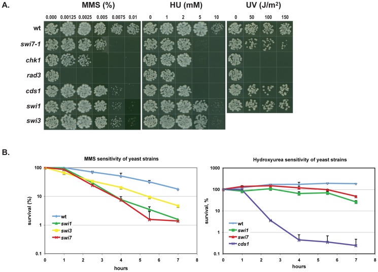
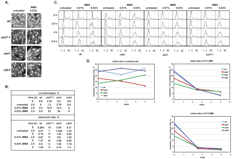
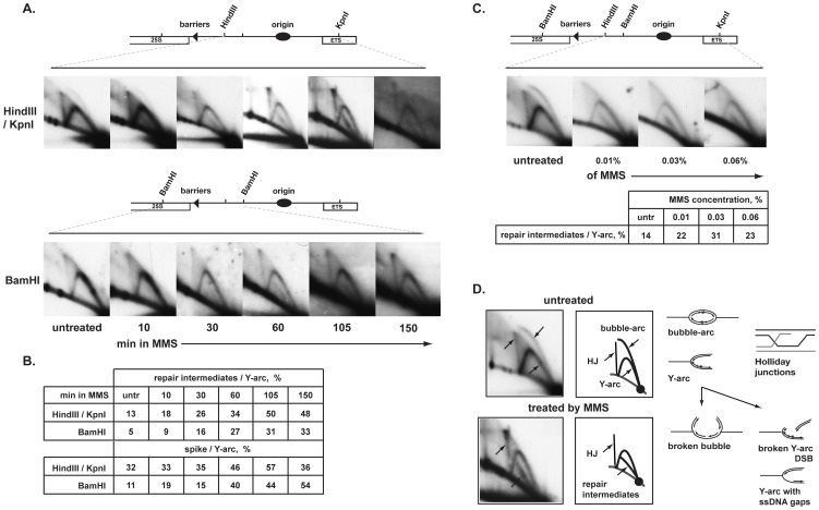
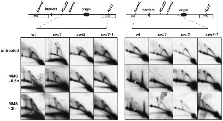
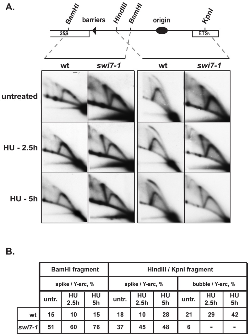
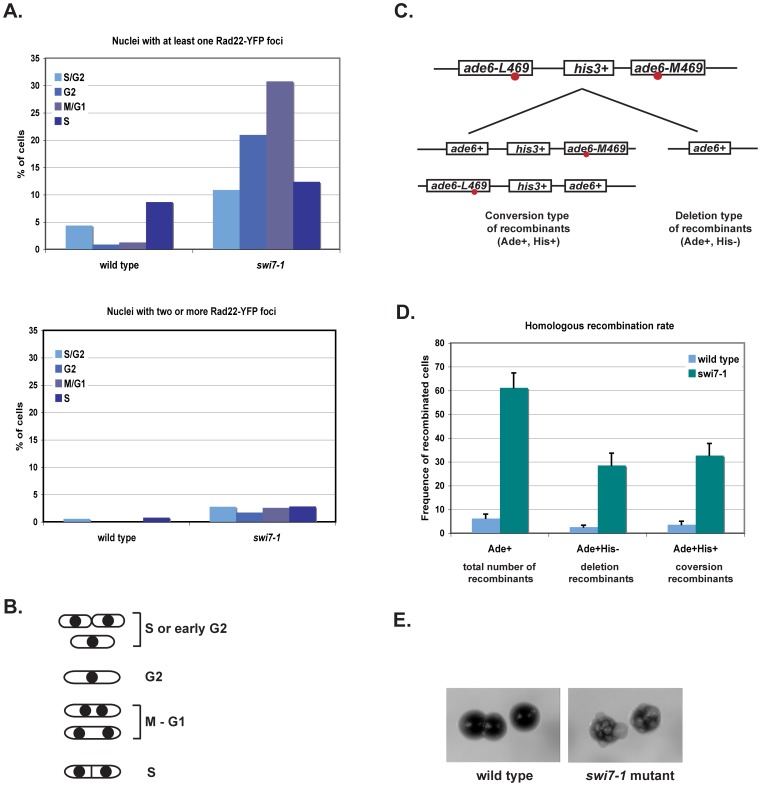
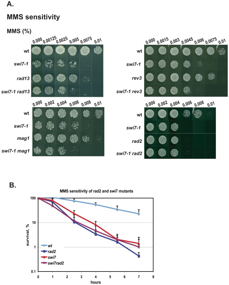
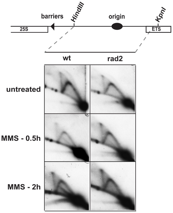
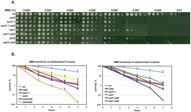
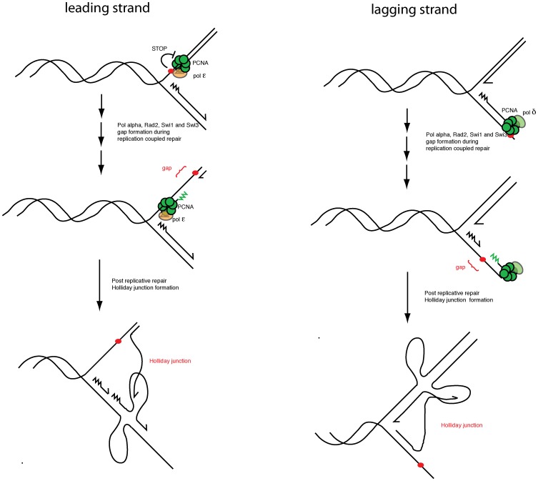
Polymerase α is an essential enzyme mainly mediating Okazaki fragment synthesis during lagging strand replication. A specific point mutation in Schizosaccharomyces pombe polymerase α named swi7-1, abolishes imprinting required for mating-type switching. Here we investigate whether this mutation confers any genome-wide defects. We show that the swi7-1 mutation renders cells hypersensitive to the DNA damaging agents methyl methansulfonate (MMS), hydroxyurea (HU) and UV and incapacitates activation of the intra-S checkpoint in response to DNA damage. In addition we show that, in the swi7-1 background, cells are characterized by an elevated level of repair foci and recombination, indicative of increased genetic instability. Furthermore, we detect novel Swi1-, -Swi3- and Pol α- dependent alkylation damage repair intermediates with mobility on 2D-gel that suggests presence of single-stranded regions. Genetic interaction studies showed that the flap endonuclease Fen1 works in the same pathway as Pol α in terms of alkylation damage response. Fen1 was also required for formation of alkylation- damage specific repair intermediates. We propose a model to explain how Pol α, Swi1, Swi3 and Fen1 might act together to detect and repair alkylation damage during S-phase.


Conflict of interest statement

Competing Interests: The authors have declared that no competing interests exist.

Figures

Figure 1
Figure 1. S. pombe swi7-1 mutant is sensitive to DNA damaging agents.
A. Drop-assay analysis. Drops containing approximately 100 cells from mutant and wild type strains were placed on medium, containing methyl methanesulfonate (MMS) or hydrohyurea (HU) at concentrations given, or were exposed to UV at the given intensities. The plates were incubated at 33° until colonies were formed. B. Survival of yeast cells exposed to MMS or HU. Logarithmically growing cultures were treated with either 0.03% MMS or 12 mM of HU. Samples were taken at indicated time points, and dilution series of cells for each culture were plated on YEA media and incubated at 33°C until colonies were formed. Strains shown in panel A and B are wt (JZ60), swi7-1 (JZ468), chk1 (JZ473), rad3 (JZ474), cds1 (JZ475), swi1 (E111), swi3 (E146).
Figure 2
Figure 2. swi7-1 mutant might have a role in S-phase but not in mitotic checkpoint.
A. Images of the wt (JZ60), swi1 (E111), swi7-1 (JZ468) and cds1 (JZ475) cells stained with DAPI and Calcofluor were taken at 2.5 h and 5 h time point for both the untreated and MMS treated cultures. Gray arrows indicate cells with the cut phenotype. Five hundred or more cells were analyzed for each strain. B. Quantification of the percentage of cells displaying the “cut” phenotype and abnormal nuclei for the indicated strains, for both treated and untreated cultures. C. The swi7-1 mutant shows defect in S-phase progression in response to MMS damage. Asynchronous cultures of wt (JZ60), cds1 (JZ475), swi1 (E111) and swi7-1 (JZ468) strains were incubated in absence or presence of 0.01% or 0.03% MMS. Cell samples were taken for flow cytometry analysis at indicated time points. D. Mitosis index of wt, cds1, swi1 and swi7-1 cells. Changes in the percentage of binucleated and septated cells were determined as a function of time without and after the addition of 0.01% or 0.03% MMS.
Figure 3
Figure 3. Analysis of replication/repair intermediates purified from wild type MMS treated cultures.
A. 2D-gel analysis of replication/repair intermediates from wild type cells (JZ60) during a time course of incubation with 0.03% MMS. Times of MMS incubation are given below the panels. Two different restriction fragments from the rDNA region were analysed. The position of restriction sites and genetic elements of the rDNA region is outlined in the line drawing above the panels. B. Quantification of the ratio (%) of the intensity of repair intermediates and spike intermediates versus the intensity of the Y-arc for the panels shown in A. C. 2D-gel analysis of replication/repair intermediates from wild type cells (JZ60) treated for 1 h with increasing amount of MMS. D. Interpretation of replication/repair intermediates profiles from untreated or MMS-treated wild type cell cultures.
Figure 4
Figure 4. Analysis of replication/repair intermediates purified from wild type, swi1, swi3 and swi7-1 mutant yeast cells treated by MMS.
2D-gel analysis of replication/repair intermediates from wild type (JZ60), swi1 (E111), swi3 (E146) and swi7-1 (JZ468) cells treated with MMS. The yeast genomic DNA was purified from cell cultures incubated without or in presence of 0.03% MMS for 0.5 and 2 h. The repair intermediates in the wild type panel are indicated with the gray arrows.
Figure 5
Figure 5. Analysis of replication/recombination intermediates purified from hydroxyurea treated wild type and swi7-1 mutant yeast cells.
A. 2D-gel analysis of replication/recombination intermediates from wild type cells (JZ60) and swi7 (JZ468) during a time course of incubation with 12 mM HU. Times of HU incubation are given to the left of the panels. Two different restriction fragments from the rDNA region were analysed. The position of restriction sites and genetic elements of the rDNA region is outlined in the line drawing above the panels. B. Calculation in ratios (%) of bubble arcs (replication intermediates) and spikes (recombination intermediates) versus Y-arc intermediates.
Figure 6
Figure 6. swi7-1 mutation causes genetic instability.
A. Quantification of Rad22-YFP foci in swi7-1 mutant and wild-type cells. Wild type (ENY670) and swi7 (MK140) mutant cells expressing Rad22-YFP were grown at room temperature until the mid-log phase. A small aliquot of cell culture was used for analysis by microscopy. About six hundred cells were analysed for each strain. B. Line drawing displaying the different cell morphologies through the cell cycle in fission yeast. C. Assay for quantification of recombination frequencies. Schematic drawing of the intrachromosomal recombination substrate and of the possible outcomes of recombination events. The red circles indicate the approximate positions of the mutations. D. Quantification of the recombination rate in wild-type (JZ518) and swi7 (MK226) mutant strains. The histograms displays the recombination rates for the substrate shown in panel E. Rates of total number, conversion- and deletion-type recombination events are shown. The given values are the means of recombination frequencies for independent colonies. E. Visualization of recombination rates by growth on media containing limited adenine. swi7 mutant displays increased sectoring on YE medium due to recombination at the direct repeats of ade6 in the substrate. Cells with ade6 mutations in the intact substrate turn red on YE medium due to accumulation of a slightly toxic red pigment. Cells with the recombined wild type ade6+ gene do not accumulate the pigment and are white (sectors in swi7 colony).
Figure 7
Figure 7. Genetic interactions of S. pombe swi7-1 mutant in the response to DNA damage.
A, B and C. Analysis of sensitivities of single and double mutant strains to increasing concentrations of MMS (A). Approximately 100 cells from mutant and wild type strains were plated on the medium, containing methyl methanesulfonate (MMS) or hydroxyurea (HU) at concentrations given above the columns, or were exposed to UV irradiation at the indicated intensities. The plates were incubated at 33°C until colonies were formed. Strain names are as follows wt (JZ60), swi7-1 (JZ487 or JZ468), rad13 (SV66), mag1 (AM006), rev3 (SV54), rad2 (SV59), swi7rad13 (MK130), swi7mag1 (MK175), swi7-1 rev3 (MK190), swi7-1 rad2 (MK125). B. Survival of wt, swi7-1, rad2 and swi7-1 rad2 strains exposed to MMS. Logarithmically growing cultures were incubated in the presence or absence of 0.03% MMS. Samples were taken at indicated time points, and dilution series of cells for each culture were plated on YEA media and incubated at 33°C until colonies formed. The survival is shown as the relative percentage compared to the values obtained using the untreated starting cultures.
Figure 8
Figure 8. The rad2 mutation abolishes the presence of the novel alkylation damage repair intermediates.
2D-gel analysis of untreated and MMS treated wild-type and rad2 strains. The HindIII/KpnI fragment of the rDNA was analyzed in the experiment, see Figure 2 line drawing. Cells were treated with 0.03% MMS for 0.5 and 2 h as shown.
Figure 9
Figure 9. Swi1 and Swi3 possess additional roles in the alkylation damage response.
Analysis of sensitivities of wild-type (JZ60), single swi7-1 (JZ468), rad2 (SV59), swi1 (E111), swi3 (E146) and double mutant swi7-1 rad2 (MK125), swi1 rad13 (MK239), swi3 rad2 (MK243) strains to increasing concentrations of MMS or hydroxyurea (HU). Approximately 100 cells from mutant and wild type strains were plated on the medium, containing methyl methanesulfonate (MMS) or hydroxyurea (HU) at concentrations given above the columns. The plates were incubated at 33°C until colonies were formed. B. Survival curves for yeast cells exposed to MMS. Logarithmically growing cultures were treated with 0.03% MMS, samples were taken at indicated time points, and dilution series of cells for each culture were plated on YEA media and incubated at 33°C until colonies formed. Strain names are given in A.
Figure 10
Figure 10. Model figure.
Alkylation damage on the leading (left half) and lagging (right half) strand template is repaired by a rad2, swi1, swi3 and swi7-1 dependent pathway leading to the formation of single-stranded regions behind but in close proximity to the progressing fork. These intermediates are detected by 2D-gel analysis below the y-arc for the wild-type strain (Figure 2). The single-stranded regions lead to Holliday junction (HJ) formation either by formation of double-stranded breaks or by fork regression.

Similar articles

Cited by

References

    1. Wang TS, Wong SW, Korn D (1989) Human DNA polymerase alpha: predicted functional domains and relationships with viral DNA polymerases. Faseb J 3: 14–21. - PubMed
    1. Singh J, Klar AJ (1993) DNA polymerase-alpha is essential for mating-type switching in fission yeast. Nature 361: 271–273. - PubMed
    1. Dalgaard JZ, Klar AJ (1999) Orientation of DNA replication establishes mating-type switching pattern in S. pombe . Nature 400: 181–184. - PubMed
    1. Kelly M, Burke J, Smith M, Klar A, Beach D (1988) Four mating-type genes control sexual differentiation in the fission yeast. Embo J 7: 1537–1547. - PMC - PubMed
    1. Holmes AM, Kaykov A, Arcangioli B (2005) Molecular and cellular dissection of mating-type switching steps in Schizosaccharomyces pombe . Mol Cell Biol 25: 303–311. - PMC - PubMed

Publication types

MeSH terms