. 2012;7(10):e47162.
doi: 10.1371/journal.pone.0047162. Epub 2012 Oct 11.

Chromatin modification by PSC occurs at one PSC per nucleosome and does not require the acidic patch of histone H2A

Affiliations

Chromatin modification by PSC occurs at one PSC per nucleosome and does not require the acidic patch of histone H2A

Stanley M Lo et al. PLoS One. 2012.

Abstract

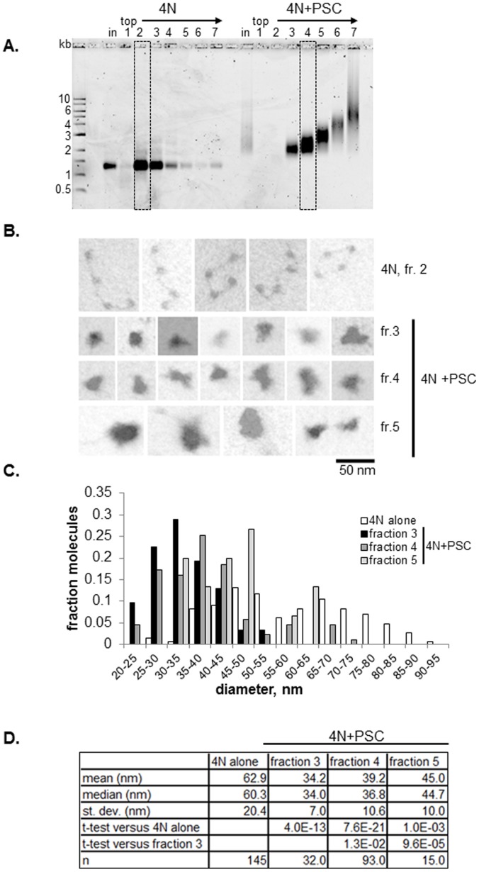
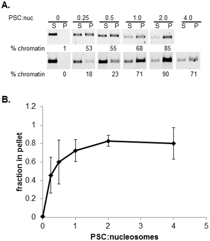
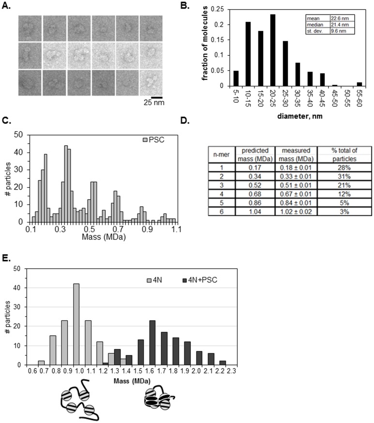
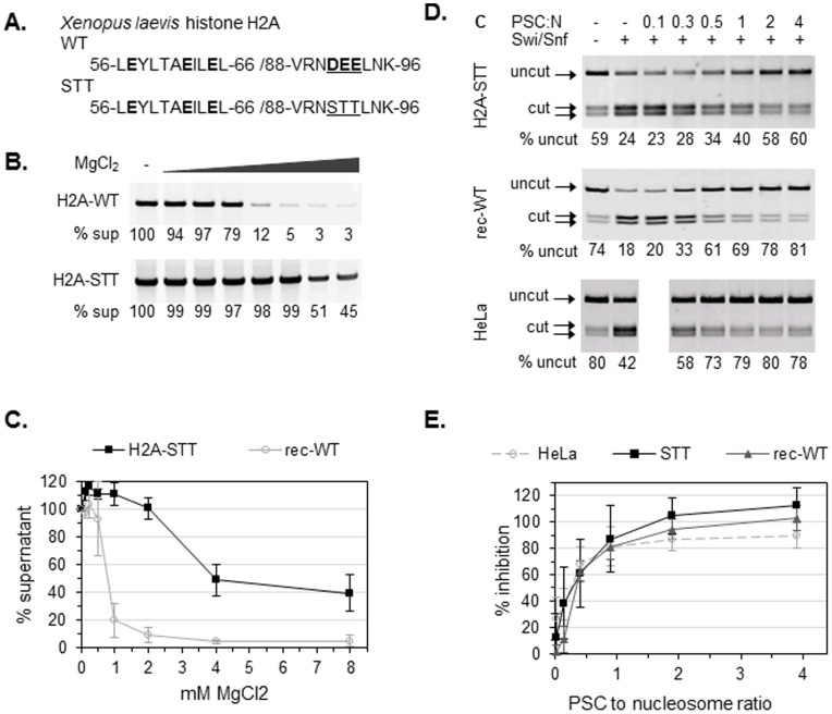
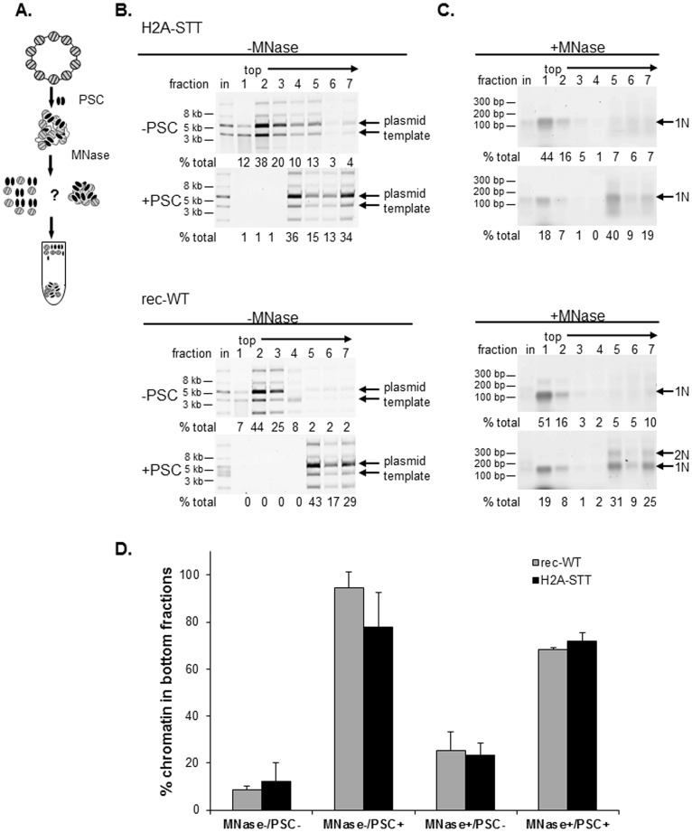
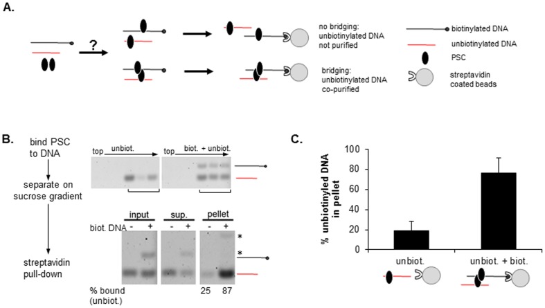
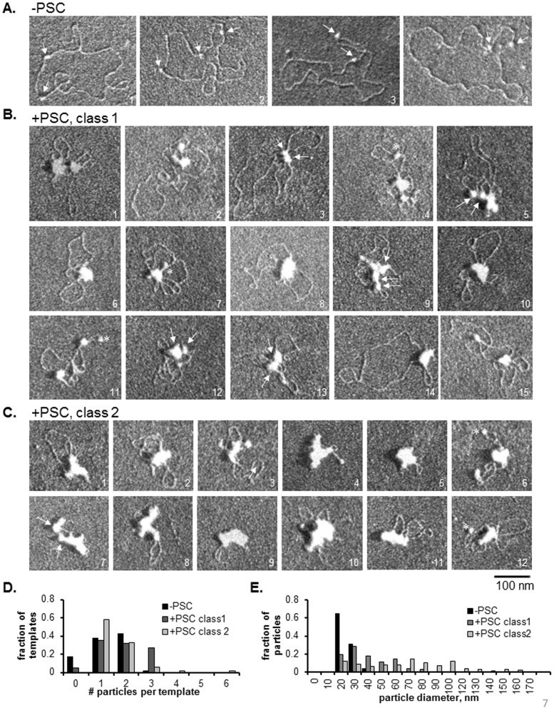
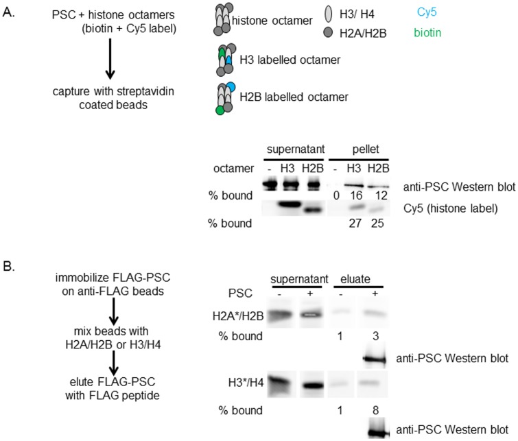
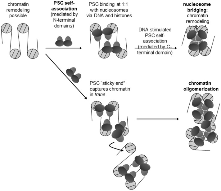
Chromatin architecture is regulated through both enzymatic and non-enzymatic activities. For example, the Polycomb Group (PcG) proteins maintain developmental gene silencing using an array of chromatin-based mechanisms. The essential Drosophila PcG protein, Posterior Sex Combs (PSC), compacts chromatin and inhibits chromatin remodeling and transcription through a non-enzymatic mechanism involving nucleosome bridging. Nucleosome bridging is achieved through a combination of nucleosome binding and self-interaction. Precisely how PSC interacts with chromatin to bridge nucleosomes is not known and is the subject of this work. We determine the stoichiometry of PSC-chromatin interactions in compact chromatin (in which nucleosomes are bridged) using Scanning Transmission Electron Microscopy (STEM). We find that full compaction occurs with one PSC per nucleosome. In addition to compacting chromatin, we show that PSC oligomerizes nucleosome arrays. PSC-mediated oligomerization of chromatin occurs at similar stoichiometry as compaction suggesting it may also involve nucleosome bridging. Interactions between the tail of histone H4 and the acidic patch of histone H2A are important for chromatin folding and oligomerization, and several chromatin proteins bind the histone H2A acidic patch. However, mutation of the acidic patch of histone H2A does not affect PSC's ability to inhibit chromatin remodeling or bridge nucleosomes. In fact, PSC does not require nucleosomes for bridging activity but can bridge naked DNA segments. PSC clusters nucleosomes on sparsely assembled templates, suggesting it interacts preferentially with nucleosomes over bare DNA. This may be due to the ability of PSC to bind free histones. Our data are consistent with a model in which each PSC binds a nucleosome and at least one other PSC to directly bridge nucleosomes and compact chromatin, but also suggest that naked DNA can be included in compacted structures. We discuss how our data highlight the diversity of mechanisms used to modify chromatin architecture.


Conflict of interest statement

Competing Interests: The authors have declared that no competing interests exist.

Figures

Figure 1
Figure 1. PSC compacts and oligomerizes 4-nucleosome arrays.
(a) Representative glycerol gradient purification of nucleosomal templates with and without PSC for EM and STEM. Boxes indicate fractions selected for analysis. (b) Representative EM images from indicated fractions. Photographs were taken in dark field and are inverted to enhance contrast. (c) Distribution of maximum diameters of particles determined from micrographs like those in (b). Note that less than 10% of the 4N alone arrays have diameters larger than 95 nm and are not shown on the graph. (d) Summary of diameter measurements. Note that all fractions of 4N arrays incubated with PSC were significantly smaller than 4N arrays alone, and fractions 4 and 5 are different from 3. Table shows p-values for student’s t-test (unpaired, assuming equal variance in samples).
Figure 2
Figure 2. Centrifugation assay demonstrates oligomerization of 12-nucleosome arrays by PSC.
(a) Nucleosomal arrays were mixed with PSC at the indicated ratios and reactions pelleted by centrifugation in a microfuge. Proteinase K digested samples were separated by agarose gel electrophoresis, stained with SYBR gold and quantified. Two different examples of the assay are shown; considerable variability was observed in this assay although the trend was constant. (b) Summary of PSC-induced oligomerization. Error bars are standard deviation in this and all other figures. n = 5.
Figure 3
Figure 3. PSC compacts chromatin at a ratio of 1∶1 with nucleosomes.
(a) Representative EM images of negatively stained PSC. (b) Distribution of diameters of negatively stained PSC (n = 235). (c) Mass distributions of STEM analysis of PSC alone (n = 515). (d) Summary of measured masses of PSC. (e) Mass distributions of STEM analyses. Measured masses for 4N and 4N + PSC are 0.97±0.01 MDa (n = 130) and 1.67±0.03 MDa (n = 86) respectively.
Figure 4
Figure 4. PSC does not require histone modifications or the acidic patch of H2A to inhibit chromatin remodeling.
(a) Amino acid sequences for the wild-type H2A acidic patch (WT) and uncharged mutant (STT). Acidic residues are highlighted, and mutated residues are underlined. (b) MgCl2 dependent oligomerization of wild type and H2A-STT containing chromatin. Chromatin was incubated with the indicated concentrations of MgCl2 and centrifuged in a microfuge. Supernatants were electrophoresed on agarose gels and stained with SYBR gold; the % of the template remaining in the supernatant was determined by comparison with the 0 mM MgCl2 control. (c) Summary of chromatin oligomerization assays. (d) Restriction enzyme accessibility (REA) assays on chromatin templates with 12 nucleosomes. The chromatin template contains a unique restriction site (HhaI) that is normally occluded by nucleosomes but is exposed upon Swi/Snf-mediated chromatin remodeling. The first two lanes are negative and positive controls (with or without Swi/Snf, no PSC) demonstrating that the HhaI site becomes more accessible in the presence of Swi/Snf. (e) Summary of REA assay on chromatin templates assembled with rec-WT and H2A-STT histones. Percent inhibition is calculated as formula image.
Figure 5
Figure 5. Bridging of nucleosomes by PSC does not require the acidic patch on histone H2A.
(a) Schematic diagram of nucleosome bridging assay. (b) Representative control reactions for nucleosome bridging (mock MNase digested) showing that PSC binds both rec-WT and H2A-STT chromatin. Arrows point to the main plasmid forms (nicked and supercoiled); the array of minor isoforms observed here is atypical but the isoforms behave similarly. (c) Representative MNase digested nucleosome bridging reactions demonstrating that PSC bridges both rec-WT and H2A-STT nucleosomes. (d) Summary of nucleosome bridging assays on chromatin templates assembled with recombinant wild-type (rec-WT) and H2A acidic patch mutant histones (H2A-STT). Values from fractions 5–7 (bottom fractions) of sucrose gradients were summed and plotted.
Figure 6
Figure 6. PSC can bridge bare DNA.
(a) Schematic diagram of DNA bridging assay. (b) Representative analysis of bridging of naked DNA by PSC. Top panels show sucrose gradient fractions that were pooled for streptavidin pull-down. Bottom panels show streptavidin pull-down results. The per cent bound refers to how much of the unbiotinylated fragment is present in the pellet as a fraction of the total (pellet + supernatant). Asterisks indicate position of biotinylated fragment; note that in pellet fractions, biotinylated fragment is incompletely recovered by Proteinase K treatment of streptavidin coated beads, and migrates slowly likely due to bound streptavidin. (c) Summary of streptavidin pull-down experiments. Graphs show average per cent of the unbiotinylated fragment associated with streptavidin beads.
Figure 7
Figure 7. PSC clusters nucleosomes and DNA on sparsely assembled plasmids.
(a) Representative EM images of plasmids with two 601 nucleosome positioning sequences assembled at low ratio of histones to DNA. Note that fully assembled plasmids would contain 17 nucleosomes and 601 sequences are separated by 385 base pairs. Plasmids were assembled in the presence of E.coli Topoisomerse I so that plasmids are relaxed. Arrows point to nucleosomes. Note that template 2 was the only observed example of more than 2 nucleosomes on the plasmid (3), out of the 103 molecules that were analyzed (Table 1). (b) Sparsely assembled plasmids with PSC. Class 1 molecules (see text) are more extended, and likely have fewer copies of PSC bound than Class 2 molecules (c), which are highly compacted. Arrows point to particles (likely to be PSC bound nucleosomes) that have come together and may represent the bridged configuration. Note that in some cases, more than one template may be clustered (such as molecule 3). Asterisks indicate particles that may be unbound nucleosomes (based on their size), although they could also be bound PSC. (c) Sparsely assembled plasmids with PSC with Class 2 configurations. Note that the molecules represent a series between the most extended Class 1 molecules and the most highly compacted Class 2 molecules. (d) Summary of the number of particles per template. The finding that Class 1 molecules frequently have more than 2 particles indicates that PSC must bind to naked DNA (as well as nucleosomes) on some templates. See Table 1 for summary of measurements from this and a similar experiment.
Figure 8
Figure 8. PSC can bind histone proteins.
(a) Representative assay of PSC binding to histone octamers. PSC was mixed with histone octamers that contain one biotinylated and one fluorescent copy of either H3 (H3 labeled octamers) or H2B (H2B labeled octamers) (see Methods for detailed description). Mixtures were incubated with streptavidin coated beads and the amount of captured PSC determined by Western blotting. Fluorescence (Cy5) was used to monitor octamer capture. Similar results were observed in two additional assays. (b) Representative assay of PSC binding to H2A/H2B dimers or H3/H4 tetramers. Dimers and tetramers were fluorophore labeled on the indicated (asterisk) subunit. High levels of background binding to beads was observed for both dimers and tetramers, as shown, but in each of three assays, more H2A/H2B and H3/H4 were eluted from Flag beads that have immobilized PSC than control beads with no immobilized protein. PSC in the elution is detected by Western blotting.
Figure 9
Figure 9. Model for nucleosome bridging and chromatin oligomerization by PSC.
See discussion for details.

Similar articles

Cited by

References

    1. McBryant SJ, Adams VH, Hansen JC (2006) Chromatin architectural proteins. Chromosome Res 14(1): 39–51. - PubMed
    1. McBryant SJ, Krause C, Woodcock CL, Hansen JC (2008) The silent information regulator 3 protein, SIR3p, binds to chromatin fibers and assembles a hypercondensed chromatin architecture in the presence of salt. Mol Cell Biol 28(11): 3563–3572. - PMC - PubMed
    1. Ghosh RP, Horowitz-Scherer RA, Nikitina T, Shlyakhtenko LS, Woodcock CL (2010) MeCP2 binds cooperatively to its substrate and competes with histone H1 for chromatin binding sites. Mol Cell Biol 30(19): 4656–4670. - PMC - PubMed
    1. Canzio D, Chang EY, Shankar S, Kuchenbecker KM, Simon MD, et al. (2011) Chromodomain-mediated oligomerization of HP1 suggests a nucleosome-bridging mechanism for heterochromatin assembly. Mol Cell 41(1): 67–81. - PMC - PubMed
    1. Lewis EB (1978) A gene complex controlling segmentation in Drosophila. Nature 276: 565–570. - PubMed

Publication types

MeSH terms

LinkOut - more resources