Case Reports
. 2012 Jul;27(4):319-22.
doi: 10.5001/omj.2012.79.

Anomalous systemic artery to a normal lung: a rare cause of hemoptysis in adults

Affiliations
Case Reports

Anomalous systemic artery to a normal lung: a rare cause of hemoptysis in adults

Ashu Seith Bhalla et al. Oman Med J. 2012 Jul.

Abstract

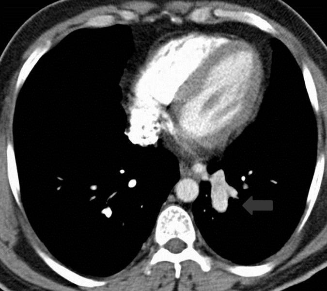
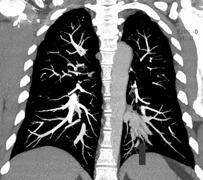
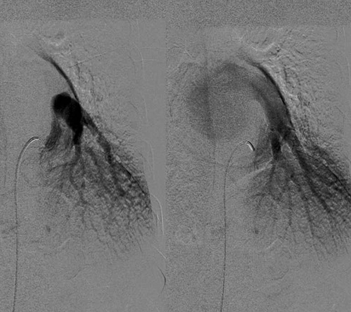
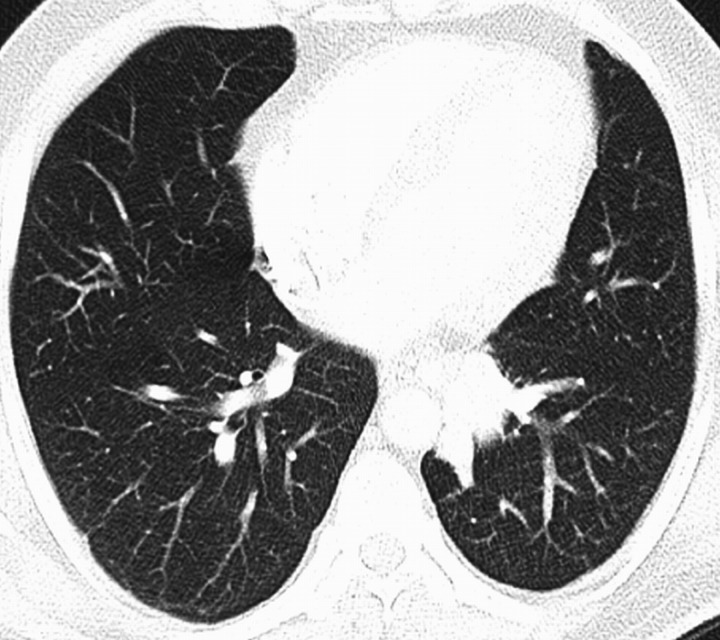
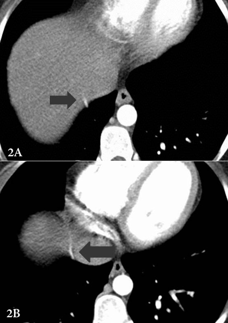
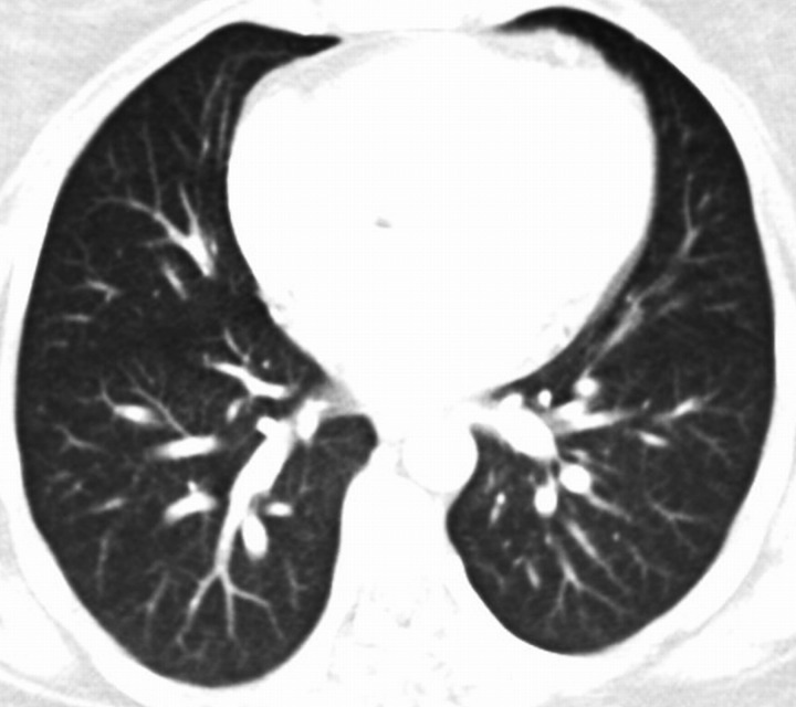
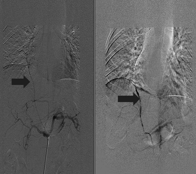
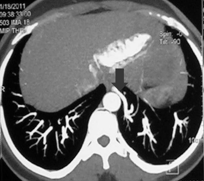
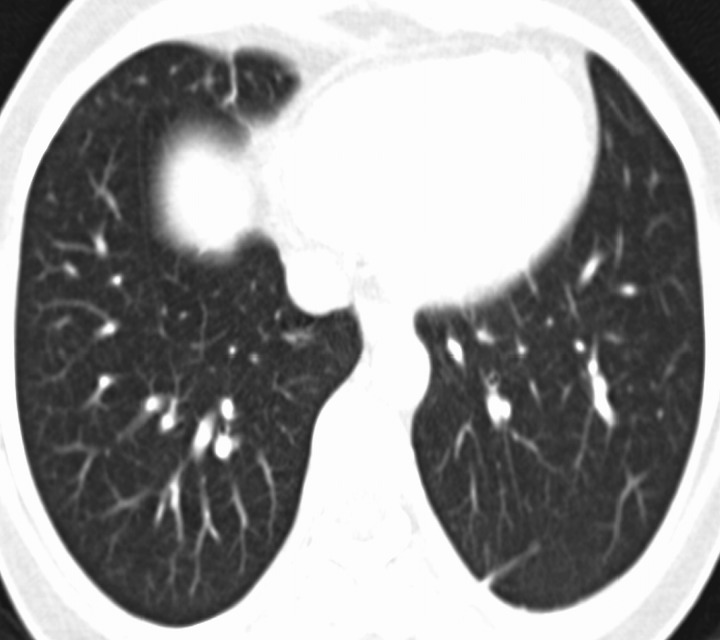
Bronchopulmonary sequestration represents a spectrum of abnormalities. One of these abnormalities is an aberrant systemic arterial supply to a normal lung with no bronchial sequestration. These lesions were originally classified by Pryce as type 1. Most of these patients are asymptomatic but with time, many patients develop localized pulmonary hypertension, hemoptysis, and eventually high output cardiac failure. Multidetector computed tomography (MDCT) plays an important role in the diagnosis and planning of definitive treatment by identifying the origin and course of the aberrant artery. Definitive treatment can be surgical (lobectomy or segmentectomy) or endovascular.

Keywords: Hemoptysis; Pryce type 1; Sequestration.


Figures

Figure 1a
Figure 1a
A scout image from of the chest CT shows ill-defined retrocardiac opacity (arrow). This retrocardiac opacity correlates with the aberrant artery as shown in Fig. 1d.
Figure 1b
Figure 1b
An axial CT section from CTPA in mediastinal window settings at the level of inferior pulmonary veins shows a nipple like structure (arrow) with enhancement similar to that of descending thoracic aorta at the mediastinal edge of the left basal lung segments, which on subsequent figures is shown to be an aberrant artery arising from the descending thoracic aorta. This pattern of origin of the aberrant artery from descending thoracic aorta and supply to the basal segment of left lower lobe represents the most common pattern.
Figure 1d
Figure 1d
Coronal reformatted MIP image from CTPA shows that the basal segments of the left lung receive their arterial supply from descending thoracic aorta via an aberrant artery (arrow).
Figure 1e
Figure 1e
DSA images in arterial and venous phases confirm the origin of the aberrant artery, in addition to the normal pulmonary arterial supply, to the left lower lobe from the lower descending thoracic aorta and venous drainage into the left atrium via inferior pulmonary vein. This figure again highlights the most common pattern of origin of the aberrant artery and the venous drainage of the sequestrated segment. Patient subsequently underwent surgery.
Figure 1c
Figure 1c
The corresponding axial section in lung window setting shows normal lung parenchyma.
Figures 2a & 2b
Figures 2a & 2b
Serial axial CT sections in mediastinal window setting show an aberrant vessel coursing along the diaphragm upwards suggesting an infra-diaphragmatic origin (arrow). However the exact origin of this vessel and lung segment which it supplies is not apparent on these two axial sections; this is well demonstrated on the DSA images shown subsequently.
Figure 2c
Figure 2c
An axial CT section in lung window setting shows no significant pulmonary parenchymal abnormality.
Figure 2d
Figure 2d
Pre-embolization and post-embolization DSA images.
Figure 3a
Figure 3a
An axial MIP image from CTPA reveals an aberrant artery arising from the lower part of the descending thoracic aorta and coursing towards the posterior basal segment of the left lower lobe with paucity of pulmonary vessels in this lung segment (arrow).
Figure 3b
Figure 3b
The corresponding lung window axial section image demonstrates paucity of vessels in the basal segments of the left lower lobe. Apart from this, the lung parenchyma is normal.

References

    1. Pryce DM. Lower accessory pulmonary artery with intralobar sequestration of lung; a report of seven cases. J Pathol Bacteriol 1946. Jul;58(3):457-467 10.1002/path.1700580316 - DOI - PubMed
    1. Irodi A, Cherian R, Keshava SN, James P. Dual arterial supply to normal lung: within the sequestration spectrum. Br J Radiol 2010. May;83(989):e86-e89 10.1259/bjr/30458107 - DOI - PMC - PubMed
    1. Sade RM, Clouse M, Ellis FH., Jr The spectrum of pulmonary sequestration. Ann Thorac Surg 1974. Dec;18(6):644-658 10.1016/S0003-4975(10)64417-7 - DOI - PubMed
    1. Ellis K. Fleischner lecture. Developmental abnormalities in the systemic blood supply to the lungs. AJR Am J Roentgenol 1991. Apr;156(4):669-679 - PubMed
    1. Kurosaki Y, Kurosaki A, Irimoto M, Kuramoto K, Itai Y. Systemic arterial supply to normal basal segments of left lower lobe: CT findings. J Comput Assist Tomogr 1993. Nov-Dec;17(6):857-861 10.1097/00004728-199311000-00004 - DOI - PubMed

Publication types

LinkOut - more resources