Review
. 2013 Jan 24:8:105-37.
doi: 10.1146/annurev-pathol-020712-163918. Epub 2012 Oct 31.

Autophagy as a stress-response and quality-control mechanism: implications for cell injury and human disease

Affiliations
Review

Autophagy as a stress-response and quality-control mechanism: implications for cell injury and human disease

Lyndsay Murrow et al. Annu Rev Pathol. .

Abstract

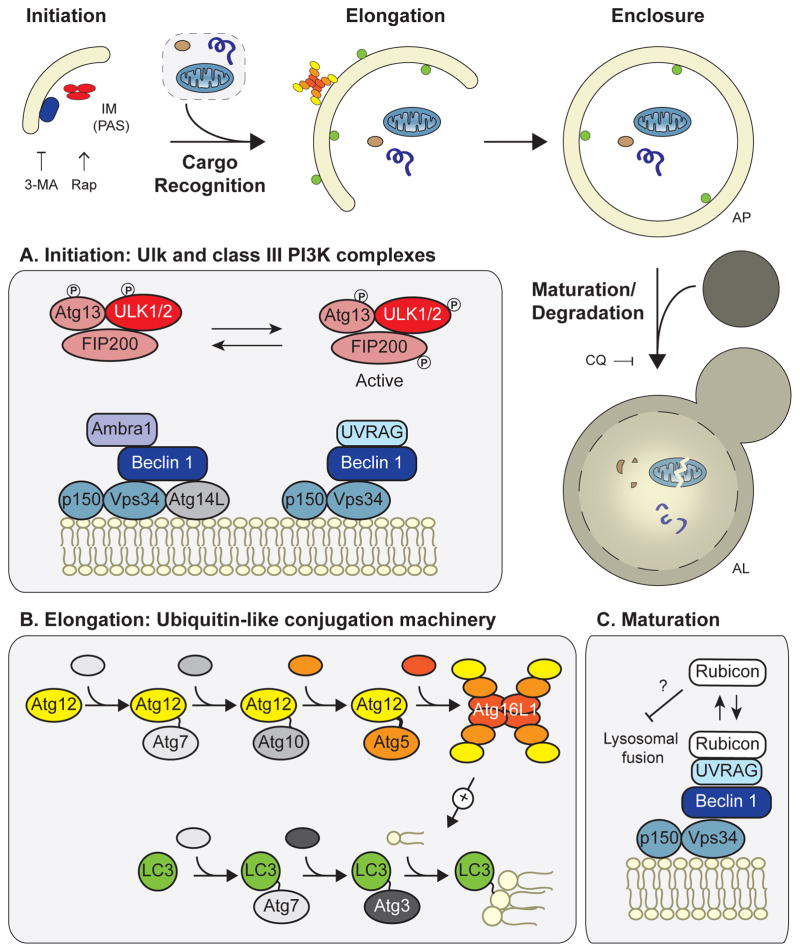
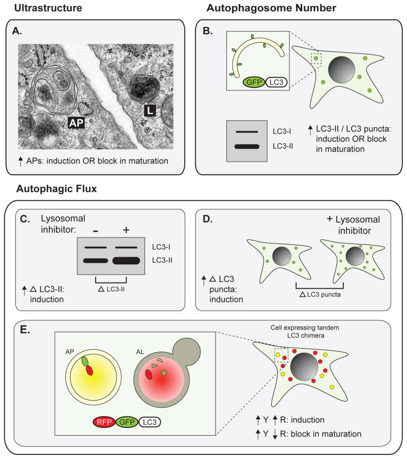
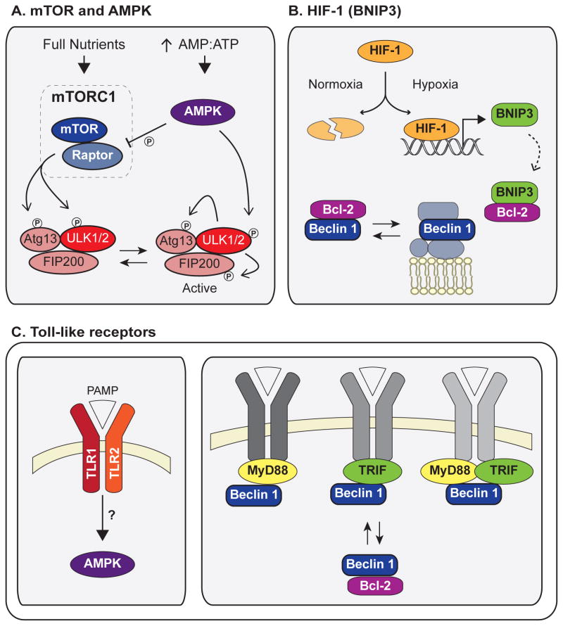
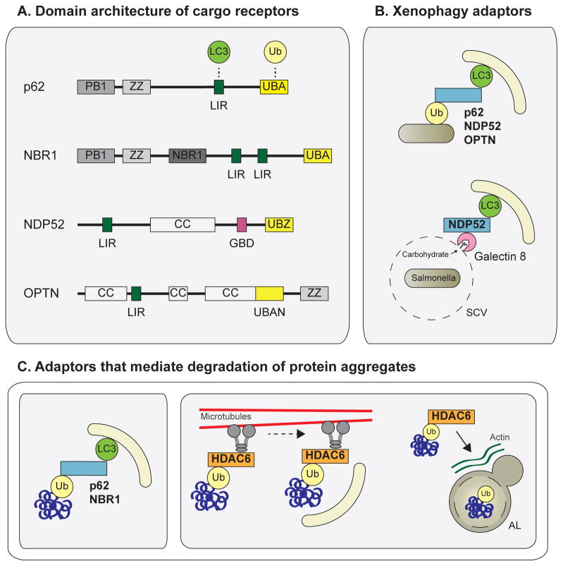
Autophagy, a vital catabolic process that degrades cytoplasmic components within the lysosome, is an essential cytoprotective response to pathologic stresses that occur during diseases such as cancer, ischemia, and infection. In addition to its role as a stress-response pathway, autophagy plays an essential quality-control function in the cell by promoting basal turnover of long-lived proteins and organelles, as well as by selectively degrading damaged cellular components. This homeostatic function protects against a wide variety of diseases, including neurodegeneration, myopathy, liver disease, and diabetes. This review discusses our current understanding of these two principal functions of autophagy and describes in detail how alterations in autophagy promote human disease.


Figures

Figure 1
Figure 1. Overview of the mammalian autophagy machinery
Autophagy occurs in a series of distinct stages: 1) initiation of the isolation membrane, also known as the phagopore assembly site; 2) cargo recognition; 3) elongation; 4) enclosure of the double-membrane structure to form the autophagosome; and 5) maturation/degradation, in which the AP fuses to the lysosome to form an autolysosome, upon which its contents are degraded. The effects of autophagy inhibitors 3-MA and chloroquine and the autophagy inducer rapamycin are shown. (A) The Ulk and class III PI3K complexes are necessary for autophagy initiation. Beclin 1 forms two distinct autophagy-promoting complexes with the PI3K, Vps34. (B) The ubiquitin-like conjugation machinery attaches Atg12 to Atg5 and LC3 to the lipid phosphotidylethanolamine; this machinery mediates autophagosome elongation. Loss of any of these core components (e.g. Beclin 1, Atg5, etc.) leads to autophagy deficiency. (C) In addition to its role in autophagy induction, Beclin 1 promotes autophagic maturation via interactions with Rubicon. Abbreviations: IM, isolation membrane; PAS, phagopore assembly site; AP, autophagosome; AL, autolysosome; 3-MA, 3-methyladenine; CQ, chloroquine; Rap, rapamycin; PI3K, phosphatidylinositol 3-kinase.
Figure 2
Figure 2. Methods for monitoring autophagy
Early studies implicating autophagy in disease primarily measured total autophagosome number, either (A) by identification of APs and ALs by electron microscopy or (B) by measuring total levels of lipidated LC3 (LC3-II) or number of LC3 puncta (which mark APs) in cells expressing GFP-tagged LC3 (GFP-LC3). Importantly, in both of these cases, an increase in APs can indicate either autophagy induction or a block in AP maturation. Autophagic flux assays more directly measure total autophagic activity. In the presence of lysosomal inhibitors, (C) increased accumulation of lipidated LC3 or (D) increased accumulation of GFP-LC3 puncta indicates autophagic induction. (E) In cells expressing tandem RFP-GFP-tagged LC3, APs are identified as yellow puncta and ALs are detected as red puncta following quenching of GFP fluorescence in the lysosome. An increase in both signals indicates autophagic induction, whereas an increase in yellow with a decrease in red indicates a block in maturation. Abbreviations: AP, autophagosome; AL, autolysosome; LC3-I, non-lipidated LC3; LC3-II, lipidated LC3; Δ LC3-II, accumulation of lipidated LC3; Δ LC3 puncta, accumulation of GFP-LC3 puncta; Y, yellow; R, red.
Figure 3
Figure 3. Major stress pathways that induce autophagy
(A) As part of the mTORC1 complex, active mTOR phosphorylates and inactivates Ulk1. Starvation inactivates mTOR, leading to formation of an active Ulk1 complex in which Ulk1 phosphorylates Atg13 and FIP200. During conditions of low cellular energy, AMPK is activated by high AMP:ATP and induces autophagy both by phosphorylation and activation of Ulk1 and by inhibition of mTORC1 via phosphorylation of Raptor. (B) HIF-1 and BNIP3 induce autophagy following hypoxia. HIF-1 is stabilized under hypoxic conditions, leading to increased BNIP3 transcription. BNIP3 binds Bcl-2 and disrupts its inhibitory interaction with Beclin 1, leading to autophagy induction. (C) Following infection, activation of toll-like receptors (TLRs) by pathogen-associated molecular patterns (PAMPs) leads to autophagy induction. Induction downstream of TLR1/2 stimulation depends on AMPK signaling. The downstream TLR signaling molecules MyD88 and TRIF interact with Beclin 1 following TLR activation and disrupt the inhibitory Bcl-2/Beclin 1 complex. Abbreviations: PAMP, pathogen-associated molecular pattern.
Figure 4
Figure 4. Degradation of ubiquitinated autophagy substrates
(A) Domain architecture of the autophagy cargo receptors p62, NBR1, NDP52, and Optineurin. These autophagy adaptors contain an ubiquitin-binding domain (UBA, UBZ, UBAN) and an LC3-interacting region (LIR), mediating recruitment of LC3-containing APs to ubiquitinated cargo (B) The adaptors p62, NDP52, and OPTN mediate degradation of ubiquitinated pathogens via ubiquitin-binding domain and LIR interactions. During Salmonella infection, Galectin 8 binds host carbohydrates exposed on ruptured Salmonella-containing vesicles and recruits NDP52 to these sites, mediating AP recruitment (C) p62 and NBR1 also mediate degradation of ubiquitinated protein aggregates via UBA and LIR interactions. HDAC6 is important for clearance of ubiquitinated aggregates and mitochondria. HDAC6 interacts with ubiquitin, dynein motors, and the actin remodeling machinery to promote dynein-mediated transport of ubiquitinated substrates to APs and enhanced AP-lysosome fusion at these sites. Abbreviations: OPTN, optineurin; Ub, ubiquitin; UBA/UBZ/UBAN, ubiquitin-binding domain; LIR, LC3-interacting region; SCV, Salmonella-containing vesicle
Figure 5
Figure 5. Mitophagy
(A) Domain architecture of the mitophagy adaptors Nix and FUNDC1. Both proteins contain a transmembrane domain embedded in the outer mitochondrial membrane (OMM) as well as a LIR, which mediates recruitment of APs to mitochondria. (B). Following mitochondrial stress, Nix promotes mitochondrial depolarization and ROS formation, leading to autophagy induction. Nix also mediates targeting of APs to mitochondria via its LIR. (C) In healthy polarized mitochondria, PINK1 is constitutively degraded by the inner mitochondrial membrane protease PARL. Following depolarization, full length PINK1 accumulates on the OMM and recruits the E3 ligase Parkin. Parkin ubiquitinates multiple OMM proteins, leading to proteasomal degradation of OMM components and p62/NBR1-mediated AP recruitment to mitochondria. Abbreviations: TM, transmembrane domain; Ψm, mitochondrial membrane potential; ROS, reactive oxygen species.
Figure 6
Figure 6. Summary of autophagy alterations in disease and potential treatments
The major autophagy alterations in human disease and examples of diseases for each class are listed. (A) In diseases with “overactive” autophagy, inhibition of autophagy (via 3-MA or CQ) may provide therapeutic benefit. In diseases that lead to a defect in (B) autophagy induction/initiation or (C) cargo recognition, patients may benefit from drugs such as rapamycin (Rap) that induce autophagy. (D) In diseases that lead to a block in autophagic maturation, Rap treatment may partially restore flux and provide a cytoprotective effect. Alternatively, in diseases with a severe block in autophagic maturation, blocking autophagy at an early step (e.g., via 3-MA) may be therapeutically useful because they mitigate the cytotoxic accumulation of autophagosomes and lysosomes.

References

    1. Mizushima N, Levine B, Cuervo AM, Klionsky DJ. Autophagy fights disease through cellular self-digestion. Nature. 2008;451:1069–75. - PMC - PubMed
    1. Klionsky DJ, Cregg JM, Dunn WA, Jr, Emr SD, Sakai Y, Sandoval IV, Sibirny A, Subramani S, Thumm M, Veenhuis M. A Unified Nomenclature for Yeast Autophagy-Related Genes. Developmental cell. 2003;5:539–45. - PubMed
    1. Klionsky DJ, Abeliovich H, Agostinis P, Agrawal DK, Aliev G, Askew DS, Baba M, Baehrecke EH, Bahr BA, Ballabio A. Guidelines for the use and interpretation of assays for monitoring autophagy in higher eukaryotes. Autophagy. 2008;4:151. - PMC - PubMed
    1. Kroemer G, Mariño G, Levine B. Autophagy and the integrated stress response. Molecular cell. 2010;40:280–93. - PMC - PubMed
    1. Rabinowitz JD, White E. Autophagy and metabolism. Science. 2010;330:1344. - PMC - PubMed

Publication types

MeSH terms