. 2012 Oct 16;9(1):27.
doi: 10.1186/1742-9994-9-27.

The nervous system of Isodiametra pulchra (Acoela) with a discussion on the neuroanatomy of the Xenacoelomorpha and its evolutionary implications

Affiliations

The nervous system of Isodiametra pulchra (Acoela) with a discussion on the neuroanatomy of the Xenacoelomorpha and its evolutionary implications

Johannes Georg Achatz et al. Front Zool. .

Abstract

Introduction: Acoels are microscopic marine worms that have become the focus of renewed debate and research due to their placement at the base of the Bilateria by molecular phylogenies. To date, Isodiametra pulchra is the most promising "model acoel" as it can be cultured and gene knockdown can be performed with double-stranded RNA. Despite its well-known morphology data on the nervous system are scarce. Therefore we examined this organ using various microscopic techniques, including histology, conventional histochemistry, electron microscopy, and immunocytochemistry in combination with CLSM and discuss our results in light of recently established phylogenies.

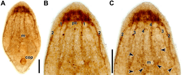
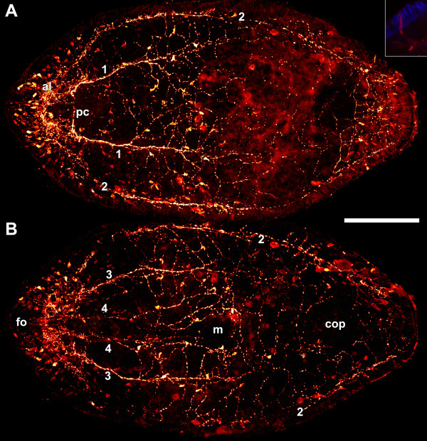
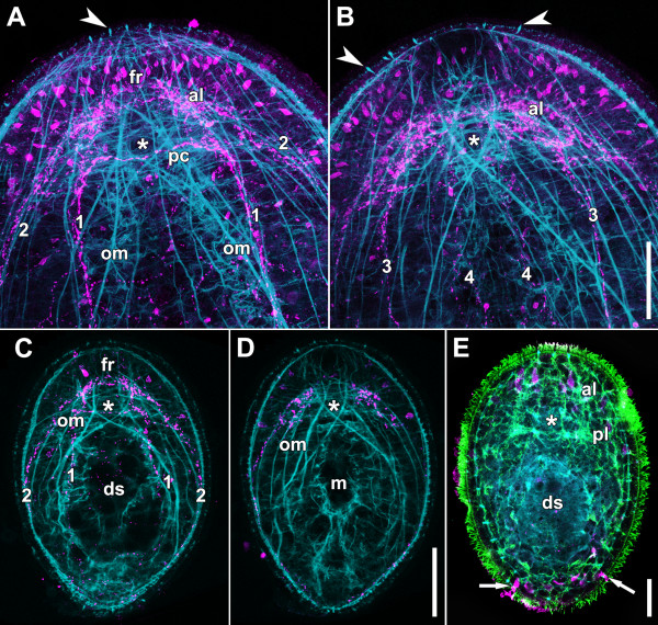
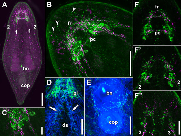
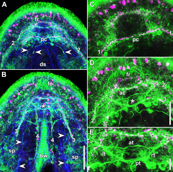
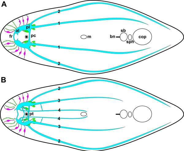
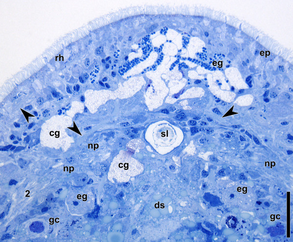
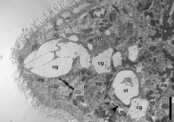
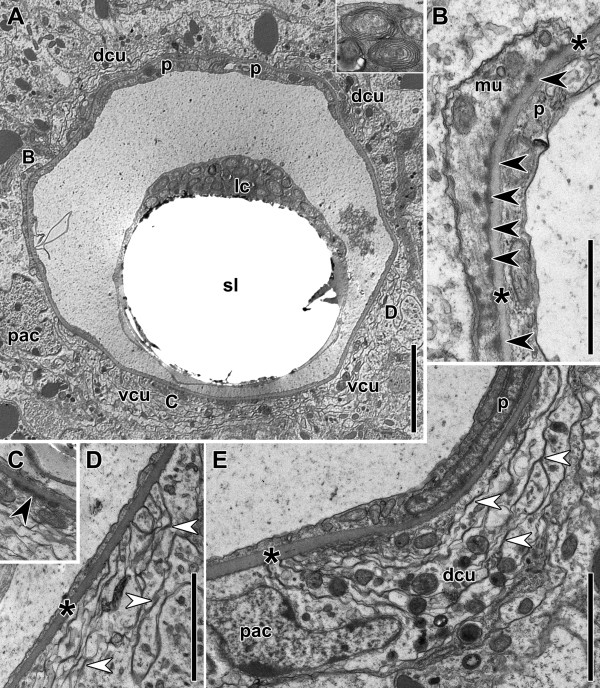
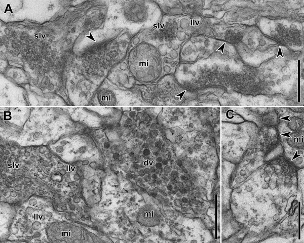
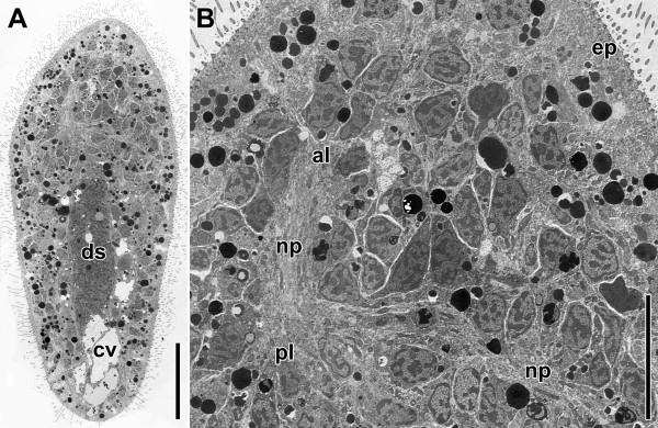
Results: The nervous system of Isodiametra pulchra consists of a bilobed brain with a dorsal posterior commissure, a frontal ring and tracts, four pairs of longitudinal neurite bundles, as well as a supramuscular and submuscular plexus. Serotonin-like immunoreactivity (SLI) is displayed in parts of the brain, the longitudinal neurite bundles and a large part of the supramuscular plexus, while FMRFamide-like immunoreactivity (RFLI) is displayed in parts of the brain and a distinct set of neurons, the longitudinal neurite bundles and the submuscular plexus. Despite this overlap SLI and RFLI are never colocalized. Most remarkable though is the presence of a distinct functional neuro-muscular system consisting of the statocyst, tracts, motor neurons and inner muscles, as well as the presence of various muscles that differ with regard to their ultrastructure and innervation.

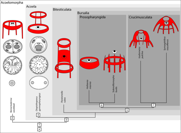
Conclusions: The nervous system of Isodiametra pulchra consists of an insunk, bilobed brain, a peripheral part for perception and innervation of the smooth body-wall musculature as well as tracts and motor neurons that together with pseudostriated inner muscles are responsible for steering and quick movements. The insunk, bilobed brains with two to three commissures found in numerous acoels are homologous and evolved from a ring-commissural brain that was present in the stem species of acoelomorphs. The acoelomorph brain is bipartite, consisting of a Six3/6-dependend animal pole nervous system that persists throughout adulthood and an axial nervous system that does not develop by exhibiting a staggered pattern of conserved regulatory genes as in other bilaterians but by a nested pattern of these genes. This indicates that acoelomorphs stem from an ancestor with a simple brain or with a biphasic life cycle.


Figures

Figure 1
Figure 1
Whole mount of mature specimen stained by direct-coloring method of Karnovsky and Roots[36].A. Entire specimen. B. Anterior half, dorsal side in focus. C. Anterior half, ventral side in focus. Arrowheads point to neurons in an anterior-posterior orientation and around the posterior rim of the mouth. Abbreviations: 1 dorsal neurite bundle; 2 lateral neurite bundle; 3 ventral neurite bundle; 4 medio-ventral neurite bundle; cop male copulatory organ; m mouth; pc dorsal posterior commissure. Scale bars: A, B, C 100 μm.
Figure 2
Figure 2
Whole mount stained with polyclonal antibodies against serotonin.A. Projection of dorsal sections. B. Projection of ventral sections. Inset: Projection of body wall showing cilia of epidermal cells in blue and receptor cell with single cilium with SLI in red. Note that the soma of the cell was out of the section plane and its intensity is therefore weaker. Abbreviations: 1 dorsal neurite bundle; 2 lateral neurite bundle; 3 ventral neurite bundle; 4 medio-ventral neurite bundle; al anterior lobe; cop male copulatory organ; fo frontal organ; m mouth; pc dorsal posterior commissure. Scale bar: 100 μm.
Figure 3
Figure 3
Whole mounts stained with polyclonal antibodies against serotonin (magenta: A, B, C, D, E), tyrosinated tubulin (green: E), and fluorophore-tagged phalloidin (cyan). Asterisks mark position of statocyst. A. Anterior end of adult specimen, projection of dorsal sections. The dorsal body-wall musculature has been omitted for clarity. Arrowhead points to swallow’s nest receptor cell. Note the position of the X-muscle ventral to the dorsal posterior commissure. B. Anterior end of adult specimen, projection of ventral sections. Arrowheads point to swallow’s nest receptor cells. Note the position of the X-muscle ventral to the statocyst. C. Dorsal projection of juvenile. Note the position of the X-muscle ventral to the dorsal posterior commissure. D. Ventral projection of juvenile. Note the position of the X-muscle ventral to the statocyst. E. Dorsal projection of juvenile. Arrows point to gland cells at posterior tip. Abbreviations: 1 dorsal neurite bundle; 2 lateral neurite bundle; 3 ventral neurite bundle; 4 medio-ventral neurite bundle; al anterior lobe; ds digestive syncytium; fr frontal ring; m mouth; om oblique inner muscle; pc dorsal posterior commissure; pl posterior lobe. Scale bars: A, B 50 μm; C, D 50 μm; E 20 μm.
Figure 4
Figure 4
Whole mounts of adult specimens stained with antibodies against FMRFamide (green: A, B, C, F, F’, F”), serotonin (monoclonal; magenta: A, B, C, F, F’, F”), and fluorophore-tagged phalloidin (blue; D, E).A. Dorsal side of entire specimen. B. Anterior end. Arrows point to lateral varicosities of neurites that probably connect the lobes with receptor cells in the epidermis. C. Detail of B. D. Anterior half of a specimen showing RFL immunoreactive parts of the brain and neurites surrounding the dorso-ventral muscles of the ventral groove. E. Musculature of copulatory organs innervated by RFL immunoreactive neurites. F. Dorsal projection of brain. F’. Central projection of brain. F”. Ventral projection of brain. Abbreviations: 1 dorsal neurite bundle; 2 lateral neurite bundle; 3 ventral neurite bundle; bn bursal nozzle; cop male copulatory organ; ds digestive syncytium; fr frontal ring; pc dorsal posterior commissure. Scale bars: A 100 μm; B 50 μm; C 10 μm; D 50 μm; E 50 μm; F-F” 25 μm.
Figure 5
Figure 5
Whole mount stained with antibodies against tyrosinated tubulin (green: A, B, C, D, E), serotonin (polyclonal; magenta: A, B, C, D, E), and fluorophore-tagged phalloidin (blue: A, B).A. Projection of central sections. Arrowheads point to neurites that follow distinct inner muscles. B. Projection of ventral sections. Arrowheads point to neurites that follow distinct inner muscles. Asterisk marks position of statocyst. C. Projection of dorsal sections. D. Projection of central sections. Asterisk marks position of statocyst. E. Magnification of D. Abbreviations: 1 dorsal neurite bundle; 2 lateral neurite bundle; 3 ventral neurite bundle; 4 medio-ventral neurite bundle; at anterior tract; bw body wall; ds digestive syncytium; fo frontal organ; fr frontal ring; lt left tract; pc dorsal posterior commissure; pt posterior tract; rt right tract; sp spermatids and sperm. Scale bars: A, B 50 μm; C, D 30 μm; E 10 μm.
Figure 6
Figure 6
Schematic drawing of the nervous system of I. pulchra (green: RFLI; magenta: SLI; cyan: central nervous system).A. Dorsal view. B. Venral view. Abbreviations: 1 dorsal neurite bundle; 2 lateral neurite bundle; 3 ventral neurite bundle; 4 medio-ventral neurite bundle; al anterior lobe; bn bursal nozzle; cop male copulatory organ; fr frontal ring; m mouth; pc dorsal posterior commissure; pl posterior lobe; pt posterior tract; sb seminal bursa; sph sphincter.
Figure 7
Figure 7
Horizontal semithin section through anterior end of adult specimen stained with Richardson's. Arrowheads point to muscles. Note nuclei around the statocyst and the anterior neuropil, pseudostriation in highlighted inner muscles, and metaphase chromosomes in germ cells. Abbreviations: 2 lateral neurite bundle; cg cyanophilic gland cells; ds digestive syncytium; eg eosinophilic gland cells; ep epidermis; gc germ cells; np neuropil; rh rhabdoids; sl statolith. Scale bar: 25 μm.
Figure 8
Figure 8
Electron micrograph of sagittal section through anterior end of adult specimen. Arrowhead points to ventral polar cell. Note various tissues extending through brain and lipid droplets ventral to statocyst. Abbreviations: cg cyanophilic gland cells; ds digestive syncytium; eg eosinophilic gland cells; fr frontal ring; pc dorsal posterior commissure; pt posterior tract; rh rhabdoids; st statocyst. Scale bar: 10 μm.
Figure 9
Figure 9
Electron micrographs of cross sections through the statocyst of an adult specimen. Asterisks mark the ECM of the statocyst. A. Cross section through the statocyst. Inset: multilaminar bodies of the lithocyte. B. Magnification of A. Black arrowheads mark dense plucks of muscle cell attached to statocyst capsule. C. Magnification of A showing a dense junction of cushion with statocyst capsule. D. Magnification of A showing part of the ventral cushion. White arrowheads point to synapse-like structures. E. Dorso-lateral cushion. Picture is rotated clock-wise; position of parietal cell nucleus is usually dorso-lateral. White arrowheads point to synapse-like structures. Abbreviations: dcu dorsal nerve cushion; mu muscle; lc lithocyte; pac parenchymal cell; p parietal cell; sl statolith; vc ventral nerve cushion. Scale bars: A 5 μm; B 1 μm; C, D, E 2 μm.
Figure 10
Figure 10
Electron micrographs of horizontal sections through the brain of an adult specimen. Arrowheads point to synapses. A, B. Area of neuropil showing various vesicles and synapses. C. Synapses. Abbreviations: dv dense vesicles; llv large lucent vesicles; mi mitochondrion; slv small lucent vesicles. Scale bars: A, B, C 500 nm.
Figure 11
Figure 11
Electron micrographs of a horizontal section through a juvenile specimen.A. Whole specimen. B. Anterior end. Abbreviations: al anterior lobe; cv chordoid vacuole; ds digestive syncytium; ep epidermis; np neuropil; pl posterior lobe. Scale bars: A 25 μm; B 10 μm.
Figure 12
Figure 12
Phylogeny of depicted acoelomorphs amended with schemes of corresponding nervous system (red), statocysts, digestive tracts and spermatozoa (from top to bottom). (*) Hypothetical ancestor either with a bipartite brain that was not crossed by the alimentary tract and not staggered into forebrain, midbrain and hindbrain along the A-P axis but into anterior pole and axial nervous system or with a biphasic life cycle and an actively swimming and feeding larva, acoelomorphs being progenetic descendants. (1) Ring-commissure and small number of posterior neurite bundles. Adaptations to interstitial lifestyle: statocyst, frontal glands, multiciliary epidermis with special rootlet system and stepped tips of cilia, copulation. (2) Statocyst with two statoliths. Adaptation to internal fertilization: cork-screw-shaped sperm. (3) Nervous system looses basiepidermal position and tendency to develop an additional anterior ring commissure. Statocyst with one statolith, digestive system with unpolarized cells. Adaptation to internal fertilization: biflagellate sperm. (4) Posterior pharynx. (5) Female accessory organs. (6) Subterminal anterior pharynx. (7) Circular pattern of brain is abandoned and results in bilobed brains with one to three commissures. Branch-lengths estimated after [14,17], schemes of sperm, digestive tracts and statocysts from [9,45], pattern of nervous system from the following sources: N. westbladi[47]; D. gymnopharyngeus[48]; P. rubra[49]; H. miamia[50]; S. funilis[51]; I. pulchra [this paper]; S. roscoffensis[10-12].

Similar articles

Cited by

References

    1. Hyman LH. The Invertebrates: Platyhelminthes and Rhynchocoela. The acoelomate Bilateria. Volume 2. McGraw Hill Book Co, New York; 1951.
    1. Westblad E. Studien über Skandinavische Turbellaria Acoela. 5. Arkiv för Zoologi. 1948;41A(7):1–82.
    1. Raikova OI, Reuter M, Kotikova EA, Gustafsson MKS. A commissural brain! The pattern of 5-HT immunoreactivity in Acoela (Plathelminthes) [Platyhelminthes] Zoomorphology. 1998;118(2):69–77. doi: 10.1007/s004350050058. - DOI
    1. Reuter M, Raikova OI, Gustafsson MKS. An endocrine brain? The pattern of FMRF-amide immunoreactivity in Acoela (Plathelminthes) Tissue Cell. 1998;30(1):57–63. doi: 10.1016/S0040-8166(98)80006-2. - DOI - PubMed
    1. Reuter M, Raikova OI, Gustafsson MKS. Patterns in the nervous and muscle systems in lower flatworms. Belgian J Zool. 2001;131:47–53.