. 2013 Feb;130(2-3):122-31.
doi: 10.1016/j.mod.2012.10.002. Epub 2012 Oct 13.

Global analysis of the haematopoietic and endothelial transcriptome during zebrafish development

Affiliations

Global analysis of the haematopoietic and endothelial transcriptome during zebrafish development

J E Cannon et al. Mech Dev. 2013 Feb.

Abstract

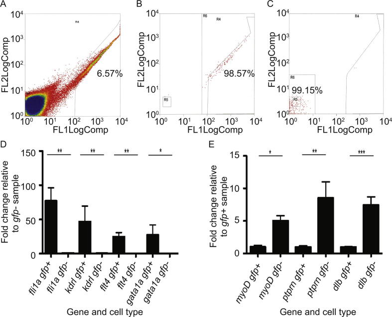
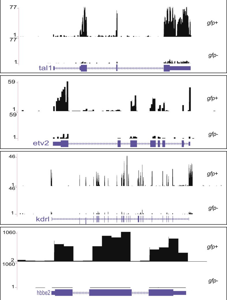
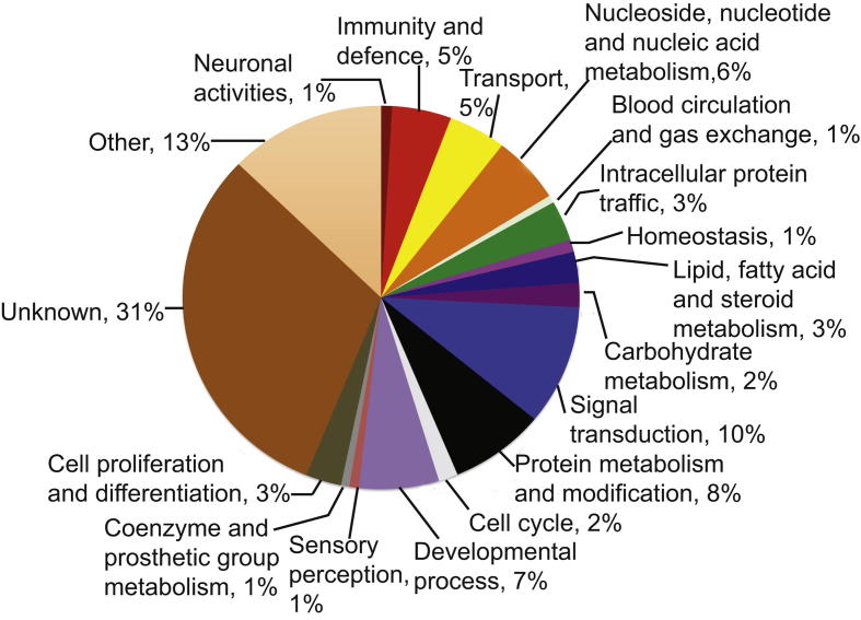
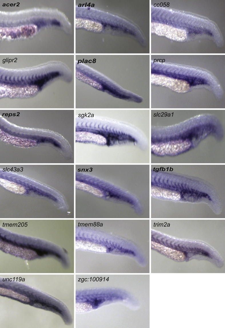
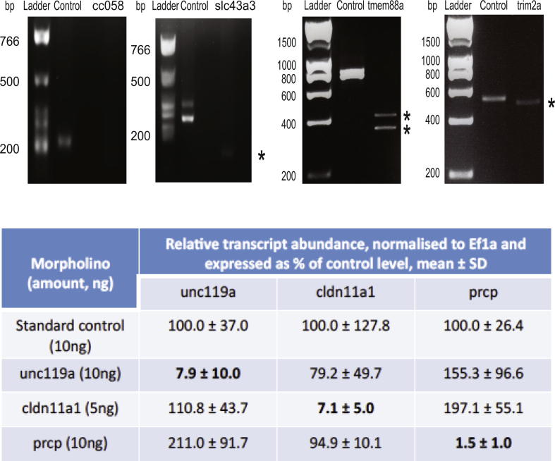
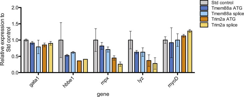
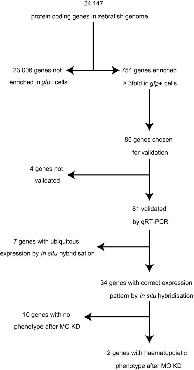
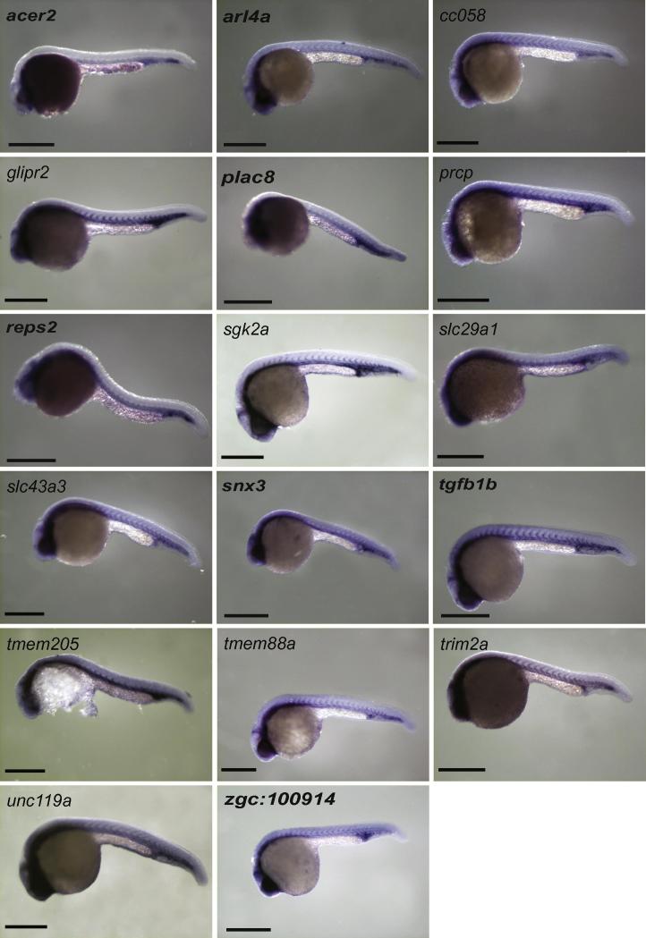
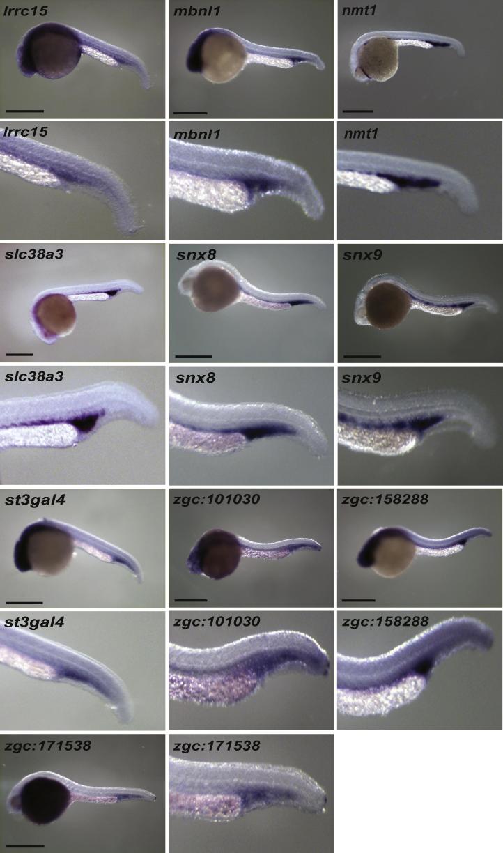
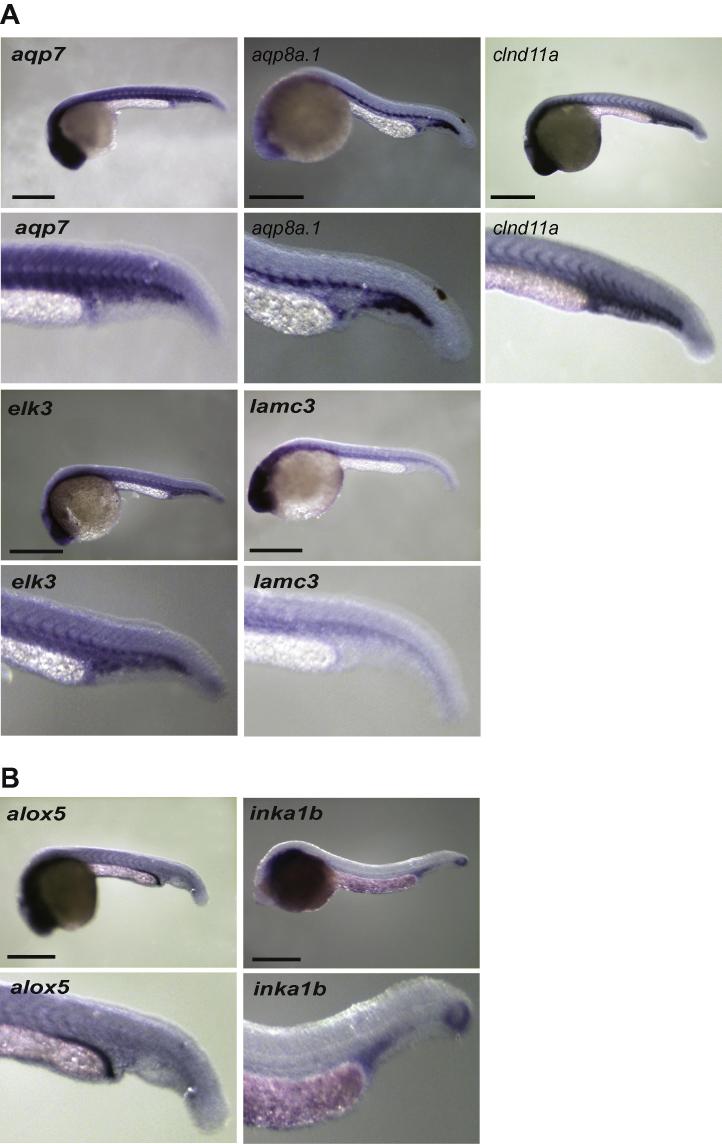
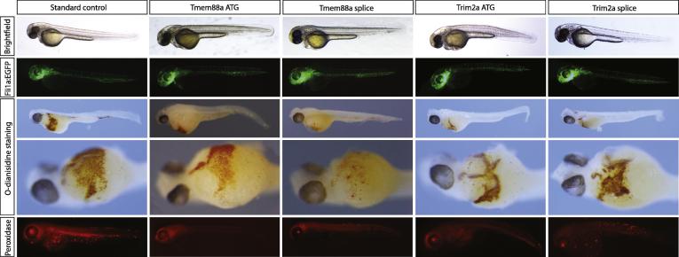
In this paper, we use zebrafish embryos to characterise the transcriptome of the developing blood and endothelium, two cell types that are closely associated during development. High-throughput sequencing identified 754 genes whose transcripts are enriched threefold or more in blood and/or vascular endothelial cells compared with the rest of the embryo at 26-28 h post fertilisation. Of these genes, 388 were classified as novel to these cell types after cross-reference with PubMed and the zebrafish information network (ZFIN). Analysis by quantitative PCR and in situ hybridisation showed that 83% (n=41) of these novel genes are expressed in blood or vascular endothelium. Of 10 novel genes selected for knockdown by antisense morpholino oligonucleotides, we confirmed that two, tmem88a and trim2a, are required for primitive erythropoiesis and myelopoiesis. Our results provide a catalogue of genes whose expression is enriched in the developing blood and endothelium in zebrafish, many of which will be required for the development of those cell types, both in fish and in mammals.


Figures

Supplementary Fig. 1
Isolation of pure populations of gfp+ and gfp− cells from dissociated Tg(fli1a:egfp)y1 embryos. (A) Summary of FACS sort. gfp+ cells were in R4 and gfp- cells in R5. Approximately 6.6% of cells were gfp+. Re-analysis of sorted cells demonstrated that 98.6% and 99.8% were true positive (B) or true negative respectively (C). Summary of relative expression of genes expected to be enriched in either gfp+ (D) or gfp− (E) cells by qRT-PCR. Expression levels were normalised to ef1α prior to expressing fold changes relative to the gfp− (D) or gfp+ (E) values. Error bars indicate SD. n = 3.
Supplementary Fig. 2
RNA-Seq read alignment to Zv8 zebrafish genome on UCSC genome browser. Illustrative alignments of reads from the first biological replicate of gfp+ and gfp− libraries. The genes (RefSeq data) are found at the bottom of each box and consist of the gene name, intron (purple line containing arrows showing the direction of the gene), UTR (thin block) and exons (thicker block). The scale for the number of reads for each gene are on the left side.
Supplementary Fig. 3
The biological functions of the 754 enriched genes using the Panther Ontology.
Supplementary Fig. 4
High powered tail view of embryos with blood and vascular cell in situ hybridisation expression patterns in 24–28 hpf embryos. All embryos are lateral views with anterior to left.
Supplementary Fig. 5
Confirmation of morpholino efficiency. Top panel shows splicing morpholinos designed to cause exon deletion detectable by RT-PCR. Bottom panel shows qPCR results for genes where morpholinos are designed to cause intron inclusion.
Supplementary Fig. 6
Quantitative PCR for selected genes in tmem88a and trim2a morphants. Graphs show relative expression compared to standard control injected embryos. hbbe1 – haemoglobin beta embryonic 1, mpx – myelperoxidase, lyz – lysozyme, myoD – myogenic differentiation.
Fig. 1
Fig. 1
Overview detailing the analysis and validation of the high-throughput sequencing data. MO = morpholino, KD = knockdown.
Fig. 2
Fig. 2
Genes with blood and vascular in situ hybridisation expression pattern in 24–28hpf embryos. All embryos are lateral views with anterior to left. Scale bars indicate 500 μm. High powered views of tail region of these embryos are shown in Supplementary Fig. 4.
Fig. 3
Fig. 3
Genes with blood in situ hybridisation expression pattern in 24–28hpf embryos. All embryos are lateral views with anterior to left. Scale bars indicate 500 μm.
Fig. 4
Fig. 4
Remaining in situ hybridisation expression patterns in 24–28hpf embryos. (A) Vascular expression (B) Other. All embryos are lateral views with anterior to left. Scale bars indicate 500 μm.
Fig. 5
Fig. 5
Tmem88a and trim2a morphants have reduced erythrocytes and myeloid cells as shown by O-dianisidine and peroxidase staining respectively at 48 h post fertilisation. In control embryos erythrocytes are present in axial vessels and returning to the heart across the yolk. Myeloid cells are found across the yolk and randomly around the remaining parts of the embryos. There is loss of erythrocytes and myeloid cells in both trim2a and tmem88a morphants (translation and splice blocking morpholinos) without any defect in vascular development. Panel row 1 shows representative brightfield images, row 2 epifluorescent images of Tg(fli1a:egfp) embryos, rows 3 and 4 show embryos post O-dianisidine staining and row 5 show embryos post peroxidase staining. All images are lateral views with anterior to left with the exception of row 4 which are ventral views.

References

    1. Adams R.H., Wilkinson G.A., Weiss C., Diella F., Gale N.W., Deutsch U., Risau W., Klein R. Roles of ephrinB ligands and EphB receptors in cardiovascular development: demarcation of arterial/venous domains, vascular morphogenesis, and sprouting angiogenesis. Genes Dev. 1999;13:295–306. - PMC - PubMed
    1. Balastik M., Ferraguti F., Pires-da Silva A., Lee T.H., Alvarez-Bolado G., Lu K.P., Gruss P. Deficiency in ubiquitin ligase TRIM2 causes accumulation of neurofilament light chain and neurodegeneration. Proc. Natl. Acad. Sci. USA. 2008;105:12016–12021. - PMC - PubMed
    1. Baldessari D., Mione M. How to create the vascular tree? (Latest) help from the zebrafish. Pharmacol. Ther. 2008;118:206–230. - PubMed
    1. Bertrand J.Y., Chi N.C., Santoso B., Teng S., Stainier D.Y.R., Traver D. Haematopoietic stem cells derive directly from aortic endothelium during development. Nature. 2010;464:108–111. - PMC - PubMed
    1. Bertrand J.Y., Kim A.D., Violette E.P., Stachura D.L., Cisson J.L., Traver D. Definitive hematopoiesis initiates through a committed erythromyeloid progenitor in the zebrafish embryo. Development. 2007;134:4147–4156. - PMC - PubMed

Publication types

Substances

LinkOut - more resources