

Corneal endothelial cell changes 5 years after laser in situ keratomileusis: femtosecond laser versus mechanical microkeratome
- PMID: 23073480
- PMCID: PMC3511658
- DOI: 10.1016/j.jcrs.2012.07.034
Corneal endothelial cell changes 5 years after laser in situ keratomileusis: femtosecond laser versus mechanical microkeratome
Abstract
Purpose: To compare corneal endothelial cell density (ECD) and morphology between flap creation with a femtosecond laser and flap creation with a mechanical microkeratome 5 years after laser in situ keratomileusis (LASIK).
Setting: Mayo Clinic, Rochester, Minnesota, USA.
Design: Prospective randomized masked paired-eye study.
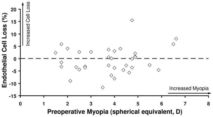
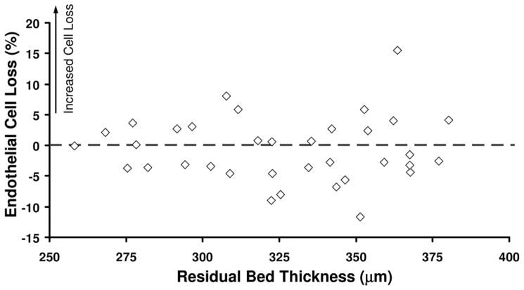
Methods: In this study of LASIK for myopia or myopic astigmatism, fellow eyes were randomized by ocular dominance to flap creation by a femtosecond laser or by a mechanical microkeratome. Central endothelial images were analyzed before and 3 years and 5 years after LASIK; endothelial cell variables were compared between treatments at each examination. Relationships between endothelial cell loss and contact lens wear, residual bed thickness, and preoperative refractive error were evaluated.
Results: There were no differences in the ECD, percentage of hexagonal cells, or coefficient of variation of cell area between treatments at any examination (all P = .99); the smallest detectable differences were 120 cells/mm(2), 5%, and 2%, respectively. The mean annual rate of corneal endothelial cell loss was -0.1% ± 1.2% (SD) and -0.1% ± 1.0% for the femtosecond laser and the mechanical microkeratome, respectively. Endothelial cell loss was not associated with contact lens wear, residual bed thickness, or preoperative refractive error.
Conclusions: The energy delivered to the cornea during femtosecond laser flap creation did not affect the corneal endothelium 5 years after LASIK when compared with flap creation with a mechanical microkeratome. Corneas that have had either method of flap creation could be accepted as donor tissue for endothelial keratoplasty from the standpoint of endothelial health.
Financial disclosure: No author has a financial or proprietary interest in any material or method mentioned.
Copyright © 2012 ASCRS and ESCRS. Published by Elsevier Inc. All rights reserved.
Figures
References
-
- Pallikaris IG, Siganos DS. Excimer laser in situ keratomileusis and photorefractive keratectomy for correction of high myopia. J Refract Corneal Surg. 1994;10:498–510. - PubMed
-
- Patel SV, Bourne WM. Corneal endothelial cell loss 9 years after excimer laser keratorefractive surgery. [Accessed August 12, 2012];Arch Ophthalmol. 2009 127:1423–1427. Available at: http://archopht.jamanetwork.com/data/Journals/OPHTH/10132/ecs90022_1423_.... - PMC - PubMed
-
- Juhasz T, Kastis GA, Suárez C, Bor Z, Bron WE. Time-resolved observations of shock waves and cavitation bubbles generated by femtosecond laser pulses in corneal tissue and water. Lasers Surg Med. 1996;19:23–31. - PubMed
-
- Lubatschowski H, Maatz G, Heisterkamp A, Hetzel U, Drommer W, Welling H, Ertmer W. Application of ultrashort laser pulses for intrastromal refractive surgery. Graefes Arch Clin Exp Ophthalmol. 2000;238:33–39. - PubMed
Other Cited Material
-
- A. National Institutes of Health Clinical Trials. [Accessed August 12, 2012];Long-term effects of laser refractive surgery. :NCT00350246. Available at: http://www.clinicaltrials.gov/ct2/show/NCT00350246?term=NCT00350246&rank=1.
