. 2012 Oct 16;3(5):e00267-12.
doi: 10.1128/mBio.00267-12.

Herpes simplex virus γ34.5 interferes with autophagosome maturation and antigen presentation in dendritic cells

Affiliations

Herpes simplex virus γ34.5 interferes with autophagosome maturation and antigen presentation in dendritic cells

Philipe A M Gobeil et al. mBio. .

Abstract

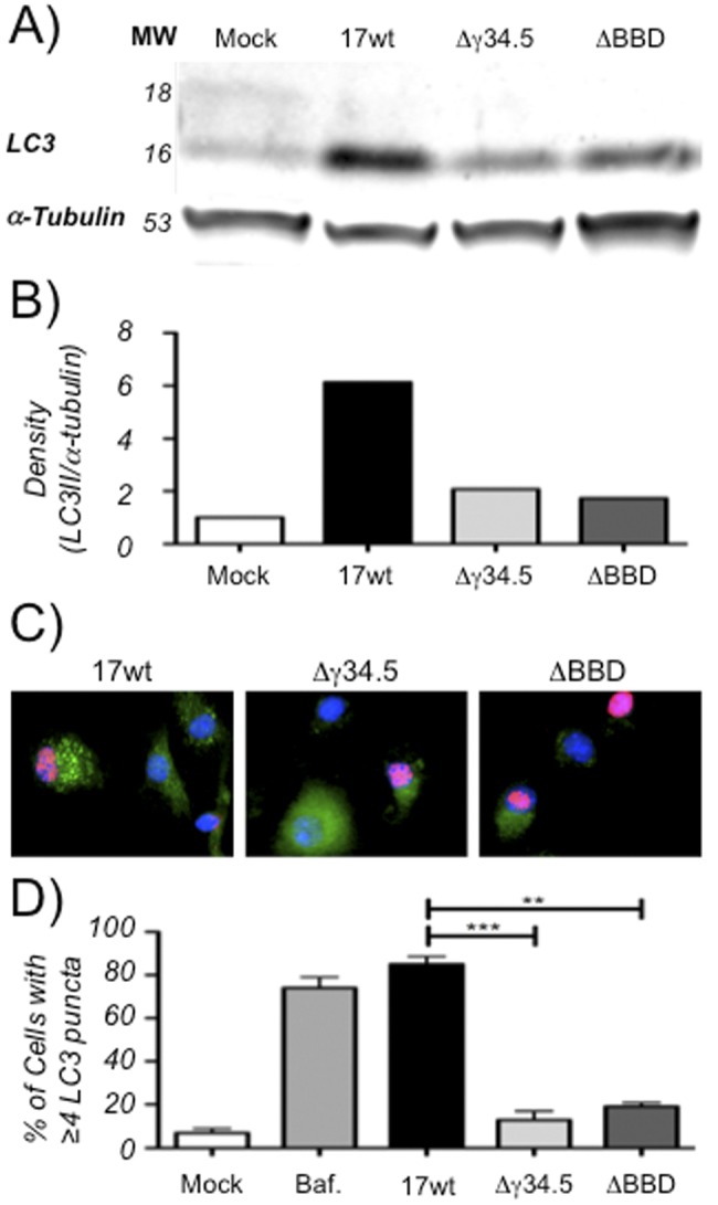
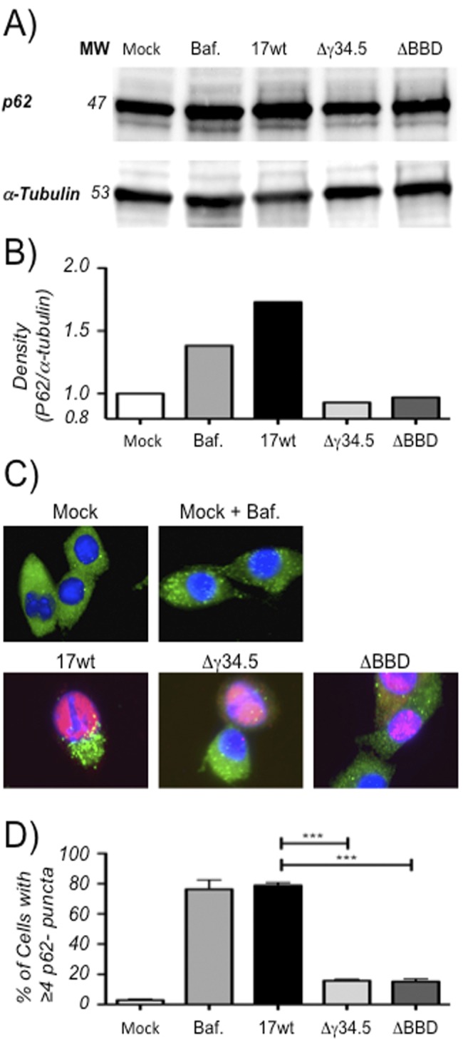
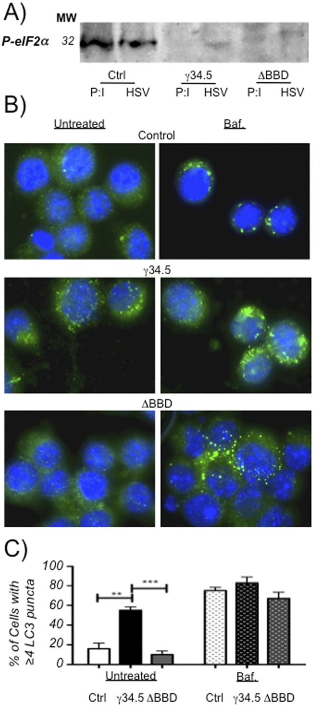
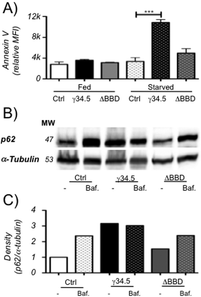
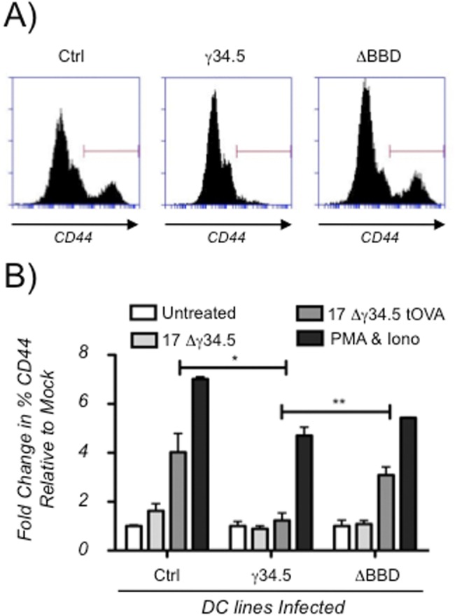
The cellular autophagy response induced by herpes simplex virus 1 (HSV-1) is countered by the viral γ34.5 protein. γ34.5 modulates autophagy by binding to the host autophagy protein Beclin-1 and through this binding inhibits the formation of autophagosomes in fibroblasts and neurons. In contrast, in this study dendritic cells (DCs) infected with HSV-1 showed an accumulation of autophagosomes and of the long-lived protein p62. No such accumulations were observed in DCs infected with a γ34.5-null virus or a virus lacking the Beclin-binding domain (BBD) of γ34.5. To explore this further, we established stably transduced DC lines to show that γ34.5 expression alone induced autophagosome accumulation yet prevented p62 degradation. In contrast, DCs expressing a BBD-deleted mutant of γ34.5 were unable to modulate autophagy. DCs expressing γ34.5 were less capable of stimulating T-cell activation and proliferation in response to intracellular antigens, demonstrating an immunological consequence of inhibiting autophagy. Taken together, these data show that in DCs, γ34.5 antagonizes the maturation of autophagosomes and T cell activation in a BBD-dependent manner, illustrating a unique interface between HSV and autophagy in antigen-presenting cells. IMPORTANCE Herpes simplex virus 1 (HSV-1) is a highly prevalent pathogen causing widespread morbidity and some mortality. HSV infections are lifelong, and there are no vaccines or antivirals to cure HSV infections. The ability of HSV to modulate host immunity is critical for its virulence. HSV inhibits host autophagy, a pathway with importance in many areas of health and disease. Autophagy is triggered by many microbes, some of which harness autophagy for replication; others evade autophagy or prevent it from occurring. Autophagy is critical for host defense, either by directly degrading the invading pathogen ("xenophagy") or by facilitating antigen presentation to T cells. In this study, we show that HSV manipulates autophagy through an unsuspected mechanism with a functional consequence of reducing T cell stimulation. These data further our understanding of how HSV evades host immunity to persist for the lifetime of its host, facilitating its spread in the human population.


Figures

FIG 1
FIG 1
Infection of DCs with HSV leads to autophagosome accumulation. (A) Representative Western blot (of two experiments) of lysates from DC2.4 cells that were mock infected or infected at an MOI of 8.0 with WT, Δγ34.5, or ΔBBD virus for 8 h. Blots were probed with anti-LC3 or anti-α-tubulin. (B) Quantification of representative Western blot showing relative band density for LC3-II (the 16-kDa band) normalized to α-tubulin. (C) Immunofluorescence micrographs of cells infected at an MOI of 2.0 with WT, Δγ34.5, or ΔBBD virus for 8 h and probed for autophagosomes with anti-LC3 and for viral infection with anti-ICP8. Bystander cells without ICP8 stain serve as internal, uninfected controls. (D) Quantification of cells displaying 4 or more bright LC3 puncta for various treatments following analysis of at least three experiments involving 100 cells in four or more fields for each treatment. Positive cells are graphed as a function of total cells using DAPI for mock-infected cultures or as a function of ICP8-positive stain for infected cultures. Error bars indicate standard deviations between visualized fields. **, P < 0.005; ***, P < 0.001 (t test).
FIG 2
FIG 2
p62 accumulates in DC2.4 cells infected with HSV-1. (A) Representative Western blots (from two experiments) from lysates of DC2.4 cells that were mock infected or infected at an MOI of 8.0 with WT, Δγ34.5, or ΔBBD virus for 8 h and probed with anti-p62 or anti-α-tubulin antibodies. (B) Graph from representative Western blot showing relative band density for p62 normalized to α-tubulin. (C) Immunofluorescence micrographs of cells infected at an MOI of 2.0 with WT, Δγ34.5, or ΔBBD virus for 16 h and probed for p62 and HSV ICP0 expression. (D) Quantification of cells from fluorescence micrographs. Cells displaying 4 or more p62 puncta were counted as positive and graphed as a function of total cells using DAPI for mock-infected cells or as a function of ICP0-positive stain for infected cells. Analysis included at least 60 cells in three or more fields for each treatment in two experiments. Error bars indicate standard deviations between visualized fields. ***, P < 0.001 (t test).
FIG 3
FIG 3
Stably transduced DC2.4 cells express functional γ34.5 and accumulate autophagosomes. (A) Western blot (representative of two experiments) of cell lysates from γ34.5 or ΔBBD mutant-expressing DC2.4 cells or control DC2.4 cells transfected with poly(I:C) at 20 µg/ml or infected with Δγ34.5 HSV-1 at an MOI of 8.0 for 12 h. Western blots were probed with an antibody for phospho-eIF2α. (B) Fluorescence micrographs of γ34.5- or ΔBBD mutant-expressing or control cells probed with an anti-LC3 antibody. Cells were untreated or treated with 100 µM bafilomycin for 6 h. (C) Quantification of cells from panel B displaying 4 or more LC3 puncta. At least 100 cells in 4 or more fields were counted for each cell type and each treatment in 2 experiments. Positive cells are graphed as a function of total cells using DAPI stain, and the error bars indicate standard deviations between visualized fields. **, P < 0.005; ***, P < 0.001 (t test).
FIG 4
FIG 4
Stably transduced DC2.4 cells expressing γ34.5 are sensitive to starvation and accumulate long-lived proteins. (A) Annexin V staining of DC2.4 cells stably expressing γ34.5 or the ΔBBD mutant or control cells. Cells were untreated or starved and analyzed by flow cytometry, and mean fluorescence intensity was recorded. Graph shows the mean from two experiments, each consisting of at least 2 biological duplicates, and error bars indicate standard deviations. ***, P < 0.001 (t test). (B) Representative Western blot (of three experiments) of p62 or α-tubulin on cell lysates of control cells or cells expressing γ34.5 or the ΔBBD mutant. Cells were untreated or incubated with 100 µM bafilomycin for 6 h. (C) Quantification of a representative Western blot showing band density of p62 normalized to α-tubulin band density.
FIG 5
FIG 5
γ-34.5 expression in dendritic cells antagonizes activation of CD4+ T cells via Beclin-1 binding. (A) Representative flow cytometry histograms from DC2.4 cell lines infected with the 17Δ34.5tOva strain. Histograms represent one of two experiments analyzing 2 biological replicates. The γ34.5 or ΔBBD strains expressing DC2.4 cells or control cells were mock infected or infected with the Δ34.5 or Δ34.5tOva strain. Cells were cocultured with whole splenocytes from OT-II mice, stained cells were gated for CD4+ T cells, and CD44 staining was measured. (B) Graph showing fold changes in percentages of CD4+ T cell populations staining high for CD44 relative to mock-infected controls. Results are means from two experiments, each consisting of biological duplicates. Error bars indicate standard deviations. *, P < 0.05; **, P < 0.005 (t test).

References

    1. Nicoll MP, Proença JT, Efstathiou S. 2012. The molecular basis of herpes simplex virus latency. FEMS Microbiol. Rev. 36:684–705 - PMC - PubMed
    1. Kaufman RJ, Murtha P. 1987. Translational control mediated by eucaryotic initiation factor-2 is restricted to specific mRNAs in transfected cells. Mol. Cell. Biol. 7:1568–1571 - PMC - PubMed
    1. Samuel CE, Brody MS. 1990. Biosynthesis of reovirus-specified polypeptides. 2-Aminopurine increases the efficiency of translation of reovirus s1 mRNA but not s4 mRNA in transfected cells. Virology 176:106–113 - PubMed
    1. Chou J, Roizman B. 1992. The gamma 1(34.5) gene of herpes simplex virus 1 precludes neuroblastoma cells from triggering total shutoff of protein synthesis characteristic of programed cell death in neuronal cells. Proc. Natl. Acad. Sci. U. S. A. 89:3266–3270 - PMC - PubMed
    1. Chou J, Chen JJ, Gross M, Roizman B. 1995. Association of a M(r) 90,000 phosphoprotein with protein kinase PKR in cells exhibiting enhanced phosphorylation of translation initiation factor eIF-2 alpha and premature shutoff of protein synthesis after infection with gamma 134.5- mutants of herpes simplex virus 1. Proc. Natl. Acad. Sci. U. S. A. 92:10516–10520 - PMC - PubMed

Publication types

MeSH terms

Substances

LinkOut - more resources