. 2012 Oct 30;109(44):18096-101.
doi: 10.1073/pnas.1213332109. Epub 2012 Oct 16.

BapE DNA endonuclease induces an apoptotic-like response to DNA damage in Caulobacter

Affiliations

BapE DNA endonuclease induces an apoptotic-like response to DNA damage in Caulobacter

Julia Bos et al. Proc Natl Acad Sci U S A. .

Abstract

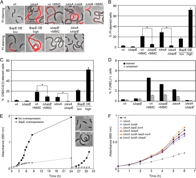
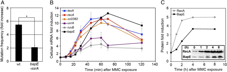
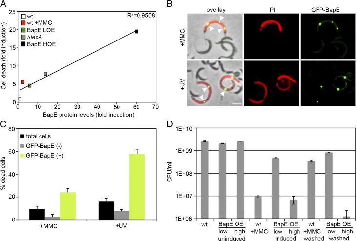
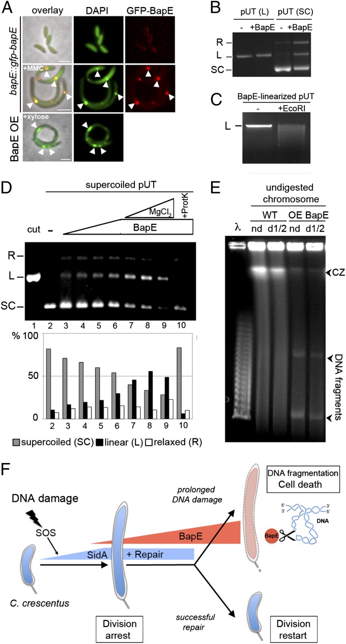
In the presence of extensive DNA damage, eukaryotes activate endonucleases to fragment their chromosomes and induce apoptotic cell death. Apoptotic-like responses have recently been described in bacteria, but primarily in specialized mutant backgrounds, and the factors responsible for DNA damage-induced chromosome fragmentation and death have not been identified. Here we find that wild-type Caulobacter cells induce apoptotic-like cell death in response to extensive DNA damage. The bacterial apoptosis endonuclease (BapE) protein is induced by damage but not involved in DNA repair itself, and mediates this cell fate decision. BapE fragments chromosomes by cleaving supercoiled DNA in a sequence-nonspecific manner, thereby perturbing chromosome integrity both in vivo and in vitro. This damage-induced chromosome fragmentation pathway resembles that of eukaryotic apoptosis. We propose that damage-induced programmed cell death can be a primary stress response for some bacterial species, providing isogenic bacterial communities with advantages similar to those that apoptosis provides to multicellular organisms.


Conflict of interest statement

The authors declare no conflict of interest.

Figures

Fig. 1.
Fig. 1.
DNA-damage–induced BapE protein mediates apoptotic-like death. (A and B) Visualization and quantification of cell shape and PI-stained cells in mutant and MMC-treated cells. Asterisks denote significant differences of the percentage of PI-stained cells between MMC-treated wild-type and bapE mutant strains (P = 0.005) and ΔlexA and ΔlexA ΔbapE strains (P = 0.004). (C) Quantification of DIBAC4(3)-stained cells in mutant and MMC-treated cells. Asterisks denote significant differences of the percentage of DIBAC4(3)-stained cells between MMC-treated wild-type and bapE mutant strains (P = 0.01) and between ΔlexA and ΔlexA ΔbapE strains (P = 0.01). Microscopy images of DIBAC4(3)-stained cells are shown in Fig. S1A. (D) Quantification of TUNEL assays performed by fluorescence flow cytometry in mutant and MMC-treated cells. Due to a high-fluorescence background of nonapoptotic cells, control samples with no d-Transferase enzyme (unstained) were performed in parallel to samples treated with d-Transferase enzyme (stained). Flow cytometry plots are shown in Fig. S1B. (E) Growth curves of a BapE high-overexpression strain with and without xylose induction. ZG484 was grown in PYE supplemented with 0.3% xylose to induce BapE. After 24 h of growth, cultures were rediluted to an OD660 of 0.05 in fresh PYE medium with or without xylose. (Inset) Phase-contrast images from both cultures at time (t) = 24 h. (F) Growth curves of wild-type and ΔlexA mutant strains. (Scale bar, 2 μm in all images.) In all panels, error bars indicate SE of proportion.
Fig. 2.
Fig. 2.
BapE is induced after prolonged DNA damage but does not affect DNA repair. (A) Mutation frequency (fold increase) of wild-type and bapE-ssrA strains after MMC treatment. Error bars indicate SD. Asterisk denotes significant difference between MMC-treated wild-type and bapE-ssrA mutant strains (P = 0.04) (B) Dynamics of SOS-induced gene induction as measured by qRT-PCR. Fold induction of each gene as a function of time after the addition of MMC. Error bars represent SD. (C) The endogenous levels of BapE and RecA protein were determined by Western blotting after 0, 1, 2, 4, and 8 h of MMC treatment (Inset shows blot and plot shows fold induction).
Fig. 3.
Fig. 3.
BapE induces cell death in a concentration-dependent manner. (A) Plot of cell death extent (based on PI staining) as a function of BapE protein levels in various strains (raw data for each are shown in Figs. S3 and S5). (B) Images of cells expressing GFP-BapE from its endogenous promoter (ZG683), grown either in the presence of MMC or after UV irradiation (12 h) and stained with PI to assess death. Arrowheads point to GFP-BapE foci in dead cells. (C) Quantification of the number of dead cells described in B after MMC treatment (n = 119) or after UV irradiation (n = 542) among all cells (black bars), among cells without localized GFP-BapE (gray bars), and among cells with GFP-BapE localized to discrete foci (green bars). Error bars represent SE. (Scale bar, 2 μm.) (D) Cell viability counts for the wild-type, low-BapE overexpressing and high-BapE overexpressing strains after 4 h of growth in the presence of either MMC or xylose to induce BapE expression (induced) and after the strains are shifted in fresh medium with no inducer (washed) for 2 h. Control cultures (uninduced) were grown in parallel. All error bars represent SD.
Fig. 4.
Fig. 4.
BapE affects DNA integrity in vivo and in vitro. (A) Examination of chromosome morphology (DAPI staining) in cells expressing GFP-BapE from its endogenous promoter (bapE::gfp-bapE) grown either with or without MMC and in cells overexpressing BapE. White arrowheads indicate colocalization of GFP-BapE and bright foci of DNA. (B) Cut pUT plasmid (linearized with EcoRI) and uncut plasmid (supercoiled) DNA samples were incubated either with or without BapE protein. The topological states of pUT plasmid are indicated: SC, supercoiled; L, linear; and R, relaxed. (C) Purifed BapE-linearized pUT plasmid was incubated either with or without EcoRI restriction enzyme. The linear (L) state of pUT plasmid is indicated. (D) Supercoiled plasmid DNA (pUT) was incubated alone (lane 2), with increasing concentrations of BapE protein (0.16, 0.32, 0.54, and 0.82 μM) (lanes 3–6), with BapE protein (0.82 μM), and with increasing concentrations of MgCl2 (0.1, 0.5, and 1 mM) (lanes 7–9), and with BapE (0.82 μM) and proteinase K (lane 10). Cut (linear) plasmid is shown as a control (lane 1). Quantifications of the resulting plasmid topological states (SC, supercoiled; L, linear; and R, relaxed) are shown below the gel. (E) Analysis of in vivo chromosome fragmentation by pulsed-field gel electrophoresis of undigested Caulobacter chromosomes [nondiluted (nd) or 1/2 diluted (d1/2)] from cells with and without BapE overexpression. Arrowheads at the bottom denote linearized DNA fragments that are specifically obtained when BapE is overexpressed; arrowhead at the top indicates the compression zone (CZ) of intact high-molecular-weight DNA likely due to overloading of DNA. Lambda concatemers are used as molecular size markers. (F) Model of BapE-dependent DNA damage cell fate determination. In response to DNA damage, C. crescentus induces a graded expression of LexA-dependent genes. Early in the SOS response, cells induce the expression of the division inhibitor SidA (9) and DNA repair pathways to arrest cell division and give cells time to attempt to fix the damage. If DNA damage persists, cells induce the expression of the unique endonuclease, BapE, which functions as a cell fate determinant in a concentration-dependent manner: low levels of BapE reinforce the SidA-mediated reversible division arrest until cells succeed in repairing the damage and re-enter the cell cycle, whereas high levels of BapE promote apoptotic-like cell death by stimulating chromosome fragmentation in conditions of prolonged DNA damage.

Comment in

References

    1. Vousden KH. Outcomes of p53 activation—spoilt for choice. J Cell Sci. 2006;119(Pt 24):5015–5020. - PubMed
    1. Lakin ND, Jackson SP. Regulation of p53 in response to DNA damage. Oncogene. 1999;18(53):7644–7655. - PubMed
    1. Little JW, Mount DW. The SOS regulatory system of Escherichia coli. Cell. 1982;29(1):11–22. - PubMed
    1. Erental A, Sharon I, Engelberg-Kulka H. Two programmed cell death systems in Escherichia coli: An apoptotic-like death is inhibited by the mazEF-mediated death pathway. PLoS Biol. 2012;10(3):e1001281. - PMC - PubMed
    1. Dwyer DJ, Camacho DM, Kohanski MA, Callura JM, Collins JJ. Antibiotic-induced bacterial cell death exhibits physiological and biochemical hallmarks of apoptosis. Mol Cell. 2012;46(5):561–572. - PMC - PubMed

Publication types

Substances

LinkOut - more resources