. 2012 Dec 6;120(24):4829-39.
doi: 10.1182/blood-2012-06-434894. Epub 2012 Oct 16.

CaMKII γ, a critical regulator of CML stem/progenitor cells, is a target of the natural product berbamine

Affiliations

CaMKII γ, a critical regulator of CML stem/progenitor cells, is a target of the natural product berbamine

Ying Gu et al. Blood. .

Abstract

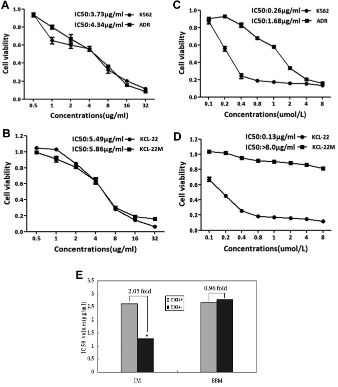
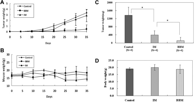
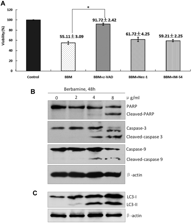
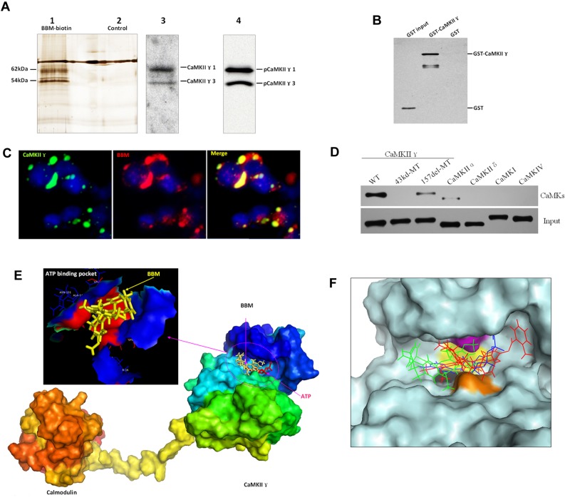
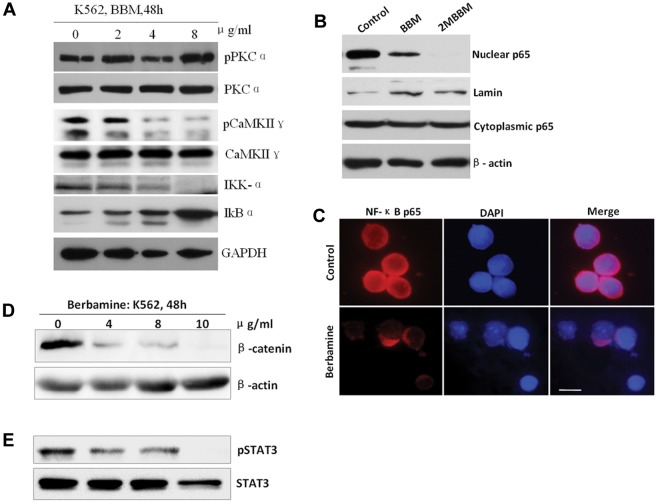
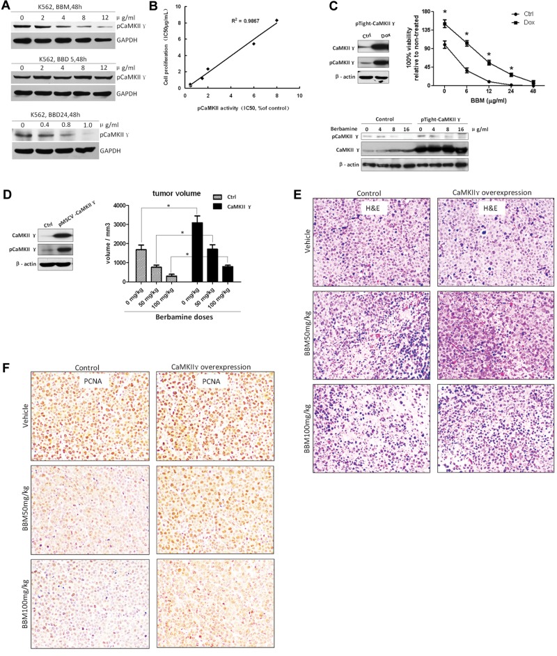
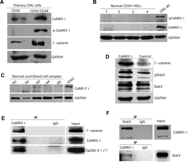
Bcr-Abl tyrosine kinase inhibitors (TKIs) have been a remarkable success for the treatment of Ph(+) chronic myeloid leukemia (CML). However, a significant proportion of patients treated with TKIs develop resistance because of leukemia stem cells (LSCs) and T315I mutant Bcr-Abl. Here we describe the unknown activity of the natural product berbamine that efficiently eradicates LSCs and T315I mutant Bcr-Abl clones. Unexpectedly, we identify CaMKII γ as a specific and critical target of berbamine for its antileukemia activity. Berbamine specifically binds to the ATP-binding pocket of CaMKII γ, inhibits its phosphorylation and triggers apoptosis of leukemia cells. More importantly, CaMKII γ is highly activated in LSCs but not in normal hematopoietic stem cells and coactivates LSC-related β-catenin and Stat3 signaling networks. The identification of CaMKII γ as a specific target of berbamine and as a critical molecular switch regulating multiple LSC-related signaling pathways can explain the unique antileukemia activity of berbamine. These findings also suggest that berbamine may be the first ATP-competitive inhibitor of CaMKII γ, and potentially, can serve as a new type of molecular targeted agent through inhibition of the CaMKII γ activity for treatment of leukemia.


Figures

Figure 1
Figure 1
Berbamine overrides TKI-resistance to leukemia stem cells and T315I mutant–Bcr-Abl of CML. (A-D) Effect of berbamine (A-B) and IM (C-D) on the growth of IM-resistant K562 cells containing LSCs and IM-resistant T315I mutant Bcr-Abl cells (KCL-22M). Two pairs of CML cell lines: K562 cell and IM-resistant K562 cells (containing 2.5% LSCs), KCL-22 and IM-resistant KCL-22M (T315I mutant Bcr-Abl clone) were treated with berbamine or IM at various concentrations for 72 hours and cell viability were measured using MTT assay. IM-sensitive K562 and KCL-22 cells were used as controls. (E) Effect of BBM and IM on the growth of CD34+ and CD34 leukemia cells. CML CD34+ stem cells and CD34 leukemia cells were treated with BBM or IM at various concentrations. After 72 hours in culture, cell viability was measured using MTT assay and IC50 values were calculated. IM was used as control (*P < .01).
Figure 2
Figure 2
Berbamine inhibits the growth of TKI-resistant CML cells and primary CML cells in immunocompromised mice. BBM or imatinib was administered at 100 mg/kg body weight orally 3 times daily for 10 consecutive days, 24 hours after the subcutaneous injection of 2 × 10⁷ TKI-resistant K562 cells or primary CML cells. (A) Effects of BBM and IM on the growth of TKI-resistant tumors. (B) Effects of BBM and IM on body weight of tumor-bearing mice. (C) Effects of BBM and IM on the growth of primary CML cells at the end of experiment (day 15). (D) Effects of BBM and IM on body weight of tumor-bearing mice (n = 4; *P < .01).
Figure 3
Figure 3
Berbamine induces apoptotic and autophagic death of leukemia cells. (A) Effect of berbamine on the viability of CML cells in the presence of various cell death inhibitors. CML cells were treated with BBM at 4 μg/mL in the presence of z-VAD (50μM), Nec-1 (25μM), or IM-54 (5μM) for 24 hours, and then collected for analysis of cell viability using FCM (*P < .01). (B-C) Berbamine treatment induced cleaved PARP, cleaved-caspase-3, cleaved-caspase-9, and LC3-II levels of CML cells in dose-dependent manners. CML cells were treated with BBM at the indicated concentrations for 48 hours, followed by Western blot analysis for PARP, caspase-3, caspase-9, and LC3-II. β-actin was used as a loading control.
Figure 4
Figure 4
Berbamine specifically binds to the ATP-binding pocket of CaMKII γ and inhibits its kinase activity. (A) BBM physically interacts with CaMKII γ proteins. Two proteins of ∼ 62 kDa and 54 kDa were captured specifically by BBM as shown by silver staining (lane 1). These 2 bands were identified as CaMKIIγ isoform 1 (62 kDa) and isoform 3 (54 kDa), respectively, by mass spectrometry (MS) and confirmed by Western blot with total and phospho-CaMKIIγ antibodies (lanes 3-4). (B) BBM directly binds to CaMKII γ protein. Purified GST-CaMKIIγ protein was incubated with biotinylated BBM and the complex was isolated with streptavidin agarose. Western blot with anti-GST antibody revealed that BBM specifically pulled down CaMKIIγ proteins (lane 1), but not GST protein (lane 2). GST protein was used as negative control. (C) Confocal images of BBM colocalization with CaMKII γ in 293T cells. Green: EGFP-tagged CaMKIIγ protein. Red: biotin-labled BBM visualized by Rhodamine-labeled streptavidin. Yellow: BBM was predominantly colocalized with CaMKIIγ protein in 293T cells. (D) Berbamine selectively binds to ATP binding pocket of CaMKIIγ. (E) BBM molecule is located within the ATP binding pocket of CaMKIIγ protein. The model of the CaMKIIγ in complex with calmodulin was built using Modeller based on the x-ray crystal of CaMKIIδ/calmodulin complex. (F) Best docking poses of ATP (blue), XBA24 (red), and berbamine (green) at the ATP-binding pocket of CaMKIIγ. The ligand is clamped by 4 surface residues in CaMKIIγ docking groove: K43 (purple), V74 (black), F90 (yellow), and D157 (brown).
Figure 5
Figure 5
Berbamine inhibits CaMKII γ-dependent NF-κB, β-catenin, and Stat3 without affecting its upstream target PKCα. (A) Effect of berbamine on PKCα, CaMKIIγ, IKK-α, and IkBα. (B-C) Berbamine analogs blocked nuclear translocation of NF-κB p65 realed by Western blot (B) and immunofluorescent staining (C). (C) Immunofluorescent image results exhibited a significant decrease of nuclear NF-κB p65 of leukemia cells after treatment of berbamine. Cells were immobilized on coverslips, fixed, permeabilized, and subjected to immunostaining of p65 (red fluorescence) and nuclear counterstaining (blue). Scale bars, 10 μm. (D-E) Berbamine inhibited CaMKIIγ downstream targetsβ-catenin (D) and Stat3 (E) of leukemia cells. Leukemia cells were treated with berbamine at various concentrations for 48 hours and harvested for analysis of upstream (PKCα) and downstream (IKK-α, IkBα,NF-κB p65, β-catenin and Stat3) targets of CaMKIIγ using Western blotting or immunofluorescent staining.
Figure 6
Figure 6
Correlation between inhibition of CaMKIIγ kinase activity and inhibition of leukemia cell proliferation by berbamine analogs. (A) Berbamine (BBM) and 2-methylbenzoyl berbamine (BBD24) inhibited phosphorylation of CaMKIIγ of leukemia cells. BBM or BBD24 treatment reduced phosphor-CaMKIIγ protein level, but not total CaMKIIγ protein level of K562 cells. Leukemia cells were treated with BBM or BBD24 at the indicated concentrations for 48 hours, and then total proteins were extracted for Western blot analysis for phosphor and total CaMKIIγ proteins. BBD5 was served as an inactive berbamine analog. (B) Correlation between inhibition of CaMKIIγ kinase and inhibition of cell proliferation by analogs of berbamine (BBM, BBD3, BBD12, BBD15, BBD24, and CP15; R2 = 0.9867). (C) Overexpression of CaMKII γ attenuated berbamine-induced growth inhibition of leukemia cells in vitro. Control or CaMKII γ overexpression cells were treated with BBM at the indicated concentrations for 48 hours, and then collected for analysis of cell viability by MTT assay, and phosphor and total CaMKIIγ proteins by Western blot. β-actin was used as loading control (* P < .01). (D) Overexpression of CaMKII γ reduced berbamine-induced growth inhibition of xenograft tumors in a dose-dependent manner (*P < .01). (E-F) hematoxylin-eosin (E) and PCNA (F) stain of xenograft tumors from NOD-SCID mice dosed with vehicle (top), BBM 50 mg/kg (middle), and BBM 100mg/kg (bottom). Left: A representative of control groups. Right: A representative of CaMKIIγ overexpression groups.
Figure 7
Figure 7
High expression of CaMKIIγ is associated with leukemia stem cells of CML. (A-C) CaMKIIγ was highly activated in leukemia stem cells (LSCs) with a concomitant increased β-catenin(g) but not in normal hematopoietic cells (HSCs) and normal blood cells. LSCs and HSCs were sorted from primary CML samples of CML patients at blast crisis and normal cord blood samples, respectively, using FACS. (D) CaMKIIγ increased β-catenin and phospho-Stat3 proteins of leukemia cells. K562 cells were transfected with CaMKIIγ expression vector for 48 hours and then analyzed for β-catenin and Stat3 proteins by Western blot. (E-F) β-catenin and Stat3 protein physically interacted with CaMKIIγ in leukemia cells. Leukemia cell lysates were incubated with CaMKIIγ or Stat3 antibody, and the immune complexes were then purified, separated by SDS-PAGE, and immunoblotted with β-catenin, Stat3, or CaMKII γ antibody.

References

    1. Hantschel O, Superti-Furga G. Regulation of the c-Abl and Bcr-Abl tyrosine kinases. Nat Rev Mol Cell Biol. 2004;5(1):33–44. - PubMed
    1. Rowley JD. Letter: A new consistent chromosomal abnormality in chronic myelogenous leukaemia identified by quinacrine fluorescence and Giemsa staining. Nature. 1973;243(5405):290–293. - PubMed
    1. Deininger MW, Goldman JM, Melo JV. The molecular biology of chronic myeloid leukemia. Blood. 2000;96(10):3343–3356. - PubMed
    1. Stuart SA, Minami Y, Wang JY. The CML stem cell: evolution of the progenitor. Cell Cycle. 2009;8(9):1338–1343. - PMC - PubMed
    1. Li S, Li D. Stem cell and kinase activity-independent pathway in resistance of leukaemia to BCR-ABL kinase inhibitors. J Cell Mol Med. 2007;11(6):1251–1262. - PMC - PubMed

Publication types

MeSH terms