. 2013 Jan;56(2):171-9.
doi: 10.1093/cid/cis870. Epub 2012 Oct 16.

Near-universal prevalence of Pneumocystis and associated increase in mucus in the lungs of infants with sudden unexpected death

Affiliations

Near-universal prevalence of Pneumocystis and associated increase in mucus in the lungs of infants with sudden unexpected death

Sergio L Vargas et al. Clin Infect Dis. 2013 Jan.

Abstract

Background: Pneumocystis without obvious accompanying pathology is occasionally reported in autopsied infant lungs. Its prevalence and significance are unknown. Interestingly, this mild infection induces a strong activation of mucus secretion-related genes in young immunocompetent rodents that has not been explored in infants. Excess mucus is induced by multiple airway offenders through nonspecific pathways and would explain a cofactor role of Pneumocystis in respiratory disease. We undertook characterization of the prevalence of Pneumocystis and associated mucus in infant lungs.

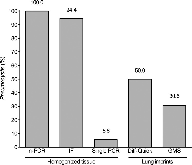
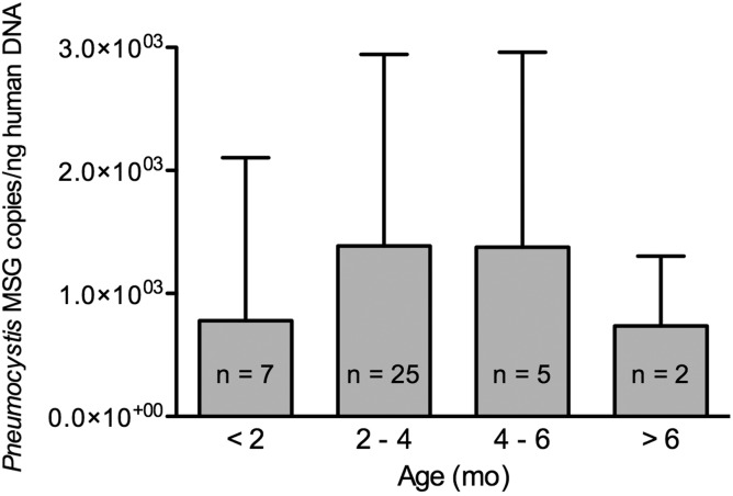
Methods: Samples from 128 infants (mean age, 101 days) who died suddenly and unexpectedly in Santiago during 1999-2004 were examined for Pneumocystis using nested polymerase chain reaction (nPCR) amplification of the P. jirovecii mtLSU ribosomal RNA gene and immunofluorescence microscopy (IF). Pneumocystis-negative infants 28 days and older and their age-closest positives were studied for MUC5AC expression and Pneumocystis burden by Western blot and quantitative PCR, respectively.

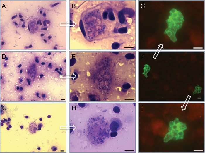
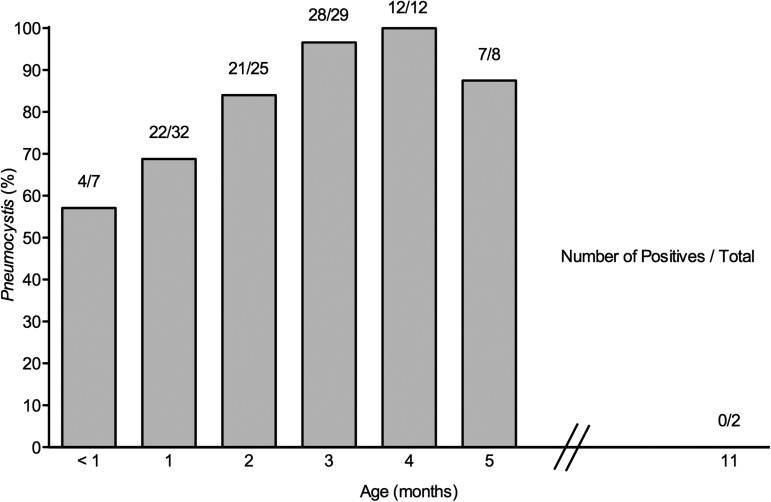
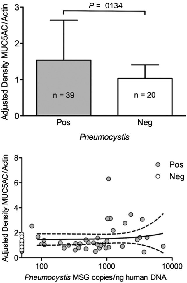
Results: Pneumocystis DNA was detected by nPCR in 105 of the 128 infants (82.0%) and Pneumocystis organisms were visualized by IF in 99 (94.3%) of the DNA-positive infants. The infection was commonest at 3-4 months with 40 of 41 (97.6%) infants of that age testing positive. MUC5AC was significantly increased in Pneumocystis-positive tissue specimens (P = .013). Death was unexplained in 113 (88.3%) infants; Pneumocystis was detected in 95 (84.0%) of them vs 10 of 15 (66.7%) with explained death (P = .28).

Conclusions: A highly focal Pneumocystis infection associated to increased mucus expression is almost universally present in the lungs of infants dying unexpectedly in the community regardless of autopsy diagnosis.


Figures

Figure 1.
Figure 1.
Diagnosis of Pneumocystis in infant biopsy specimens requires sensitive techniques applied to homogenized tissue: Percentage of Pneumocystis detection as relative to nested polymerase chain reaction (n-PCR), of immunofluorescence microscopy (IF), and single-round PCR in homogenized lung tissue specimens of 36 infants. Results of microscopy readings using rapid Giemsa (Diff-Quick) and Gomori-Grocott methenamine silver (GMS) stains in imprints of cruent-cut-surface lung tissue adjacent to the sections analyzed by n-PCR and IF are also presented. Abbreviations: IF, immunofluorescence microscopy; GMS, Gomori-Grocott methenamine silver; n-PCR, nested polymerase chain reaction; PCR, polymerase chain reaction.
Figure 2.
Figure 2.
Detection of this highly focal Pneumocystis infection by microscopy examination in homogenized preparations or imprints from lung tissue specimens. Pneumocystis forms as visualized by microscopy using immunofluorescence stain in aliquots of homogenized lung biopsy specimens (F = ×400; C and I = ×1000), or by rapid Giemsa stain (Diff-Quick) in imprints from fresh lung infant autopsy specimen sections (A, D, and G = ×400; B, F, and H = ×1000). Arrows on each ×400 picture point to their ×1000 magnifications. Bar = 10μ.
Figure 3.
Figure 3.
Pneumocystis jirovecii infection in autopsied infant lungs peaks at 3–5 months. Lung autopsy specimens from 128 infants dying in the community were analyzed for P. jirovecii using nested polymerase chain reaction (nPCR) and immunofluorescence microscopy (IF). P. jirovecii DNA was detected in 105 (82.0%), and Pneumocystis forms were confirmed by IF in 99 (94.2%) of those found positive for P. jirovecii DNA by nPCR and in 0 of 23 infants who tested negative. Each bar represents a minimum of 5 infants. Pneumocystis was additionally detected in 4 of 4, 2 of 2, 2 of 3, 2 of 3, 1 of 1, and 0 of 2 infants dying at 6, 7, 8, 9, 10, and 11 months of age, respectively.
Figure 4.
Figure 4.
Pneumocystis organisms burden increases up to 3–5 months of infant age and declines thereafter. Age progression of Pneumocystis organisms load in autopsy lung samples from 39 infants dying suddenly in the community is shown. Pneumocystis MSG quantitative polymerase chain reaction results were normalized to nanograms of human β-globin DNA for comparisons and expressed as the normalized mean of Pneumocystis MSG copies ± SD.
Figure 5.
Figure 5.
Mucus (MUC5AC) expression is increased by Pneumocystis presence and not influenced by organism load. Top: MUC5AC protein expression according to Pneumocystis status in lung tissue specimens from 39 P. jirovecii–positive and 20 P. jirovecii–negative infants (mean ± SD). Bottom: Correlation between normalized MUC5AC protein expression and normalized quantification values of P. jirovecii MSG in the same lung sample specimen for each infant (Pearson r = 0.0908, P = .5822). MUC5AC level values were normalized by human actin protein expression, and Pneumocystis MSG determinations by human β-globin levels (mean ± SD). Abbreviation: MUC5AC, mucus.

Comment in

Similar articles

Cited by

References

    1. Vestereng VH, Bishop LR, Hernandez B, Kutty G, Larsen HH, Kovacs JA. Quantitative real-time polymerase chain-reaction assay allows characterization of Pneumocystis infection in immunocompetent mice. J Infect Dis. 2004;189:1540–4. - PubMed
    1. Icenhour CR, Rebholz SL, Collins MS, Cushion MT. Early acquisition of Pneumocystis carinii in neonatal rats as evidenced by PCR and oral swabs. Eukaryot Cell. 2002;1:414–9. - PMC - PubMed
    1. Soulez B, Dei-Cas E, Charet P, Mougeot G, Caillaux M, Camus D. The young rabbit: a nonimmunosuppressed model for Pneumocystis carinii pneumonia. J Infect Dis. 1989;160:355–6. - PubMed
    1. Vargas SL, Hughes WT, Santolaya ME, et al. Search for primary infection by Pneumocystis carinii in a cohort of normal, healthy infants. Clin Infect Dis. 2001;32:855–61. - PubMed
    1. Larsen HH, von Linstow ML, Lundgren B, Hogh B, Westh H, Lundgren JD. Primary Pneumocystis infection in infants hospitalized with acute respiratory tract infection. Emerg Infect Dis. 2007;13:66–72. - PMC - PubMed

Publication types

MeSH terms