

Single photon emission computed tomography for the diagnosis of coronary artery disease: an evidence-based analysis
- PMID: 23074411
- PMCID: PMC3377554
Single photon emission computed tomography for the diagnosis of coronary artery disease: an evidence-based analysis
Abstract
In July 2009, the Medical Advisory Secretariat (MAS) began work on Non-Invasive Cardiac Imaging Technologies for the Diagnosis of Coronary Artery Disease (CAD), an evidence-based review of the literature surrounding different cardiac imaging modalities to ensure that appropriate technologies are accessed by patients suspected of having CAD. This project came about when the Health Services Branch at the Ministry of Health and Long-Term Care asked MAS to provide an evidentiary platform on effectiveness and cost-effectiveness of non-invasive cardiac imaging modalities.After an initial review of the strategy and consultation with experts, MAS identified five key non-invasive cardiac imaging technologies for the diagnosis of CAD. Evidence-based analyses have been prepared for each of these five imaging modalities: cardiac magnetic resonance imaging, single photon emission computed tomography, 64-slice computed tomographic angiography, stress echocardiography, and stress echocardiography with contrast. For each technology, an economic analysis was also completed (where appropriate). A summary decision analytic model was then developed to encapsulate the data from each of these reports (available on the OHTAC and MAS website).The Non-Invasive Cardiac Imaging Technologies for the Diagnosis of Coronary Artery Disease series is made up of the following reports, which can be publicly accessed at the MAS website at: www.health.gov.on.ca/mas or at www.health.gov.on.ca/english/providers/program/mas/mas_about.htmlSINGLE PHOTON EMISSION COMPUTED TOMOGRAPHY FOR THE DIAGNOSIS OF CORONARY ARTERY DISEASE: An Evidence-Based AnalysisSTRESS ECHOCARDIOGRAPHY FOR THE DIAGNOSIS OF CORONARY ARTERY DISEASE: An Evidence-Based AnalysisSTRESS ECHOCARDIOGRAPHY WITH CONTRAST FOR THE DIAGNOSIS OF CORONARY ARTERY DISEASE: An Evidence-Based Analysis64-Slice Computed Tomographic Angiography for the Diagnosis of Coronary Artery Disease: An Evidence-Based AnalysisCARDIAC MAGNETIC RESONANCE IMAGING FOR THE DIAGNOSIS OF CORONARY ARTERY DISEASE: An Evidence-Based AnalysisPease note that two related evidence-based analyses of non-invasive cardiac imaging technologies for the assessment of myocardial viability are also available on the MAS website:POSITRON EMISSION TOMOGRAPHY FOR THE ASSESSMENT OF MYOCARDIAL VIABILITY: An Evidence-Based AnalysisMAGNETIC RESONANCE IMAGING FOR THE ASSESSMENT OF MYOCARDIAL VIABILITY: an Evidence-Based AnalysisThe Toronto Health Economics and Technology Assessment Collaborative has also produced an associated economic report entitled:The Relative Cost-effectiveness of Five Non-invasive Cardiac Imaging Technologies for Diagnosing Coronary Artery Disease in Ontario [Internet]. Available from: http://theta.utoronto.ca/reports/?id=7
Objective: The objective of the analysis is to determine the diagnostic accuracy of single photon emission tomography (SPECT) in the diagnosis of coronary artery disease (CAD) compared to the reference standard of coronary angiography (CA). The analysis is primarily meant to allow for indirect comparisons between non-invasive strategies for the diagnosis of CAD, using CA as a reference standard. SPECT: Cardiac SPECT, or myocardial perfusion scintigraphy (MPS), is a widely used nuclear, non-invasive image acquisition technique for investigating ischemic heart disease. SPECT is currently appropriate for all aspects of detecting and managing ischemic heart disease including diagnosis, risk assessment/stratification, assessment of myocardial viability, and the evaluation of left ventricular function. Myocardial perfusion scintigraphy was originally developed as a two-dimensional planar imaging technique, but SPECT acquisition has since become the clinical standard in current practice. Cardiac SPECT for the diagnosis of CAD uses an intravenously administered radiopharmaceutical tracer to evaluate regional coronary blood flow usually at rest and after stress. The radioactive tracers thallium (201Tl) or technetium-99m (99mTc), or both, may be used to visualize the SPECT acquisition. Exercise or a pharmacologic agent is used to achieve stress. After the administration of the tracer, its distribution within the myocardium (which is dependent on myocardial blood flow) is imaged using a gamma camera. In SPECT imaging, the gamma camera rotates around the patients for 10 to 20 minutes so that multiple two-dimensional projections are acquired from various angles. The raw data are then processed using computational algorithms to obtain three-dimensional tomographic images. Since its inception, SPECT has evolved and its techniques/applications have become increasingly more complex and numerous. Accordingly, new techniques such as attenuation correction and ECG gating have been developed to correct for attenuation due to motion or soft-tissue artifact and to improve overall image clarity.
Research questions: What is the diagnostic accuracy of SPECT for the diagnosis of CAD compared to the reference standard of CA?Is SPECT cost-effective compared to other non-invasive cardiac imaging modalities for the diagnosis of CAD?What are the major safety concerns with SPECT when used for the diagnosis of CAD?
Methods: A preliminary literature search was performed across OVID MEDLINE, MEDLINE In-Process and Other Non-Indexed Citations, EMBASE, the Cochrane Library, and the International Agency for Health Technology Assessment (INAHTA) for all systematic reviews/meta-analysis published between January 1, 2004 and August 22, 2009. A comprehensive systematic review was identified from this search and used as a basis for an updated search. A second comprehensive literature search was then performed on October 30, 2009 across the same databases for studies published between January 1, 2002 and October 30, 2009. Abstracts were reviewed by a single reviewer and, for those studies meeting the eligibility criteria, full-text articles were obtained. Reference lists were also hand-searched for any additional studies. Inclusion CriteriaExclusion CriteriaSystematic reviews, meta-analyses, controlled clinical trials, and observational studiesMinimum sample size of 20 patients who completed coronary angiographyUse of CA as a reference standard for the diagnosis of CADData available to calculate true positives (TP), false positives (FP), false negatives (FN) and true negatives (TN)Accuracy data reported by patient not by segmentEnglish languageNon-systematic reviews, case reportsGrey literature and abstractsTrials using planar imaging onlyTrials conducted in patients with non-ischemic heart diseaseStudies done exclusively in special populations (e.g., patients with left branch bundle block, diabetics, minority populations) unless insufficient data available
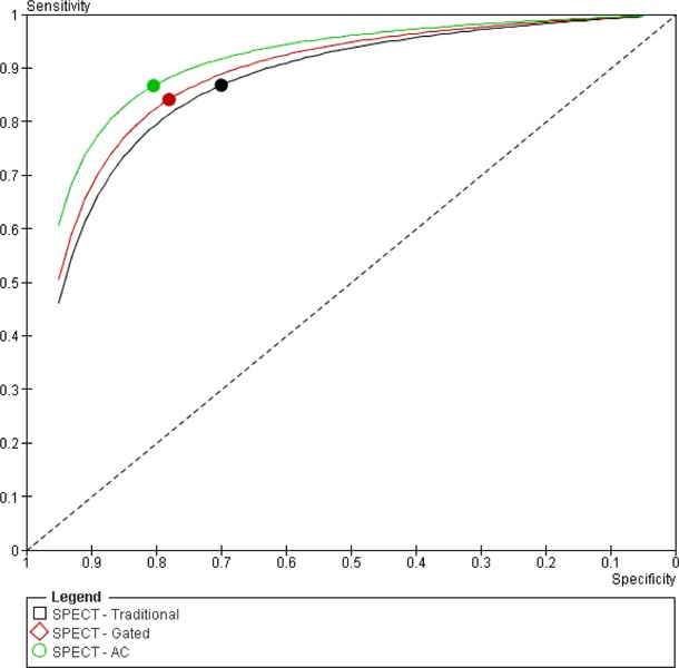
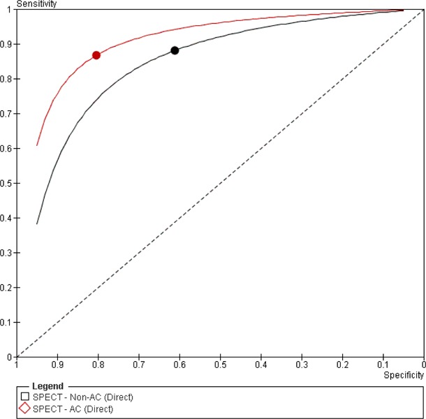
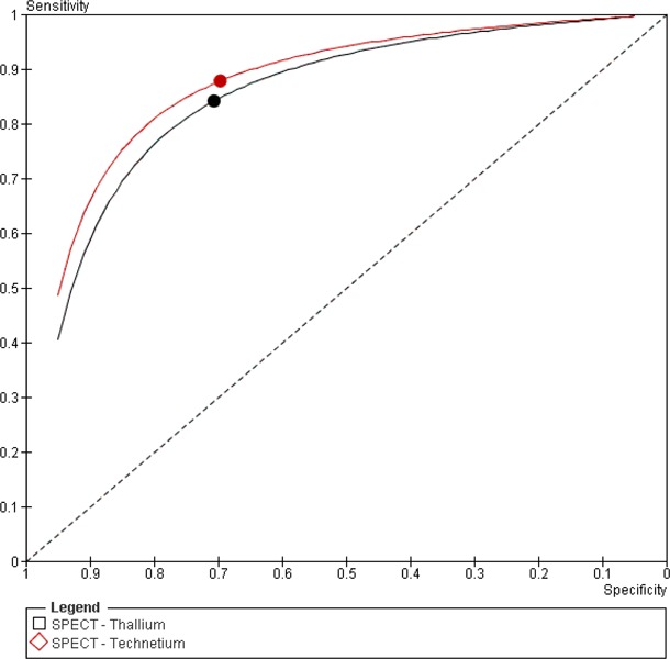
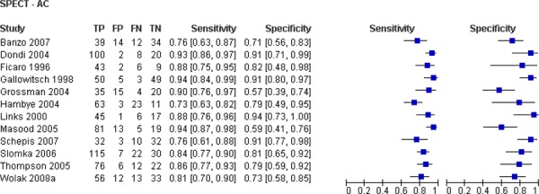
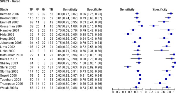
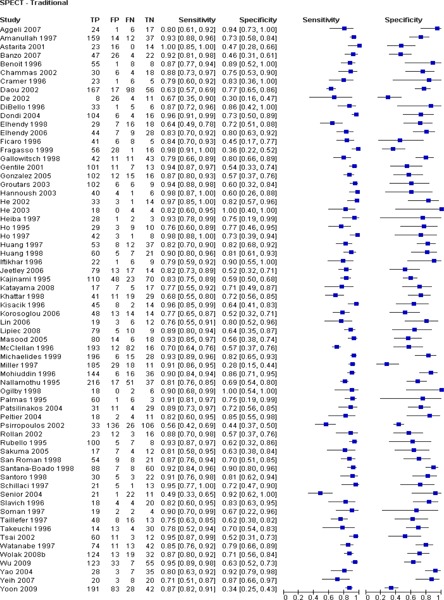
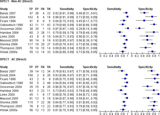
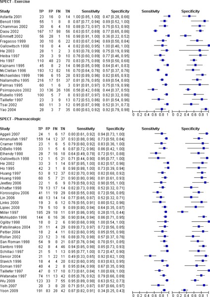
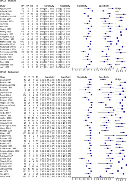
Summary of findings: Eighty-four observational studies, one non-randomized, single arm controlled clinical trial, and one poorly reported trial that appeared to be a randomized controlled trial (RCT) met the inclusion criteria for this review. All studies assessed the diagnostic accuracy of myocardial perfusion SPECT for the diagnosis of CAD using CA as a reference standard. Based on the results of these studies the following conclusions were made: According to very low quality evidence, the addition of attenuation correction to traditional or ECG-gated SPECT greatly improves the specificity of SPECT for the diagnosis of CAD although this improvement is not statistically significant. A trend towards improvement of specificity was also observed with the addition of ECG gating to traditional SPECT.According to very low quality evidence, neither the choice of stress agent (exercise or pharmacologic) nor the choice of radioactive tracer (technetium vs. thallium) significantly affect the diagnostic accuracy of SPECT for the diagnosis of CAD although a trend towards accuracy improvement was observed with the use of pharmacologic stress over exercise stress and technetium over thallium.Considerably heterogeneity was observed both within and between trials. This heterogeneity may explain why some of the differences observed between accuracy estimates for various subgroups were not statistically significant.More complex analytic techniques such as meta-regression may help to better understand which study characteristics significantly influence the diagnostic accuracy of SPECT.
Figures

Similar articles
-
Stress echocardiography with contrast for the diagnosis of coronary artery disease: an evidence-based analysis.Ont Health Technol Assess Ser. 2010;10(10):1-59. Epub 2010 Jun 1. Ont Health Technol Assess Ser. 2010. PMID: 23074387 Free PMC article.
-
Cardiac magnetic resonance imaging for the diagnosis of coronary artery disease: an evidence-based analysis.Ont Health Technol Assess Ser. 2010;10(12):1-38. Epub 2010 Jun 1. Ont Health Technol Assess Ser. 2010. PMID: 23074389 Free PMC article.
-
Stress echocardiography for the diagnosis of coronary artery disease: an evidence-based analysis.Ont Health Technol Assess Ser. 2010;10(9):1-61. Epub 2010 Jun 1. Ont Health Technol Assess Ser. 2010. PMID: 23074412 Free PMC article.
-
Noninvasive Technologies for the Diagnosis of Coronary Artery Disease in Women: Future Research Needs: Identification of Future Research Needs From Comparative Effectiveness Review No. 58 [Internet].Rockville (MD): Agency for Healthcare Research and Quality (US); 2013 Feb. Report No.: 13-EHC072-EF. Rockville (MD): Agency for Healthcare Research and Quality (US); 2013 Feb. Report No.: 13-EHC072-EF. PMID: 23905197 Free Books & Documents. Review.
-
Non-invasive imaging software to assess the functional significance of coronary stenoses: a systematic review and economic evaluation.Health Technol Assess. 2021 Sep;25(56):1-230. doi: 10.3310/hta25560. Health Technol Assess. 2021. PMID: 34588097
Cited by
-
Clinical implications of referral bias in the diagnostic performance of exercise testing for coronary artery disease.J Am Heart Assoc. 2013 Dec 13;2(6):e000505. doi: 10.1161/JAHA.113.000505. J Am Heart Assoc. 2013. PMID: 24334965 Free PMC article. Review.
-
Mapping COVID-19 functional sequelae: the perspective of nuclear medicine.Am J Nucl Med Mol Imaging. 2020 Dec 15;10(6):319-333. eCollection 2020. Am J Nucl Med Mol Imaging. 2020. PMID: 33329934 Free PMC article. Review.
-
Myocardial perfusion SPECT in Germany from 2012 to 2021: insights into development and quality indicators.Eur J Nucl Med Mol Imaging. 2023 May;50(6):1621-1628. doi: 10.1007/s00259-023-06129-z. Epub 2023 Feb 13. Eur J Nucl Med Mol Imaging. 2023. PMID: 36780003 Free PMC article.
-
Recent trends in nuclear cardiology practice.Chonnam Med J. 2013 Aug;49(2):55-64. doi: 10.4068/cmj.2013.49.2.55. Epub 2013 Aug 22. Chonnam Med J. 2013. PMID: 24010067 Free PMC article. Review.
-
Update of the Brazilian Guideline on Nuclear Cardiology - 2020.Arq Bras Cardiol. 2020 Feb;114(2):325-429. doi: 10.36660/abc.20200087. Arq Bras Cardiol. 2020. PMID: 32215507 Free PMC article. No abstract available.
References
-
- Hendel RC, Berman DS, Di Carli MF, Heidenreich PA, Henkin RE, Pellikka PA, et al. ACCF/ASNC/ACR/AHA/ASE/SCCT/SCMR/SNM 2009 Appropriate Use Criteria for Cardiac Radionuclide Imaging: A Report of the American College of Cardiology Foundation Appropriate Use Criteria Task Force, the American Society of Nuclear Cardiology, the American College of Radiology, the American Heart Association, the American Society of Echocardiography, the Society of Cardiovascular Computed Tomography, the Society for Cardiovascular Magnetic Resonance, and the Society of Nuclear Medicine. J Am Coll Cardiol. 2009;53(23):2201–29. Endorsed by the American College of Emergency Physicians. - PubMed
-
- Mowatt G, Vale L, Brazzelli M, Hernandez R, Murray A, Scott N, et al. Systematic review of the effectiveness and cost-effectiveness, and economic evaluation, of myocardial perfusion scintigraphy for the diagnosis and management of angina and myocardial infarction. Health Technol Assess. 2004;8(30):1–223. - PubMed
-
- National Health Service. Myocardial perfusion scintigraphy for the diagnosis and management of angina and myocardial infarction. [Internet] [updated 2003; cited 2009 Oct 29] Available from: http://www.nice.org.uk/TA73 .
-
- Canadian Cancer Society. Medical isotopes shortage. [Internet] [updated 2009 Dec 13; cited 2009 Dec 14] Available from: http://www.cancer.ca/Canada-wide/About%20us/Media%20centre/Issues%20watc... .
LinkOut - more resources
Full Text Sources
Research Materials
Miscellaneous