Review
. 2012 Dec;93(6):389-400.
doi: 10.1111/j.1365-2613.2012.00837.x. Epub 2012 Oct 18.

Cell sources for the regeneration of articular cartilage: the past, the horizon and the future

Affiliations
Review

Cell sources for the regeneration of articular cartilage: the past, the horizon and the future

Rachel A Oldershaw. Int J Exp Pathol. 2012 Dec.

Abstract

Avascular, aneural articular cartilage has a low capacity for self-repair and as a consequence is highly susceptible to degradative diseases such as osteoarthritis. Thus the development of cell-based therapies that repair focal defects in otherwise healthy articular cartilage is an important research target, aiming both to delay the onset of degradative diseases and to decrease the need for joint replacement surgery. This review will discuss the cell sources which are currently being investigated for the generation of chondrogenic cells. Autologous chondrocyte implantation using chondrocytes expanded ex vivo was the first chondrogenic cellular therapy to be used clinically. However, limitations in expansion potential have led to the investigation of adult mesenchymal stem cells as an alternative cell source and these therapies are beginning to enter clinical trials. The chondrogenic potential of human embryonic stem cells will also be discussed as a developmentally relevant cell source, which has the potential to generate chondrocytes with phenotype closer to that of articular cartilage. The clinical application of these chondrogenic cells is much further away as protocols and tissue engineering strategies require additional optimization. The efficacy of these cell types in the regeneration of articular cartilage tissue that is capable of withstanding biomechanical loading will be evaluated according to the developing regulatory framework to determine the most appropriate cellular therapy for adoption across an expanding patient population.


Figures

Figure 1
Figure 1
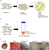
Schematic representation of autologous chondrocyte implantation (ACI). A biopsy of cartilage is taken from a non-load-bearing region of the tissue and processed by enzymatic digestion to release the chondrocytes. Ex vivo expansion of the chondrocyte culture generates sufficient cell numbers for transplantation into the focal defect within the load-bearing region of the tissue. As the ACI procedure has developed, there has been a focus on chondrocyte transplantation in combination with compatible biomaterials that improve chondrocyte retention at the site of transplantation and integration of the graft with the native tissue.
Figure 2
Figure 2
Chondrogenesis of mesenchymal stem cells (MSCs) in vivo and in vitro. (a-i) During embryonic development, initiation of chondrogenesis begins with collagen I-expressing mesenchymal cells. (a-ii) Migration of the cells and formation of mesenchymal condensations permit cell–cell contacts, and the activation of molecular signalling cascades through cell surface receptors such as N-CAM and N-cadherin. (a-iii) Mesenchymal cells undergo chondrogenic differentiation acquiring a rounded cellular morphology and depositing cartilage-specific extensive extracellular matrix (ECM) molecules (e.g. aggrecan/sGAG, collagen II) under the direction of the chondrocyte transcription factor, SOX9. (b) In vitro chondrogenic differentiation mimics in vivo chondrogenesis. (b-i) Mesenchymal stem cells are expanded ex vivo in 2D monolayer. (b-ii) Mesenchymal stem cells are placed into 3D cell aggregates and cultured in medium supplemented with TGFβ3 and dexamethasone. (b-iii) Histological evaluation of 3D cell aggregates shows evidence of chondrogenic differentiation of MSCs. From left to right – low magnification (×5) image of a safranin O (stain for sGAG) stained section shows heterogeneous tissue organization. The outer layer of the cell aggregate consists of flattened undifferentiated cells; the inner layer stains positive for sGAG-rich matrix whilst the central core is principally necrotic with no tissue deposition. Scale bar = 400 μm. Higher magnification (×40) of the inner layer of cartilage tissue shows the cells have a rounded chondrocyte morphology and have deposited an extensive ECM, which stains positively for sGAG, collagen II and aggrecan. Significantly, cell aggregates also stain positively for hypertrophic collagen X. Scale bar = 50 μm.
Figure 3
Figure 3
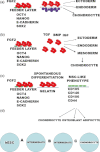
Schematic representation of the four main strategies used to generate chondrogenic cells from human embryonic stem cells (hESCs). (a) Undifferentiated pluripotent hESCs are supported by culture on a feeder cell layer in culture medium supplemented with FGF-2. Differentiation towards chondrogenic lineages is driven by co-culture with mature adult chondrocytes; (b) Undifferentiated pluripotent hESCs are supported by culture on a feeder cell layer in culture medium supplemented with FGF-2. Human embryonic stem cells are placed into suspension culture with the removal of FGF2 to form embryoid bodies. Spontaneous differentiation towards chondrogenic cell lineages is biased by supplementing the culture medium with pro-chondrogenic cytokines; (c) Undifferentiated pluripotent hESCs are supported by culture on a feeder cell layer in culture medium supplemented with FGF-2. Spontaneous differentiation is carried out by 2D monolayer culture on gelatin-coated tissue culture plastic to enrich for mesenchymal stem cell (MSC)-like cell populations. Chondrogenic differentiation is subsequently carried out using standard chondrogenic protocols. (d) Targeted differentiation strategies segment the differentiation protocol into segments, each one enriching for developmental intermediate populations to generate a chondrogenic cell population.
Figure 4
Figure 4
Targeted differentiation of human embryonic stem cells (hESCs) to chondrogenic cells. (a–l) Phase contrast images of hESCs at each stage of the targeted differentiation protocol described by Oldershaw et al. (2010a,b). (a, b) Pluripotent hES cells cultured on murine embryonic fibroblast (MEF) feeder layers form heterogeneous cultures of compact colonies. (c, d) Feeder-free hESCs (FF-hESCs) are cultured as a monolayer on a fibronectin matrix substrate. Cell–cell contact is maintained and individual cells are seen to have large nuclei with prominent nucleoli. (e, f) At the end of Stage 1, there are no obvious gross changes in cellular morphology; however, by the end of Stage 2 (g, h), the sporadic formation of cell clusters reminiscent of mesenchymal condensations is observed throughout the cultures. (i, j) At the beginning of Stage 3, the cell clusters have increased in size and the surrounding cells are seen to detach from the culture dish such that by the end of the differentiation protocol only individual cell aggregates remain in culture (k, l). (m) Individual chondrogenic cell aggregates are shown to deposit cartilage-specific collagen II. (n) Phase contrast images of chondrogenic cell aggregates before and after staining with safranin O demonstrate the deposition of sGAG. Control cultures were treated with chondroitinase prior to staining with safranin O. Weak safranin O staining in these control cultures demonstrated that chondroitin sulphate was a principal sGAG within the extensive extracellular matrix (ECM). All scale bars = 100 μm.

References

    1. Paget J. The classics. II. Healing of cartilage. Clin. Orthop. Relat. Res. 1969;64:7–8. - PubMed
    1. Adesida AB, Mulet-Sierra A, Jomha NM. Hypoxia mediated isolation and expansion enhances the chondrogenic capacity of bone marrow mesenchymal stromal cells. Stem Cell Res. Ther. 2012;3:9. - PMC - PubMed
    1. Akiyama H, Chaboissier M-C, Martin JF, Schedl A, de Crombrugghe B. The transcription factor Sox9 has essential roles in successive steps of the chondrocyte differentiation pathway and is required for expression of Sox5 and Sox6. Genes Dev. 2002;16:2813–2828. - PMC - PubMed
    1. Azarin SM, Palecek SP. Matrix revolutions: a trinity of defined substrates for long-term expansion of human ESCs. Cell Stem Cell. 2010;7:7–8. - PubMed
    1. Bai HY, Chen GA, Mao GH, Song TR, Wang YX. Three step derivation of cartilage like tissue from human embryonic stem cells by 2D-3D sequential culture in vitro and further implantation in vivo on alginate/PLGA scaffolds. J. Biomed. Mater. Res. A. 2010;94:539–546. - PubMed

Publication types

MeSH terms