

Vector competence of Culicoides sonorensis (Diptera: Ceratopogonidae) to epizootic hemorrhagic disease virus serotype 7
- PMID: 23075098
- PMCID: PMC3504516
- DOI: 10.1186/1756-3305-5-236
Vector competence of Culicoides sonorensis (Diptera: Ceratopogonidae) to epizootic hemorrhagic disease virus serotype 7
Abstract
Background: Culicoides sonorensis (Diptera: Ceratopogonidae) is a vector of epizootic hemorrhagic disease virus (EHDV) serotypes 1 and 2 in North America, where these viruses are well-known pathogens of white-tailed deer (WTD) and other wild ruminants. Although historically rare, reports of clinical EHDV infection in cattle have increased in some parts of the world over the past decade. In 2006, an EHDV-7 epizootic in cattle resulted in economic loss for the Israeli dairy industry. White-tailed deer are susceptible to EHDV-7 infection and disease; however, this serotype is exotic to the US and the susceptibility of C. sonorensis to this cattle-virulent EHDV is not known. The objective of the study was to determine if C. sonorensis is susceptible to EHDV-7 infection and is a competent vector.
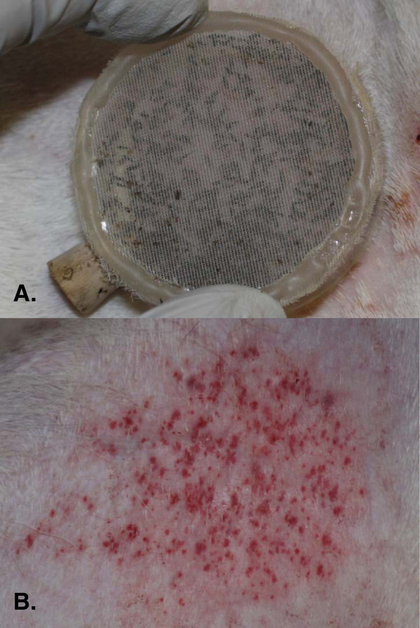
Methods: To evaluate the susceptibility of C. sonorensis, midges were fed on EHDV-7 infected WTD, held at 22 ± 1°C, and processed individually for virus isolation and titration on 4-16 days post feeding (dpf). Midges with a virus titer of ≥ 10(2.7) median tissue culture infective doses (TCID(50))/midge were considered potentially competent. To determine if infected C. sonorensis were capable of transmitting EHDV-7 to a host, a susceptible WTD was then fed on by a group of 14-16 dpf midges.
Results: From 4-16 dpf, 45% (156/350) of midges that fed on WTD with high titer viremia (>10(7) TCID(50)/ml) were virus isolation-positive, and starting from 10-16 dpf, 32% (35/109) of these virus isolation-positive midges were potentially competent (≥ 10(2.7) TCID(50)/midge). Midges that fed on infected deer transmitted the virus to a susceptible WTD at 14-16 dpf. The WTD developed viremia and severe clinical disease.
Conclusion: This study demonstrates that C. sonorensis is susceptible to EHDV-7 infection and can transmit the virus to susceptible WTD, thus, C. sonorensis should be considered a potential vector of EHDV-7. Together with previous work, this study demonstrates that North America has a susceptible ruminant and vector host for this exotic, cattle-virulent strain of EHDV-7.
Figures
References
-
- Anthony SJ, Maan S, Maan N, Kgosana L, Bachanek-Bankowska K, Batten C, Darpel KE, Sutton G, Attoui H, Mertens PPC. Genetic and phylogenetic analysis of the outer-coat proteins VP2 and VP5 of epizootic haemorrhagic disease virus (EHDV): Comparison of genetic and serological data to characterize the EHDV serogroup. Virus Res. 2009;145:200–210. doi: 10.1016/j.virusres.2009.07.012. - DOI - PubMed
-
- Allison AB, Goekjian GH, Potgieter C, Wilson W, Johnson D, Mertens PPC, Stallknecht D. Detection of a novel reassortant epizootic hemorrhagic disease virus (EHDV) in the USA containing RNA segments derived from both exotic (EHDV-6) and endemic (EHDV-2) serotypes. J Gen Virol. 2010;91:430–439. doi: 10.1099/vir.0.015651-0. - DOI - PubMed
-
- Howerth EW, Stallknecht DE, Kirkland PD. In: Infectious Diseases of Wild Mammals. 3. Williams ES, Barker IK, editor. Ames, Iowa: Iowa State Press; 2001. Bluetongue, epizootic hemorrhagic disease, and other orbivirus-related diseases; pp. 77–97.
-
- Bowen RA. Serologic responses of calves to sequential infections with epizootic hemorrhagic disease virus serotypes. Am J Vet Res. 1987;48:1449–1452. - PubMed
-
- Metcalf HE, Luedke AJ, Jochim MM. Walton TE, Osburn BI. Boca Raton, Florida: CRC Press; 1992. Epizootic hemorrhagic disease virus infection in cattle; pp. 222–237.
Publication types
MeSH terms
LinkOut - more resources
Full Text Sources
Miscellaneous
