Review
. 2013 May 1;18(13):1623-41.
doi: 10.1089/ars.2012.4973. Epub 2013 Jan 9.

Kinetics and mechanisms of thiol-disulfide exchange covering direct substitution and thiol oxidation-mediated pathways

Affiliations
Review

Kinetics and mechanisms of thiol-disulfide exchange covering direct substitution and thiol oxidation-mediated pathways

Péter Nagy. Antioxid Redox Signal. .

Abstract

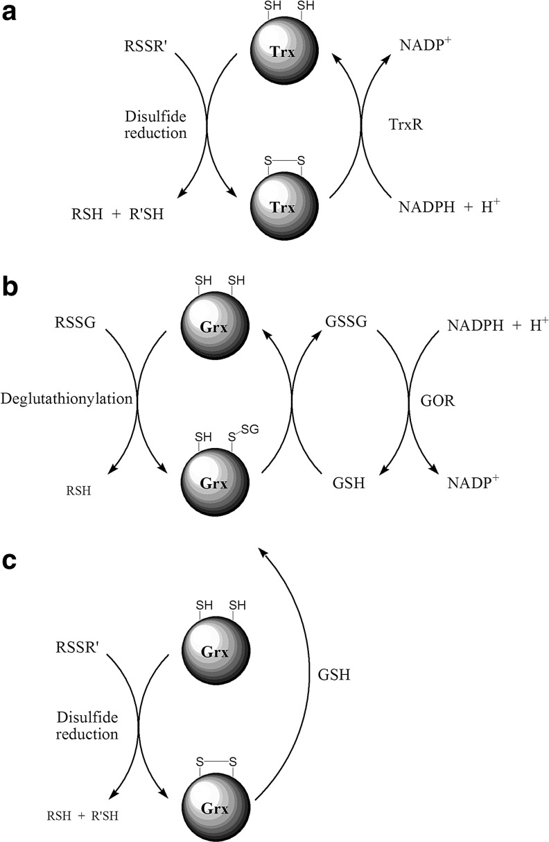
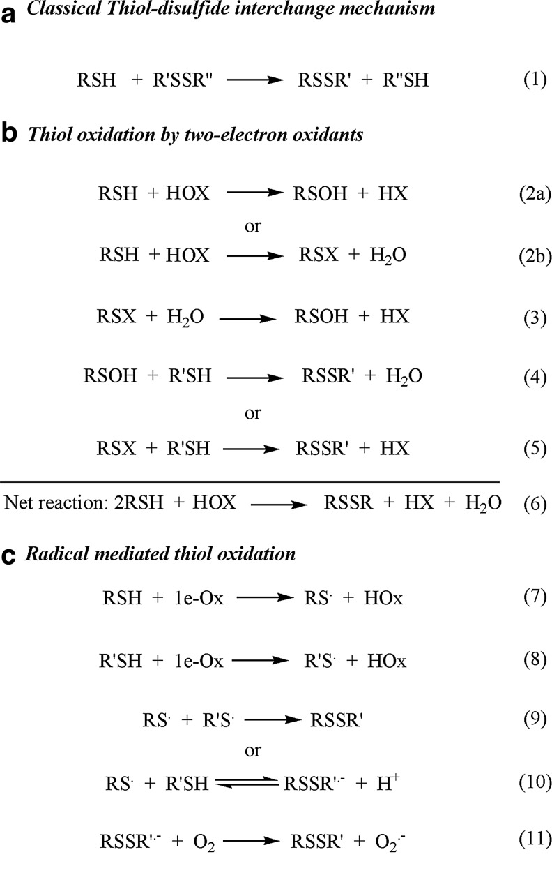
Significance: Disulfides are important building blocks in the secondary and tertiary structures of proteins, serving as inter- and intra-subunit cross links. Disulfides are also the major products of thiol oxidation, a process that has primary roles in defense mechanisms against oxidative stress and in redox regulation of cell signaling. Although disulfides are relatively stable, their reduction, isomerisation, and interconversion as well as their production reactions are catalyzed by delicate enzyme machineries, providing a dynamic system in biology. Redox homeostasis, a thermodynamic parameter that determines which reactions can occur in cellular compartments, is also balanced by the thiol-disulfide pool. However, it is the kinetic properties of the reactions that best represent cell dynamics, because the partitioning of the possible reactions depends on kinetic parameters.

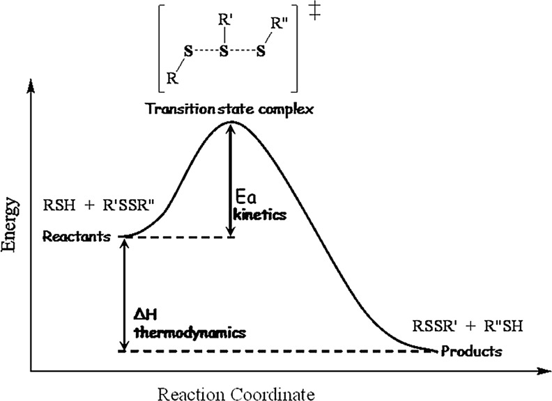
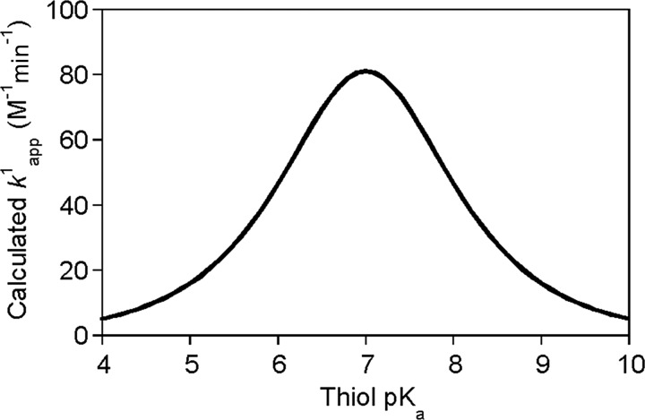
Critical issues: This review is focused on the kinetics and mechanisms of thiol-disulfide substitution and redox reactions. It summarizes the challenges and advances that are associated with kinetic investigations in small molecular and enzymatic systems from a rigorous chemical perspective using biological examples. The most important parameters that influence reaction rates are discussed in detail.

Recent advances and future directions: Kinetic studies of proteins are more challenging than small molecules, and quite often investigators are forced to sacrifice the rigor of the experimental approach to obtain the important kinetic and mechanistic information. However, recent technological advances allow a more comprehensive analysis of enzymatic systems via using the systematic kinetics apparatus that was developed for small molecule reactions, which is expected to provide further insight into the cell's machinery.


Figures

FIG. 1.
FIG. 1.
Mechanism for the folding process of polypeptides in the endoplasmic reticulum (ER) of eukaryotes. In eukaryotes folding of nonfunctional polypeptides in the ER is governed via disulfide bond formation by protein disulfide isomerase (PDI). The new protein–disulfide bond is introduced via thiol–disulfide exchange reactions between the oxidized PDI and the substrate protein thiols. In the first step, PDI forms a mixed disulfide intermediate species (not shown), which is reduced by another thiol on the substrate to give reduced PDI and the new protein-disulfide moiety. PDI is recycled via thiol–disulfide exchange reactions with the membrane-bound flavoprotein ER oxidoreductin (Ero1). Direct oxidation of PDI occurs by a disulfide that is present on a flexible loop of Ero1. Ero1 is reoxidized by oxygen reduction through its flavin adenine dinucleotide (FAD) domain, which subsequently oxidizes the so-called shuttle CXXC motif in its close proximity. The active disulfide on the loop forms via intramolecular thiol–disulfide exchange with the oxidized shuttle CXXC motif. Superoxide that forms during oxygen reduction dismutates to give H2O2 and O2. H2O2 was recently proposed to preferentially oxidize peroxiredoxin 4 (Prx)4 to its dimeric form, which can subsequently reoxidize PDI via thiol–disulfide exchange, providing a feedback model for more efficient utilization of the oxidizing equivalents to create new disulfides. An alternative way to introduce disulfide moieties into unfolded proteins is via the quiescin-sulfhydryl oxidase (QSOX) family of enzymes. These proteins are proposed to function with a similar mechanism as Ero1 reoxidizes PDI, but they directly oxidize the unfolded proteins (60). Interestingly, beside their promiscuous substrate specificity they only poorly oxidize PDI. In addition, while the reduced form of PDI was shown to be involved in the reduction and isomerization of misfolded protein disulfides (not shown), QSOX does not catalyze these processes and in the absence of PDI, incorrectly paired disulfide bonds (generated by QSOX) accumulate (60).
FIG. 2.
FIG. 2.
Mechanism for polypeptide folding in the mitochondrial inter-membrane space (IMS) of eukaryotes. Freshly synthesized polypeptides translocate from the cytosol to the IMS, where folding is facilitated by Mia40, via donating the disulfide bond of its CPC domain through a mixed disulfide intermediate species (45). Mia40 is subsequently reoxidized to its active form by the disulfide on the N-terminal shuttle domain of the flavoprotein essential for respiration and vegetative growth sulfhydryl oxidase (Erv1). In the homodimeric Erv1 protein, the active shuttle domain disulfide is reformed via intersubunit thiol–disulfide exchange with the CXXC motif of the other Erv1 in the homodimer. The CXXC is reoxidized via consecutive one-electron oxidation through the FAD domain of the same subunit. From the FAD, the electrons flow via the respiratory chain cytochrome C (Cyt C) and cytochrome C oxidase (COX) back to the stroma in the mitochondria to give H2O and avoid H2O2 production.
FIG. 3.
FIG. 3.
Protein folding in the periplasmic space of prokaryotes. (a) Unfolded polypeptides enter the periplasm, where the soluble disulfide bond protein family A (DsbA) protein catalyzes the folding process via introducing new disulfide bonds in thiol–disulfide exchange reactions through mixed disulfide intermediates. DsbA is reoxidized by DsbB. During the recycling of DsbB after an intramolecular thiol–disulfide interchange between two DsbB Cys pairs, the electrons flow (under aerobic conditions) via ubiquinone (UQ) and cytochrome oxidase (COX) of the electron transport chain to eventually reduce oxygen to H2O in the cytoplasm. (b) Reduction or isomerisation of misfolded proteins occur by the homodimeric DsbC coupled to DsbD via the flow of electrons to the opposite direction. DsbC needs to be in the reduced state, which is maintained by the membrane-bound DsbD to which the reducing equivalents are supplied by cytoplasmic NADPH via thioredoxin (Trx). The oxidizing equivalents travel from the periplasm to the cytoplasm via a cascade of intramolecular thiol–disulfide interchange reactions through the transmembrane domain of DsbD on to Trx. A major difference in the protein-folding mechanism in prokaryotes versus eukaryotes is that while a separate reduction/isomerization machinery exists in prokaryotes (DsbC-DsbD), this process is catalyzed by the PDI and Mia40 disulfide relay systems in eukaryotes. Also, despite the functional similarities of Ero1 and DsbB, they lack sequence homology. The only similarity is the Cys pairs.
FIG. 4.
FIG. 4.
General mechanism for the peroxidase function of Prx. The peroxidative Cys (Cp) reacts with the peroxide oxidant to give a CySpOH derivative. Prx-CySpOH is reduced by a reducing Cysr of another Prx to give the disulfide. Alternatively, CySpOH can be further oxidized to the corresponding CySpO2H derivative by a second equivalent of peroxide. Reduction of the disulfide occurs by Trx (using NADPH) and the CySpO2H derivative is slowly recycled by sulfiredoxins (using ATP).
FIG. 5.
FIG. 5.
General mechanisms for disulfide reduction. (a) Trx are responsible for the reduction of a wide variety of protein disulfide bonds. Reduction occurs via formation of intermediate mixed disulfides. The reduced substrate is then released via an intramolecular nucleophilic attack of the resolving Trx thiolate on the sulfur of the N-terminal Trx Cys that is engaged in the mixed disulfide bond. Trx is recycled via thioredoxin reductase (TrxR) and the reducing equivalents are supplied by NADPH. (b) Glutathionylated protein thiols are catalytically reduced by glutaredoxin (Grx). The protein-bound glutathione is transferred to Grx through a nucleophilic attack of the Grx N-terminal Cys on the GS-sulfur. An important difference in the catalytic mechanism of Grx compared to Trx is that the glutathionylated Grx is reduced in an intermolecular reaction with another reduced glutathione (GSH) as opposed to intramolecular Trx disulfide formation. Oxidized glutathione (GSSG) is recycled via Glutathione oxido-reductase (GOR) catalyzed reduction by NADPH. (c) Grx can also reduce protein disulfides by the dithiol mechanism, which includes a nucleophilic attack of the N-terminal Cys of Grx on the disulfide moiety to give a mixed disulfide. This mixed disulfide is subsequently reduced by the C-terminal Grx Cys in a similar intramolecular fashion as for Trx (see Fig. 5a). The Grx disulfide moiety is reduced by GSH, where the intermediate glutathionylated N-terminal Cys reacts preferentially with another GSH over the C-terminal Grx Cys, just like during the reduction of glutathionylated proteins.
Scheme 1.
Scheme 1.
General mechanisms for thiol–disulfide exchange via direct substitution (a) or (b-c) thiol oxidation. For the sake of simplicity, the depicted models do not take into account the different protonation states of the reactants and products. (a) The classical thiol–disulfide interchange mechanism is consistent with an SN2 type model, where in a single reaction step (1) the attacking sulfur (RS-), the nucleophile, binds to the central sulfur of the disulfide (R′S-) and the leaving thiol (R′′SH) is released via a trisulfide-like transition state structure. (b) Two-electron thiol oxidation to disulfide can occur via multiple mechanisms. The most common pathways are via a sulfenic acid (RSOH; 2a) or an alternative sulfenyl (RSX, e.g., sulfenyl-halides when X=Cl, Br, or I; 2b) intermediate. RSOH reacts rapidly with thiols to give the corresponding disulfide species (4). RSX can hydrolyze to sulfenic acid (3) or it could react directly with another thiol to give the disulfide (5). (c) One-electron oxidation of Cys generates thiyl radicals ( and 8). Recombination of two thiyl radicals results in disulfide formation (9). However, under physiological conditions, thiyl radicals are more likely to react with thiols to give the disulfide radical anion (10), which captures oxygen in a diffusion-controlled reaction to give the closed shell disulfide and superoxide (11).
FIG. 6.
FIG. 6.
Schematic energy profile of Reaction 1 as a function of the reaction coordinate. The reaction mechanism is consistent with an SN2-type nucleophilic substitution via a linear -S-S-S- like transition state. Ea represents the activation energy (the parameter that determines the rate of the reaction) and ΔH measures the free enthalpy change during the reaction (corresponding to the thermodynamic feasibility of the reaction).
FIG. 7.
FIG. 7.
Calculated formula image for thiol-GSSG interchange reactions as a function of thiol pKa at pH 7 based on the Brønsted relationship that was established by Whitesides. Using formula image (equation 21), the pH independent rate constant (k17) was calculated at different thiol pKa values. Substituting these rate constants into eq 20 (Box 1), the obtained formula image at a given pH for Reaction 1 were plotted as a function of thiol pKa (in the range of pKa=4–10) at pH 7. The figure clearly demonstrates that decreasing the attacking thiol's pKa will only result in an overall rate enhancement until the thiolate becomes the dominant species (i.e., until the thiol pKa approaches the applied pH). Beyond this, the drop in the nucleophilicity of the sulfur center with the pKa will determine the rate and result in slower kinetics.
Scheme 2.
Scheme 2.
Macroscopic (a) and microscopic (b) speciation of reduced glutathione at pH>5 with the corresponding macroscopic and microscopic equilibrium constants.
FIG. 8.
FIG. 8.
Model for the activation of H2O2 at the active site of Prx via H-bonding interactions with two highly conserved Arg residues. The figure shows the proposed orientation of H2O2 at the active site of bovine Prx3 (Protein Data Bank code 1ZYE) (16). The distances of the Arg nitrogens from the peroxidative Cys47 sulfur are ideal to engage in H-bonding interactions with the sulfur center and the reacting and leaving oxygens of the peroxide. We proposed that Arg 123 anchors the reacting oxygen to the thiolate sulfur and Arg 146 stabilizes the leaving group (77). In addition, we suggested that the two Arg residues pull the oxygens apart, which activate the peroxide for the OH+ transfer to the thiolate.

References

    1. Adams GE. McNaught GS. Michael BD. Pulse radiolysis of sulphur compounds 2. Free radical repair by hydrogen transfer from sulphydryl compounds. Trans Farad Soc. 1968;64:902–910.
    1. Ainavarapu SRK. Wiita AP. Dougan L. Uggerud E. Fernandez JM. Single-molecule force spectroscopy measurements of bond elongation during a bimolecular reaction. J Am Chem Soc. 2008;130:6479–6487. - PubMed
    1. Alegre-Cebollada J. Kosuri P. Andres Rivas-Pardo J. Fernandez JM. Direct observation of disulfide isomerization in a single protein. Nat Chem. 2011;3:882–887. - PMC - PubMed
    1. Armesto XL. Canle LM. Fernandez MI. Garcia MV. Santaballa JA. First steps in the oxidation of sulfur-containing amino acids by hypohalogenation: very fast generation of intermediate sulfenyl halides and halosulfonium cations. Tetrahedron. 2000;56:1103–1109.
    1. Armstrong DA. Applications of pulse-radiolysis for the study of short-lived sulfur species. In: Chatgilialogy C, editor; Asmas KD, editor. Sulfur-centered reactive intermediates in chemistry and biology. New York: Plenum Press; 1990. pp. 121–134.

Publication types