. 2013 Apr;19(7-8):809-23.
doi: 10.1089/ten.TEA.2012.0309. Epub 2012 Nov 30.

Impact of electrospun conduit fiber diameter and enclosing pouch pore size on vascular constructs grown within rat peritoneal cavities

Affiliations

Impact of electrospun conduit fiber diameter and enclosing pouch pore size on vascular constructs grown within rat peritoneal cavities

Chris A Bashur et al. Tissue Eng Part A. 2013 Apr.

Abstract

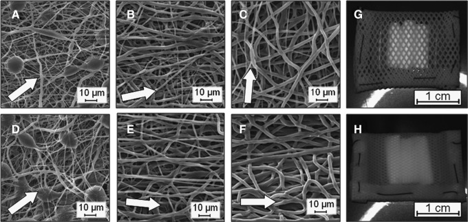
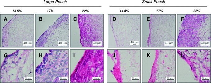
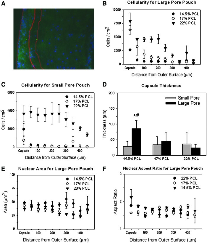
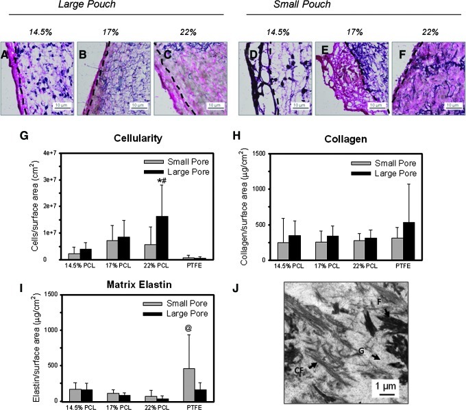
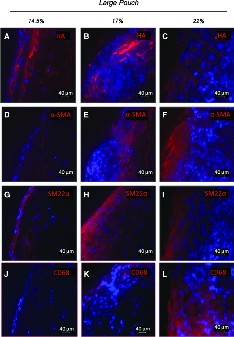
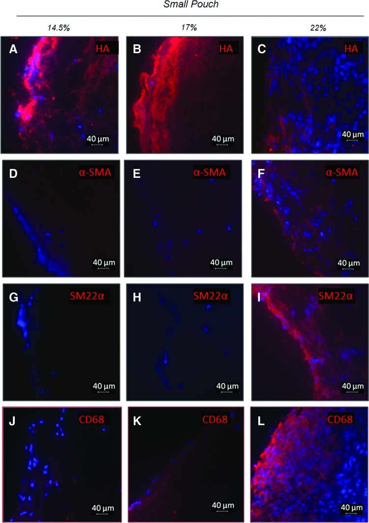
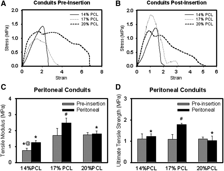
The generation of vascular grafts by recruiting autologous cells within the peritoneal cavity has shown promise. However, the microenvironment affects cell differentiation and elastic matrix production. Therefore, this study determined the impact of systematic changes in the average fiber diameter of electrospun poly(ɛ-caprolactone) conduits, and the pore size of pouches used to enclose the conduits, on recruited cells. After 2 weeks in the peritoneal cavity, fibrous capsules formed containing macrophages, α-smooth muscle actin (α-SMA)(+) and SM22α(+) myofibroblastic or smooth muscle like-cells, and what appeared to be mesothelial cells on the outer surfaces. These cells infiltrated and deposited matrix (e.g., collagen, hyaluoronan, and limited elastin) within conduit walls. Constructs enclosed within the largest pore pouches exhibited significantly better tissue generation responses (e.g., better cell infiltration, elongation, and matrix deposition). Additionally, the healing response was impacted by the conduit average fiber diameter, and consequently, the effective pore diameter, with the largest diameter fibers promoting the most positive healing response (e.g., greater total cellularity, extracellular matrix deposition, and α-SMA(+) cells). Six weeks post-intra-aortal grafting, constructs were occluded, but significant remodeling also occurred in the arterial microenvironment. Overall, these results demonstrate the importance of microenvironmental cues on recruited peritoneal cells and the necessity of developing strategies to further improve elastic matrix synthesis.


Figures

FIG. 1.
FIG. 1.
Electrospun poly(ɛ-caprolactone) (PCL) conduits exhibit a range of average fiber diameters. Shown are scanning electron microscopy images of meshes electrospun from solutions containing 14.5% w/v (A, D), 17% w/v (B, E), and 22% w/v (C, F) of PCL. Images (A–C) are of the outer surface and images (D–F) are of the luminal surface of the conduits. An arrow indicates the average direction of fiber orientation. These constructs were included within poly(tetrafluoroethylene) (PTFE) pouches with either larger (G) or smaller (H) pores to allow peritoneal cells and fluid to penetrate.
FIG. 2.
FIG. 2.
Effect of average fiber diameter and pouch pore size on the morphology of recruited peritoneal cells and generated extracellular matrix (ECM). Shown are representative high- and low-magnification hematoxylin and eosin (H&E) images of 14.5% w/v (A, D, G, J), 17% w/v (B, E, H, K), and 22% w/v (C, F, I, L) that demonstrate differences in cell infiltration and cell morphology between different electrospinning concentrations. Cell nuclei are blue and cell cytoplasms and ECM (e.g., collagen) are pink. Conduits are shown after insertion in larger (A–C, G–I) or smaller pore pouches (D–E, J–L), showing differences in cell morphology and generated matrix. The outer surface of the construct is on the left side. Color images available online at www.liebertpub.com/tea
FIG. 3.
FIG. 3.
Impact of electrospun fiber diameter and pouch pore size on peritoneal cell infiltration. Included is a large-pore pouch, 14.5% w/v PCL condition overlay image (A) with 4′,6-diamino-2-phenylindole dihydrochloride (DAPI)-stained nuclei (blue) and background autofluorescence (green) that shows the areas of interest created for the capsule and the first 50 μm within the electrospun fibers. The impact of electrospun condition on cellularity throughout the conduit wall (i.e., cell infiltration) was quantified for conduits within both large-pore (B) and small-pore (C) pouches. Results demonstrated greater cell infiltration with increasing electrospinning concentration. The capsule thickness for the different conditions were also compared (D). The nuclear area (E) and aspect ratio (F) are shown for the large-pore pouches and demonstrate the change throughout the thickness of the conduit wall. The results are for n=4 images/condition. *Statistical significance from the 22% w/v PCL condition; #significance from the small-pore pouch. Color images available online at www.liebertpub.com/tea
FIG. 4.
FIG. 4.
Effect of average fiber diameter and pouch pore size on the composition and distribution of ECM components after 14 days in the peritoneal cavity. Shown are representative high-magnification images of modified Verhoff's stained 14.5% w/v (A, D), 17% w/v (B, E), and 22% w/v (C, F) constructs. Constructs are shown after insertion in larger (A–C) or smaller pore pouches (D–F). Verhoff's Van Gieson stain shows collagen (pink/red), nuclei (light brown), elastic fibers (dark brown/black, as verified with an aorta-positive control), and PCL fibers and sporadic beads (dark purple). The outer surface of the construct is on the left side, and the start of the conduit walls are marked by dashed lines. Biochemical analysis was performed for recruited peritoneal cell densities as determined from DNA content (G), collagen content determined with a hydroxyproline assay (H), and matrix elastin content as determined with the Fastin® assay (I). *Statistical significance from the small-pore pouch; #significance from the expanded PTFE (ePTFE) control and the smallest diameter 14.5% conduit; @significance from 22% PCL w/v for n=6 samples/condition and n=2 rats/condition. These figures show that significant collagen, but only a few elastic fibers are within the conduits. Detailed matrix structure was visualized using transmission electron microscopy (J). C, collagen fibrils; F, fibrin fibrils; G, ground substance. Color images available online at www.liebertpub.com/tea
FIG. 5.
FIG. 5.
Effect of average fiber diameter on the cell phenotype, cell morphology, and ECM components for conduits enclosed within large pore pouches. Shown are representative immunofluorescent images for hyaluronan (HA) (A–C) and myofibroblast, SMC, and macrophage markers. Markers include α-smooth muscle actin (α-SMA) (D–F), SM22α (G–I), and CD68 (J–L). DAPI-stained nuclei are shown in blue. The outer surface of the construct is on the left side. Color images available online at www.liebertpub.com/tea
FIG. 6.
FIG. 6.
Effect of average fiber diameter on the cell phenotype, cell morphology, and ECM components for conduits enclosed within small-pore pouches. Shown are representative immunofluorescent images for HA (A–C) and myofibroblast, SMC, and macrophage markers. Markers include α-SMA (D–F), SM22α (G–I), and CD68 (J–L). DAPI-stained nuclei are shown in blue. The outer surface of the construct is on the left side. Color images available online at www.liebertpub.com/tea
FIG. 7.
FIG. 7.
Tensile properties of electrospun conduits/constructs. Shown are representative stress–strain curves for conduits both without implantation (A) and after 2 weeks of implantation (B) within the peritoneal cavity. The average tensile moduli (C) and ultimate tensile strength (D) for the different conditions are also shown for n=6 samples/condition preinsertion and n=3 samples/condition after 2 weeks in the peritoneal cavity. *Statistical significance from the 17% PCL condition; #significance change with 2 weeks in the peritoneal cavity; @significance from the 20% PCL condition.

References

    1. McGill H.C., Jr. McMahan C.A. Herderick E.E. Malcom G.T. Tracy R.E. Strong J.P. Origin of atherosclerosis in childhood and adolescence. Am J Clin Nutr. 2000;72:1307S. - PubMed
    1. Lloyd-Jones D. Adams R. Carnethon M. De Simone G. Ferguson T.B. Flegal K. Ford E. Furie K. Go A. Greenlund K. Haase N. Hailpern S. Ho M. Howard V. Kissela B. Kittner S. Lackland D. Lisabeth L. Marelli A. McDermott M. Meigs J. Mozaffarian D. Nichol G. O'Donnell C. Roger V. Rosamond W. Sacco R. Sorlie P. Stafford R. Steinberger J. Thom T. Wasserthiel-Smoller S. Wong N. Wylie-Rosett J. Hong Y. Heart disease and stroke statistics—2009 update: a report from the American Heart Association Statistics Committee and Stroke Statistics Subcommittee. Circulation. 2009;119:e21. - PubMed
    1. Klinkert P. Post P.N. Breslau P.J. van Bockel J.H. Saphenous vein versus PTFE for above-knee femoropopliteal bypass. A review of the literature. Eur J Vasc Endovasc Surg. 2004;27:357. - PubMed
    1. Chlupac J. Filova E. Bacakova L. Blood vessel replacement: 50 years of development and tissue engineering paradigms in vascular surgery. Physiol Res. 2009;58(Suppl 2):S119. - PubMed
    1. Kielty C.M. Stephan S. Sherratt M.J. Williamson M. Shuttleworth C.A. Applying elastic fibre biology in vascular tissue engineering. Philos Trans R Soc Lond B Biol Sci. 2007;362:1293. - PMC - PubMed

Publication types

LinkOut - more resources