Review
. 2013 Jan;1834(1):247-71.
doi: 10.1016/j.bbapap.2012.10.005. Epub 2012 Oct 14.

Recent advances in the structural mechanisms of DNA glycosylases

Affiliations
Review

Recent advances in the structural mechanisms of DNA glycosylases

Sonja C Brooks et al. Biochim Biophys Acta. 2013 Jan.

Abstract

DNA glycosylases safeguard the genome by locating and excising a diverse array of aberrant nucleobases created from oxidation, alkylation, and deamination of DNA. Since the discovery 28years ago that these enzymes employ a base flipping mechanism to trap their substrates, six different protein architectures have been identified to perform the same basic task. Work over the past several years has unraveled details for how the various DNA glycosylases survey DNA, detect damage within the duplex, select for the correct modification, and catalyze base excision. Here, we provide a broad overview of these latest advances in glycosylase mechanisms gleaned from structural enzymology, highlighting features common to all glycosylases as well as key differences that define their particular substrate specificities.


Figures

Figure 1
Figure 1
Common DNA lesions referenced in this review. (A) Oxidized nucleobases. 8-OHG, 7,8-dihydro-8-hydroxyguanine; 8oxoG, 7,8-dihydro-8oxoGuanine; FapyG, 2,6-diamino-4-hydroxy-5-formamidopyrimidine; mFapyG, N7-methylFapyG; Tg, thymine glycol; Sp, spiroiminodihydantoin; Gh, guanidinohydantoin; Ia, iminoallantion; 5-OHU, 5-hydroxyuracil; DHU, dihydrouracil; 5-OHC, 5-hydroxycytosine; DHT, dihydrothymine. (B) Alkylated nucleobases. εA, 1,N6-ethenoadenine; εC, 3,N4-ethenocytosine; 3mA, N3-methyladenine; 3mG, N3-methylguanine; 7mG, N7-methylguanine; Hx, hypoxanthine. (C) Nucleobases repaired by the UDG/TDG family of DNA glycosylases. U, uracil; T, thymine; 5mC, 5-methylcytosine; 5hmC, 5-hydroxymethylcytosine; 5fC, 5-formylcytosine; 5caC, 5-carboxylcytosine.
Figure 2
Figure 2
Chemical reaction catalyzed by DNA glycosylases. (A,B) Monofunctional glycosylases cleave the N-glycosidic bond to liberate free nucleobase (N) from the phosphoribose backbone through either associative (A) or dissociative (B) mechanisms. (C) Bifunctional mechanism, in which both the N-glycosidic bond and the DNA backbone are cleaved.
Figure 3
Figure 3
DNA glycosylase structural superfamilies. Representative crystal structures from each class shown are: EndoV, T4 pyrimidine dimer DNA glycosylase EndoV (PDB ID 1VAS); UDG, human uracil-DNA glycosylase UDG (1EMH); Helix-hairpin-Helix (HhH), human 8oxoGuanine DNA glycosylase OGG1 (1YQK); Helix-two turn-helix (H2TH), Bacillus stearothermophilus 8oxoGuanine DNA glycosylase MutM (1L1T); AAG, human alkyladenine DNA glycosylase AAG/MPG (1EWN); ALK, Bacillus cereus alkylpurine DNA glycosylase AlkD (3JXZ). Proteins are colored according to secondary structure with the HhH and H2TH domains magenta. DNA is shown as gray sticks.
Figure 4
Figure 4
Oxidative DNA glycosylases. (A–C) OGG1, represented by human OGG1 (PDB ID 1EBM), (D–F) OGG2, represented by MjOGG (PDB ID 3KNT), (G–H) Pyrobaculum aerophilum AGOG (PDB ID 1XQP) and (I–J) Geobacillus stearothermophilus MutM. The overall folds of each enzyme are shown on the top row (blue HhH motif), active sites on the second row, and opposing base on the bottom row. In the close-up views, the protein side-chains are grey and the DNA orange. Water molecules are represented by red spheres and hydrogen bonds are shown as dashed lines. (B) The human OGG1 8oxoG recognition pocket. The only 8oxoG specific contact is the hydrogen bond from the carbonyl group of Gly42 to the protonated N7 of 8oxoG. (C) The high specificity of hOGG1 for 8oxoG•C base pairs can be rationalized by the 5 hydrogen bonds between the opposite cytosine and 3 side chains. (E) In MjOGG, the 8oxoG N7 donates a hydrogen bond (red dashed line) to the C-terminal Lys207 carboxylate. (F) The opposite cytosine in MjOGG is contacted by only one side chain. (H) 8oxoG nucleoside bound inside the AGOG active site, with a unique 8oxoG-specific contact to Trp222 (red dashed line). (J) Active site of MutM (PDB ID 1R2Y) shows multiple contacts to 8oxoG but lacks the aromatic residues seen in the OGG1, OGG2, and AGOG enzymes.
Figure 5
Figure 5
EndoIII/Nth and EndoVIII/Nei. (A) Bacillus stearothermophilus Endonuclease III (BsEndoIII) (PDB ID 1P59) bound to THF-DNA (gold). The THF moiety and iron-sulfur cluster are shown as sticks in the center and right side of the figure, respectively. The HhH DNA binding motif is magenta. (B) Mimivirus NEIL1 ortholog MvNei1 (3A46) bound to THF-DNA (gold). The H2TH motif is colored magenta and the zincless finger is cyan. (C) Overlay of the zinc finger from Escherichia coli Endonuclease VIII (EcNei) (1K3W) (gold) with zincless finger motifs from human NEIL1 (1TDH, blue) and MvNei1 (cyan). The zinc ion in EcNei is depicted as a gold sphere. A red star denotes the general location of arginine residues that contact the DNA. (D) Active site of BsEndoIII (grey) bound to THF-DNA (gold). (E) Active site of MvNei1 bound to THF-DNA.
Figure 6
Figure 6
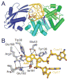
Crystal structure of Bacillus stearothermophilus MutY. (A) Overall structure of BsMutY (PDB ID 1RRQ) colored by domain (green, iron-sulfur cluster domain; cyan, catalytic domain; blue, C-terminal (8oxoG recognition) domain. The DNA is colored gold with adenine substrate in purple and opposite 8oxoG in magenta. (B) Active site details of the MutY fluorinated lesion recognition complex (FLRC) bound to adenine (3G0Q). Protein (silver) and nucleic acid (gold) atoms are shown as sticks, water molecules are shown as red spheres. Hydrogen bonds are shown as dashed lines.
Figure 7
Figure 7
Alkylpurine DNA glycosylases. Overall architectures are shown on the top row, with HhH enzymes arranged in order of increasing specificity for 3mA. (A–F) Active sites. Protein and nucleic acid atoms are shaded grey and gold, respectively, and waters are shown as red spheres. (A) Human AAG/εA-DNA substrate complex (1EWN). (B) E. coli AlkA bound to 1-azaribose-DNA (1DIZ). (C) A. fulgidus AlkA (2JHJ) with THF-DNA modeled from the S. pombe MagI/DNA complex (3S6I). (D) S. pombe Mag1/THF-DNA (3S6I). (E) H. pylori MagIII/3,9-dimethyladenine (1PU7). (F) E. coli TAG/THF-DNA/3mA product complex (2OFI).
Figure 8
Figure 8
Binding of εA and εC to AAG. The εA complex (PDB ID 1EWN) is colored blue (protein) and salmon (DNA), and the εC complex (PDB ID 3QI5) is silver and gold. Hydrogen bonds are depicted as dashed lines. A water molecule (red sphere) is in position to protonate N7 of εA, and protonated N7 would donate a hydrogen bond to Ala134 (green dashed line). εC does not have an ionizable group at this position.
Figure 9
Figure 9
Yeast 3-methyladenine DNA glycosylases MAG and Mag1. In all panels, the unbound Saccharomyces cerevisiae MAG (grey) free enzyme is superimposed onto the Schizosaccharomyces pombe Mag1/THF-DNA complex (blue/gold, PDB ID 3S6I). (A) Overall structures. (B) Active sites. Mag1 residues Phe158 and Ser159 are the only two active site residues that differ between the two enzymes. (C) Close-up of Mag1-DNA contacts at the lesion. Swapping Mag1 His64 and MAG Ser97 between the two enzymes effectively swaps their respective abilities to remove εA (see text for details).
Figure 10
Figure 10
Crystal structures of AlkD in complex with 3d3mA-DNA (PDB ID 3JX7) (A–C) and THF-DNA (3JXZ) (D–F). The protein is colored silver, DNA is gold, 3d3mA and THF lesions are blue and opposite thymines are cyan. (A,D) Overall structures showing DNA bound to the concave surface of the protein. (B,E) Side views of the base pairs flanking the lesions. (C,F) View rotated 90° with respect to panels A/D and B/E. Hydrogen bonds are shown as dashed lines.
Figure 11
Figure 11
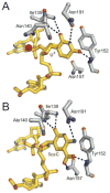
Protein-DNA contacts within the TDG active site. (A) TDG bound to DNA containing 2′-deoxy-2′-fluoroarabinouridine (UF) (PDB ID 3UFJ). Hydrogen bonds are shown as dashed lines and the putative catalytic water molecule is a red sphere. (B) TDG in complex with 5-carboxylcytosine (5caC)-DNA.
Figure 12
Figure 12
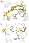
Comparison of TDG (A) and MBD4 (B) contacts to the strand opposite the lesion. The guanine base opposite the THF abasic site is marked with an asterisk. (A) TDG/THF-DNA complex (PDB ID 2RBA). (B) MBD4/THF-DNA complex (4DK9).
Figure 13
Figure 13
SUMO1 modified TDG creates steric clash with DNA. The SUMO1-modified TDG structure (blue TDG, green SUMO1, PDB ID 1WYW) is superimposed onto the TDG/THF-DNA complex (silver/gold, 2RBA). SUMO1 modification holds helix α7 in a position that would presumably clash with the DNA.
Figure 14
Figure 14
DME domain structure and distribution of critical residues. (A) Schematic of A. thaliana DEMETER (DME), showing the location of the glycosylase domain (green) and domains A (blue) and B (gold) of unknown function. IDR1, inter-domain region 1; IDR2, inter-domain region 2. Red bars mark the locations of residues identified by random mutagenesis to be critical for DME function. Putative residues important for DNA binding and catalysis and conserved in other HhH enzymes are marked by symbols below the schematic (Asn778 DNA plug, magenta triangle; Met1238 DNA wedge, blue triangle; catalytic Lys1286, yellow star). (B) Homology model of the C-terminal half of domain A and the glycosylase domain with DNA superimposed from the structure of Bacillus stearothermophilus EndoIII bound to abasic DNA (PDB ID 1P59). The putative catalytic Lys1286 (cyan) Met1238 wedge (slate) residues are contributed by the glycosylase domain (green), the putative Asn778 plug residue (magenta) is from domain A.

References

    1. Friedberg EC, Aguilera A, Gellert M, Hanawalt PC, Hays JB, Lehmann AR, Lindahl T, Lowndes N, Sarasin A, Wood RD. DNA repair: from molecular mechanism to human disease. DNA Repair (Amst) 2006;5:986–996. - PubMed
    1. Lindahl T. Suppression of spontaneous mutagenesis in human cells by DNA base excision-repair. Mutation research. 2000;462:129–135. - PubMed
    1. Dalhus B, Laerdahl JK, Backe PH, Bjørås M. DNA base repair--recognition and initiation of catalysis. FEMS Microbiol Rev. 2009;33:1044–1078. - PubMed
    1. David SS, O’Shea VL, Kundu S. Base-excision repair of oxidative DNA damage. Nature. 2007;447:941–950. - PMC - PubMed
    1. Friedman JI, Stivers JT. Detection of damaged DNA bases by DNA glycosylase enzymes. Biochemistry. 2010;49:4957–4967. - PMC - PubMed

Publication types