First demonstration of multiplexed X-ray fluorescence computed tomography (XFCT) imaging
- PMID: 23076031
- PMCID: PMC12121647
- DOI: 10.1109/TMI.2012.2223709
First demonstration of multiplexed X-ray fluorescence computed tomography (XFCT) imaging
Abstract
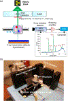
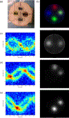
Simultaneous imaging of multiple probes or biomarkers represents a critical step toward high specificity molecular imaging. In this work, we propose to utilize the element-specific nature of the X-ray fluorescence (XRF) signal for imaging multiple elements simultaneously (multiplexing) using XRF computed tomography (XFCT). A 5-mm-diameter pencil beam produced by a polychromatic X-ray source (150 kV, 20 mA) was used to stimulate emission of XRF photons from 2% (weight/volume) gold (Au), gadolinium (Gd), and barium (Ba) embedded within a water phantom. The phantom was translated and rotated relative to the stationary pencil beam in a first-generation CT geometry. The X-ray energy spectrum was collected for 18 s at each position using a cadmium telluride detector. The spectra were then used to isolate the K shell XRF peak and to generate sinograms for the three elements of interest. The distribution and concentration of the three elements were reconstructed with the iterative maximum likelihood expectation maximization algorithm. The linearity between the XFCT intensity and the concentrations of elements of interest was investigated. We found that measured XRF spectra showed sharp peaks characteristic of Au, Gd, and Ba. The narrow full-width at half-maximum (FWHM) of the peaks strongly supports the potential of XFCT for multiplexed imaging of Au, Gd, and Ba ( FWHM(Au,Kα1) = 0.619 keV, FWHM(Au,Kα2)=1.371 keV , FWHM(Gd,Kα)=1.297 keV, FWHM(Gd,Kβ)=0.974 keV , FWHM(Ba,Kα)=0.852 keV, and FWHM(Ba,Kβ)=0.594 keV ). The distribution of Au, Gd, and Ba in the water phantom was clearly identifiable in the reconstructed XRF images. Our results showed linear relationships between the XRF intensity of each tested element and their concentrations ( R(2)(Au)=0.944 , R(Gd)(2)=0.986, and R(Ba)(2)=0.999), suggesting that XFCT is capable of quantitative imaging. Finally, a transmission CT image was obtained to show the potential of the approach for providing attenuation correction and morphological information. In conclusion, XFCT is a promising modality for multiplexed imaging of high atomic number probes.
Figures


References
-
- Miles KA, “Molecular imaging with dynamic contrast-enhanced computed tomography,” Clin. Radiol, vol. 65, pp. 549–556, Jul. 2010. - PubMed
-
- Cheong SK, Jones BL, Siddiqi AK, Liu F, Manohar N, and Cho SH, “X-ray fluorescence computed tomography (XFCT) imaging of gold nanoparticle-loaded objects using 110 kVp x-rays,” Phys. Med. Biol, vol. 55, pp. 647–662, Feb. 7, 2010. - PubMed
-
- Jones BL and Cho SH, “The feasibility of polychromatic cone-beam X-ray fluorescence computed tomography (XFCT) imaging of gold nanoparticle-loaded objects: A Monte Carlo study,” Phys. Med. Biol, vol. 56, pp. 3719–3730, Jun. 21, 2011. - PubMed
Publication types
MeSH terms
Grants and funding
LinkOut - more resources
Full Text Sources
Other Literature Sources
Medical

