Review
. 2012 Sep-Oct;6(5):397-403.
doi: 10.4161/cam.21103. Epub 2012 Sep 1.

The role of FGF signaling in guiding coordinate movement of cell groups: guidance cue and cell adhesion regulator?

Affiliations
Review

The role of FGF signaling in guiding coordinate movement of cell groups: guidance cue and cell adhesion regulator?

Young-Kyung Bae et al. Cell Adh Migr. 2012 Sep-Oct.

Abstract

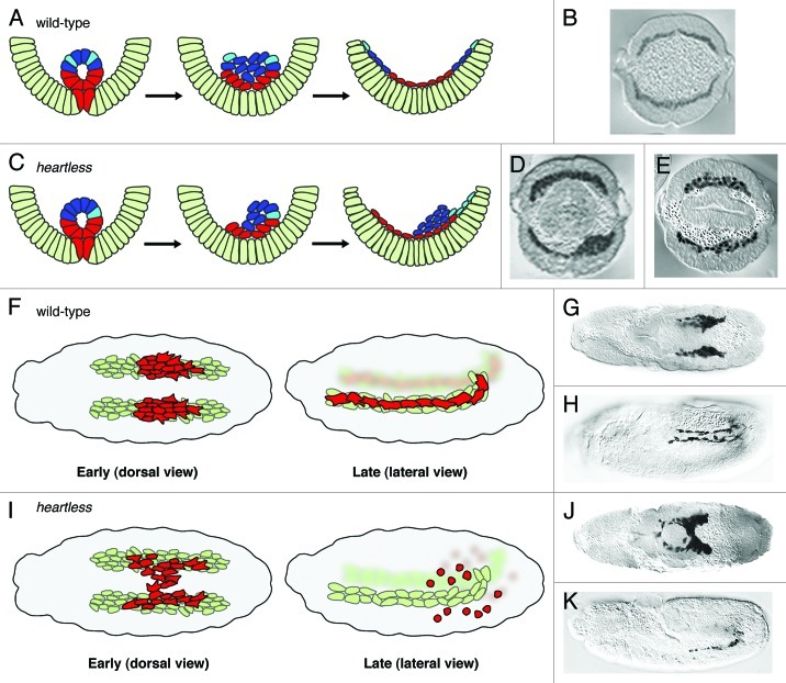
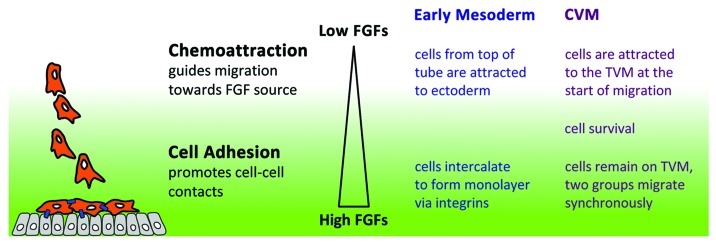
Cell migration influences cell-cell interactions to drive cell differentiation and organogenesis. To support proper development, cell migration must be regulated both temporally and spatially. Mesoderm cell migration in the Drosophila embryo serves as an excellent model system to study how cell migration is controlled and influences organogenesis. First, mesoderm spreading transforms the embryo into a multilayered form during gastrulation and, subsequently, cells originating from the caudal visceral mesoderm (CVM) migrate along the entire length of the gut. Here we review our studies, which have focused on the role of fibroblast growth factor (FGF) signaling, and compare and contrast these two different cell migration processes: mesoderm spreading and CVM migration. In both cases, FGF acts as a chemoattractant to guide cells' directional movement but is likely not the only signal that serves this role. Furthermore, FGF likely modulates cell adhesion properties since FGF mutant phenotypes share similarities with those of cell adhesion molecules. Our working hypothesis is that levels of FGF signaling differentially influence cells' response to result in either directional movement or changes in adhesive properties.


Figures

None
Figure 1. Comparison of cell movements in wild-type and heartless mutant embryos. (A and C) Schematic based on published results; (B, D and E) cross-sections of anti-Twist staining of wild-type and htl mutant embryos. (A and B) In wild-type, all mesoderm (red/blue/cyan) cells contact the ectoderm (light green) and are able to spread dorsally to form a monolayer. (C) In htl mutants, only the subset of cells (red/cyan) that contact the ectoderm undergoes directed movements. Depending on how the tube collapses, the mutant phenotype can be severe (D) or subtle (E). (F and I) Schematic based on published results; CVM reporter croc-lacZ in wild-type (G and H) and htl mutant (J and K) embryos stained with anti-βgal oriented with anterior to the left. (F and G) The dorsal view of wild-type at stage 11 shows the two distinct, symmetrical clusters of CVM cells (red) migrating on the two bands of TVM cells (light green). (F and H) At stage 13, the lateral view reveals complete CVM migration with cells evenly distributed along the TVM. (I and J) In htl mutant embryos, CVM cells are intermixed in early migration. (I and K) Later stages of mutant embryos illustrate CVM cell death and loss of contact with the TVM. (B--D) and (G and J) were reprinted with permission from references and , respectively.
None
Figure 2. Model of FGF’s dual function. Data from recent studies suggest that FGFs are able to function differentially in a concentration dependent manner. The concentration of FGFs is governed by the proximity of responding cells (orange) to the source (gray cells producing FGFs). At a distance, where levels are low, FGFs works as chemoattractant such that cells become polarized and migrate directionally. Once cells are closer to the source, the higher levels of FGFs promote cell adhesion (blue lines).

Comment on

  • Kadam S, Ghosh S, Stathopoulos A. Synchronous and symmetric migration of Drosophila caudal visceral mesoderm cells requires dual input by two FGF ligands. Development. 2012;139:699–708. doi: 10.1242/dev.068791. doi: 10.1242/dev.068791

References

    1. Ridley AJ, Schwartz MA, Burridge K, Firtel RA, Ginsberg MH, Borisy G, et al. Cell migration: integrating signals from front to back. Science. 2003;302:1704–9. doi: 10.1126/science.1092053. - DOI - PubMed
    1. Rørth P. Whence directionality: guidance mechanisms in solitary and collective cell migration. Dev Cell. 2011;20:9–18. doi: 10.1016/j.devcel.2010.12.014. - DOI - PubMed
    1. Friedl P, Gilmour D. Collective cell migration in morphogenesis, regeneration and cancer. Nat Rev Mol Cell Biol. 2009;10:445–57. doi: 10.1038/nrm2720. - DOI - PubMed
    1. Montell DJ. The social lives of migrating cells in Drosophila. Curr Opin Genet Dev. 2006;16:374–83. doi: 10.1016/j.gde.2006.06.010. - DOI - PubMed
    1. Aman A, Piotrowski T. Cell-cell signaling interactions coordinate multiple cell behaviors that drive morphogenesis of the lateral line. Cell Adh Migr. 2011;5:499–508. doi: 10.4161/cam.5.6.19113. - DOI - PMC - PubMed

Publication types

MeSH terms

Substances

LinkOut - more resources