. 2012 Nov 30;287(49):41392-404.
doi: 10.1074/jbc.M112.417360. Epub 2012 Oct 17.

Structural insights into the substrate specificity of the Rhodopseudomonas palustris protein acetyltransferase RpPat: identification of a loop critical for recognition by RpPat

Affiliations

Structural insights into the substrate specificity of the Rhodopseudomonas palustris protein acetyltransferase RpPat: identification of a loop critical for recognition by RpPat

Heidi A Crosby et al. J Biol Chem. .

Abstract

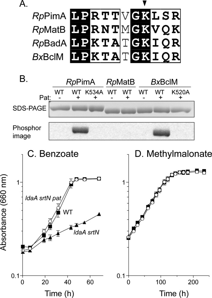
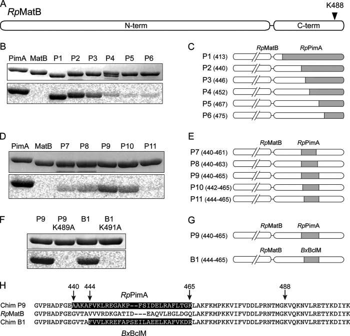
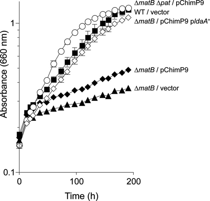
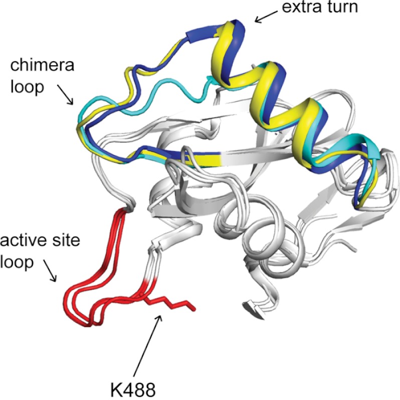
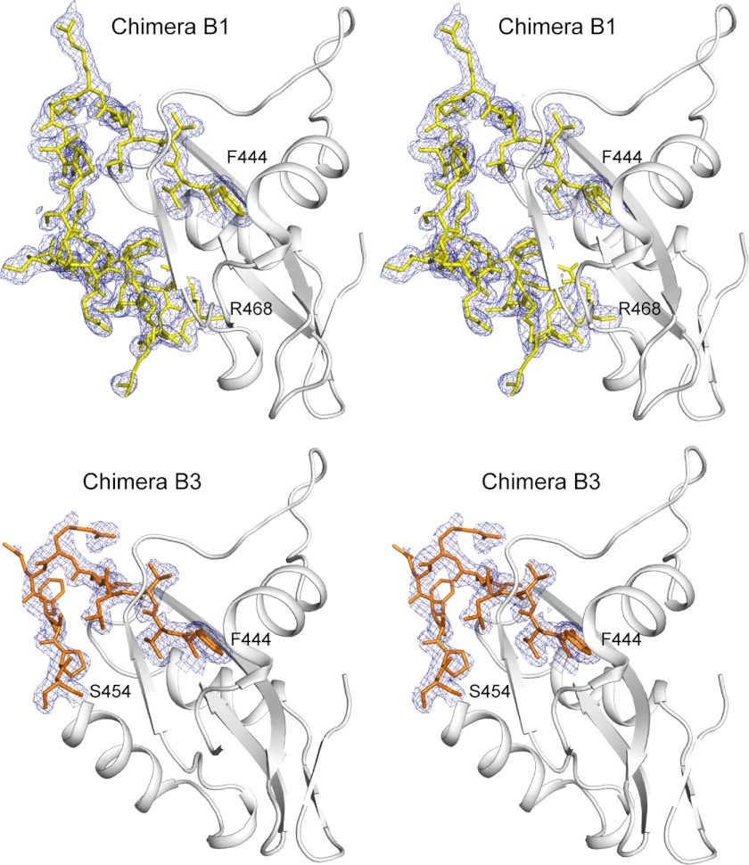
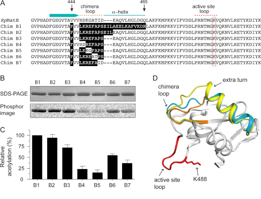
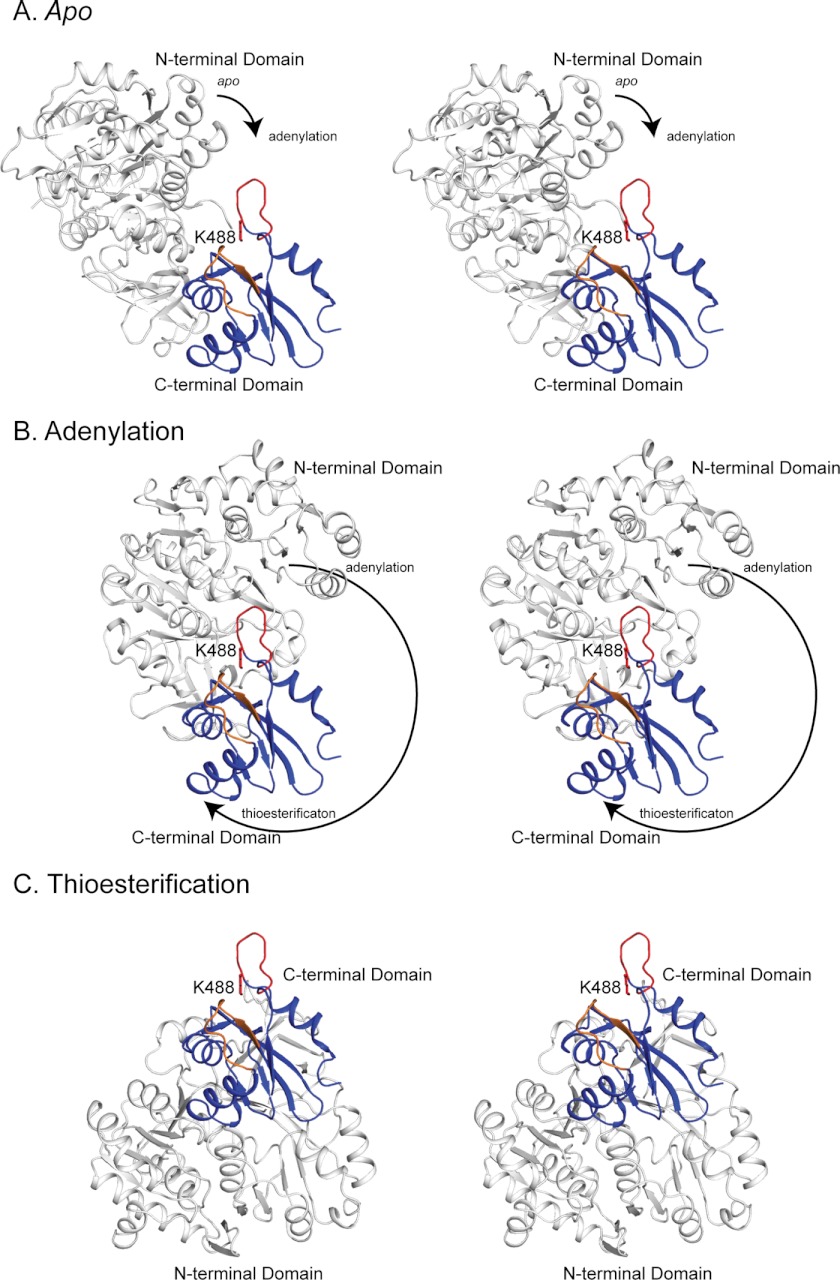
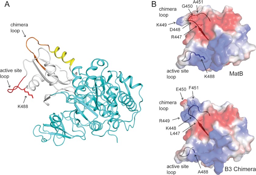
Lysine acetylation is a post-translational modification that is important for the regulation of metabolism in both prokaryotes and eukaryotes. In bacteria, the best studied protein acetyltransferase is Pat. In the purple photosynthetic bacterium Rhodopseudomonas palustris, at least 10 AMP-forming acyl-CoA synthetase enzymes are acetylated by the Pat homologue RpPat. All bona fide RpPat substrates contain the conserved motif PX(4)GK. Here, we show that the presence of such a motif is necessary but not sufficient for recognition by RpPat. RpPat failed to acetylate the methylmalonyl-CoA synthetase of this bacterium (hereafter RpMatB) in vivo and in vitro, despite the homology of RpMatB to known RpPat substrates. We used RpMatB to identify structural determinants that are recognized by RpPat. To do this, we constructed a series of RpMatB chimeras that became substrates of RpPat. In such chimeras, a short region (11-25 residues) of RpMatB located >20 residues N-terminal to the acetylation site was replaced with the corresponding sequences from other AMP-forming acyl-CoA synthetases that were known RpPat substrates. Strikingly, the enzymatic activity of RpMatB chimeras was regulated by acetylation both in vitro and in vivo. Crystal structures of two of these chimeras showed that the major difference between them and wild-type RpMatB was within a loop region ∼23 Å from the acetylation site. On the basis of these results, we suggest that RpPat likely interacts with a relatively large surface of its substrates, in addition to the PX(4)GK motif, and that RpPat probably has relatively narrow substrate specificity.


Figures

FIGURE 1.
FIGURE 1.
RpPat acetylates RpPimA and BxBclM but not RpMatB. A, alignment of the region around the lysine residue that is acetylated in RpBadA. Arrow indicates Lys-512 in RpBadA, corresponding to Lys-534 in RpPimA, Lys-488 in RpMatB, and Lys-520 in BxBclM. B, RpPimA, RpMatB, and BxBclM were incubated with [1-14C]acetyl-CoA and with or without RpPat. Changing Lys-534 of RpPimA or Lys-520 of BxBclM to alanine abolished acetylation. Top panel shows the acyl-CoA synthetases separated by SDS-PAGE, and bottom panel is the phosphorimage of the same gel. C, photosynthetic growth of R. palustris on benzoate is regulated by acetylation. A deacetylase-deficient strain (ΔldaA ΔsrtN, triangles) grew poorly because RpBadA was acetylated and rendered inactive, and deletion of patldaA ΔsrtN Δpat, circles) restored growth. D, photosynthetic growth of R. palustris on methyl malonate was not regulated by acetylation. Wild-type (squares), deacetylase-deficient (ΔldaA ΔsrtN, triangles), and deacetylase- and pat-deficient (ΔldaA ΔsrtN Δpat, circles) strains all grew equally well.
FIGURE 2.
FIGURE 2.
Construction of RpMatB chimeric proteins that can be acetylated. A, scheme of RpMatB N- and C-terminal domains drawn to scale. Arrowhead indicates location of conserved lysine residue Lys-488. B, acetylation of six RpMatB-RpPimA chimeras by RpPat using [1-14C]acetyl-CoA. Top panel shows SDS-polyacrylamide gel, and bottom panel shows the phosphorimage of the same gel. C, schematic representation of the six RpMatB-RpPimA chimeras P1–P6, in which a section of the C-terminal domain of RpMatB was replaced with the corresponding sequence from RpPimA (gray). C-terminal domains are drawn to match scale of A. Numbers in parentheses indicate the residue of RpMatB at which the fusion begins (i.e. first residue replaced by PimA). D, acetylation by RpPat of RpMatB chimeras containing an internal fragment of RpPimA. E, schematic representation of RpMatB-RpPimA chimeras P7–P11, containing an internal fragment of RpPimA (gray). Numbers in parentheses indicate residues of RpMatB that were replaced by the corresponding sequence of RpPimA. F, acetylation by RpPat of the RpMatB-BxBclM chimera B1. Changing the conserved lysine residue (Lys-488 in wild-type RpMatB numbering) in either chimera resulted in no acetylation by RpPat. G, schematic representation of the RpMatB-RpPimA chimera P9 and RpMatB-BxBclM chimera B1 that were the best identified substrates of RpPat. H, alignment of the C-terminal ends of RpMatB and the RpMatB-RpPimA P9 and RpMatB-BxBclM B1 chimeras, starting at Gly-431. The sequences derived from RpPimA and BxBclM are shaded, and RpMatB residue numbers are indicated with arrows.
FIGURE 3.
FIGURE 3.
Activity of the RpMatB-RpPimA P9 chimera is controlled by acetylation in vivo. Absorbance was monitored during photosynthetic growth of R. palustris on methyl malonate (10 mm). Data points are averages of four replicates, and error bars represent standard deviations. Squares, wild-type (CGA009) harboring plasmid pBBR1MCS-2 as a vector control; filled triangles, strain JE13568 (ΔmatB/pBBR1MCS-2); filled diamonds, strain JE13912 (ΔmatB/pRpMATB25 (encodes the RpMatB-RpPimA P9 chimera)); circles, strain JE14288 (ΔmatB Δpat/pRpMATB25 (encodes the RpMatB-RpPimA P9 chimera)); empty diamonds, strain JE14957 (ΔmatB/pRpMATB45 (encodes the RpMatB-RpPimA P9 chimera and ldaA+).
FIGURE 4.
FIGURE 4.
Crystal structure of RpMatB-BxBclM chimera B1. The C-terminal domain of the RpMatB-BxBclM B1 chimera is shown aligned with the C-terminal domains of RpMatB (PDB accession number 4FUT (36)) and BxBclM (PDB accession number 2V7B (45)). The BxBclM-derived residues of the chimera are colored yellow, and the corresponding residues of RpMatB and BxBclM are shown in cyan and dark blue, respectively. The active site lysine residue of RpMatB was modeled from the RpMatB apo structure (PDB accession number 4FUQ (36)), and the PX4GK region surrounding this residue (active site loop) is shown in red. Arrows indicate the chimera loop and the extra turn in the α-helix of BxBclM and the B1 chimera compared with RpMatB. Figure was prepared with PyMOL (60).
FIGURE 5.
FIGURE 5.
Stereo view of the C-terminal domains of RpMatB-BxBclM B1 chimera and RpMatB-BxBclM B3 chimera. The chimeric regions are depicted as sticks superimposed on an electron density map calculated with coefficients of the form 2FoFc contoured at 1σ. Density for the backbone residues is well defined, although density for side chains that do not make crystal contacts is weak (Val-465 and MLY 466 in the B1 chimera and Arg-449, Glu-450, and Phe-451 in the B3 chimera). Figure was prepared with PyMOL (60).
FIGURE 6.
FIGURE 6.
Construction of a minimal RpMatB-BxBclM chimera. A, alignment of the C-terminal end of RpMatB, starting at residues 431, with the RpMatB-BxBclM chimeras (labeled B1–B7). Relevant secondary structure elements of RpMatB are shown above the alignment, and the red box indicates the active site lysine (Lys-488). Residues in the chimeras derived from BxBclM are highlighted in black. B, acetylation of each BxBclM chimera by RpPat using [1-14C]acetyl-CoA. C, amount of acetylation in B was quantified and normalized to the total acetylation of the RpMatB-BxBclM B1 chimera. Values represent averages and standard deviations of three experiments. D, alignment of the C-terminal domains of RpMatB and the RpMatB-BxBclM B1 and B3 chimeras. BxBclM-derived residues in the B1 chimera are indicated in yellow, BxBclM-derived residues in the B3 chimera are shown in orange, and the corresponding residues in wild-type RpMatB are shown in cyan. In all three structures, the catalytic loop and active site lysine are shown in red (lysine residue is modeled from the RpMatB apo structure). D was prepared with PyMOL (60).
FIGURE 7.
FIGURE 7.
Stereo view of the accessibility of Lys-488 in the apo (A), adenylation (B), and thioesterification (C) conformations. The N-terminal domain is shown in white and the C-terminal domain in blue. The active site loop and Lys-488 are shown in red, and the chimera loop is shown in orange. The Lys-488 side chain is modeled from the MatB apo structure. The C-terminal domains are shown in the same orientation in all three conformations. The arrows indicate the direction and magnitude of rotation of the N-terminal domain relative to the C-terminal domain during the transition from one conformation to the next. In all cases, the C-terminal domain is from the RpMatB-BxBclM B3 chimera structure, and the N-terminal domains are either from wild-type MatB (apo and thioesterification, PDB accession code 4FUQ) or MatBK488A (adenylation, PDB accession code 4FUT). In each case the N- and C-terminal domains are aligned to RpMatB structures (apo, 4FUQ; adenylation, 4FUT) or to the S. coelicolor MatB structure (thioesterification, 3NYQ (54)). Figure was prepared with PyMOL (60).
FIGURE 8.
FIGURE 8.
RpPat recognition elements of the RpMatB-BxBclM B3 chimera. A, RpMatB-BxBclM B3 chimera modeled in the thioester-forming conformation. The N-terminal domain is shown in cyan, the C-terminal domain in white, the active site loop in red, the residues derived from BxBclM in orange, and additional residues changed in the RpMatB-BxBclM B1 chimera are shown in yellow. The acetylation site, Lys-488, was modeled from the RpMatB apo structure. B, surface electrostatic calculations for the RpMatB and RpMatB-BxBclM B3 chimera C-terminal domains were generated using PyMOL (60), with electropositive regions shown in blue and electronegative regions shown in red. Ribbon diagrams of the active site and chimera loop backbones are shown for reference, and residues within the chimera loop are labeled. The active site lysine residue (Lys-488) was mutated to an alanine in the RpMatB-BxBclM B3 chimera structure to aid crystallization. Figure was prepared with PyMOL (60).

References

    1. Kim G. W., Yang X. J. (2011) Comprehensive lysine acetylomes emerging from bacteria to humans. Trends. Biochem. Sci. 36, 211–220 - PubMed
    1. Thao S., Escalante-Semerena J. C. (2011) Control of protein function by reversible Nϵ-lysine acetylation in bacteria. Curr. Opin. Microbiol. 14, 200–204 - PMC - PubMed
    1. Taverna S. D., Li H., Ruthenburg A. J., Allis C. D., Patel D. J. (2007) How chromatin-binding modules interpret histone modifications. Lessons from professional pocket pickers. Nat. Struct. Mol. Biol. 14, 1025–1040 - PMC - PubMed
    1. Oberdoerffer P., Michan S., McVay M., Mostoslavsky R., Vann J., Park S. K., Hartlerode A., Stegmuller J., Hafner A., Loerch P., Wright S. M., Mills K. D., Bonni A., Yankner B. A., Scully R., Prolla T. A., Alt F. W., Sinclair D. A. (2008) SIRT1 redistribution on chromatin promotes genomic stability but alters gene expression during aging. Cell 135, 907–918 - PMC - PubMed
    1. Wang L., Tang Y., Cole P. A., Marmorstein R. (2008) Structure and chemistry of the p300/CBP and Rtt109 histone acetyltransferases. Implications for histone acetyltransferase evolution and function. Curr. Opin. Struct. Biol. 18, 741–747 - PMC - PubMed

Publication types

LinkOut - more resources