Comment
. 2013 Jan-Feb;7(1):27-32.
doi: 10.4161/cam.22126. Epub 2012 Oct 17.

Cell-to-cell communication via plasmodesmata in vascular plants

Affiliations
Comment

Cell-to-cell communication via plasmodesmata in vascular plants

Iris Sevilem et al. Cell Adh Migr. 2013 Jan-Feb.

Abstract

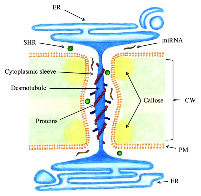
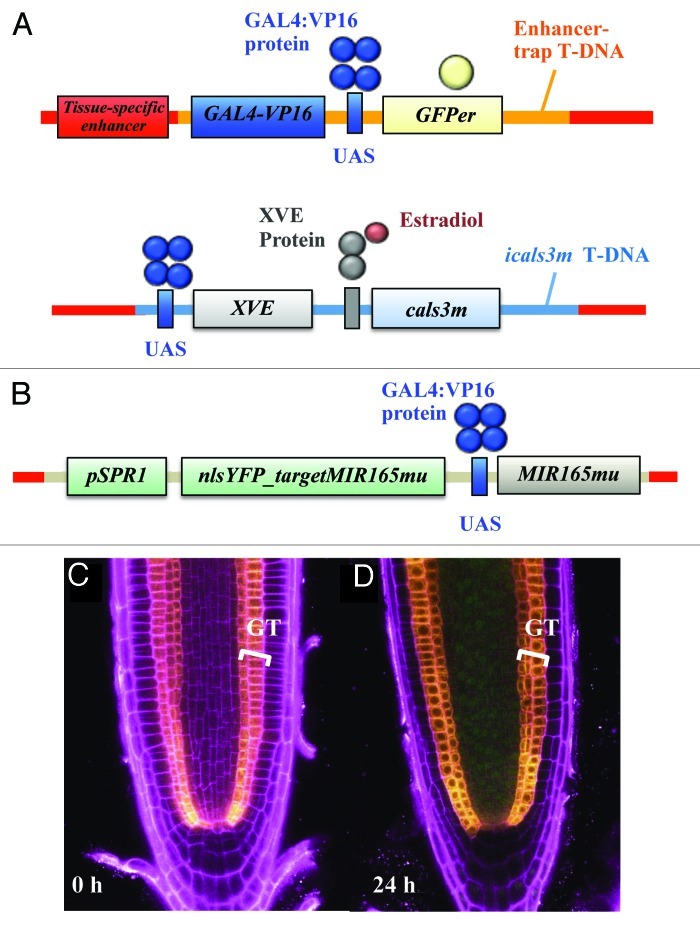
In plant development, cell-to-cell signaling is mediated by mobile signals, including transcription factors and small RNA molecules. This communication is essential for growth and patterning. Short-range movement of signals occurs in the extracellular space via the apoplastic pathway or directly from cell-to-cell via the symplastic pathway. Symplastic transport is mediated by plant specific structures called plasmodesmata, which are plasma membrane-lined pores that traverse the cell walls of adjacent cells thus connecting their cytoplasms. However, a thorough understanding of molecules moving via plasmodesmata and regulatory networks relying on symplastic signaling is lacking. Traffic via plasmodesmata is highly regulated, and callose turnover is known to be one mechanism. In Arabidopsis, plasmodesmata apertures can be regulated in a spatially and temporally specific manner with the icals3m, an inducible vector system expressing the mutated CalS3 gene encoding a plasmodesmata localized callose synthase that increases callose deposition at plasmodesmata. We discuss strategies to use the icals3m system for global analyses on symplastic signaling in plants.


Figures

None
Figure 1. Cell types in the Arabidopsis root. Radial (left) and longitudinal (right) sections of the Arabidopsis root tip. Cell types are marked by different colors. Modified from Miyashima et al. and Carlsbecker et al.
None
Figure 2. Bidirectional signaling pathway between the stele and the ground-tissue. SHR transcription occurs in the stele (1), after which the protein moves outwards from the stele to the endodermis to activate miRNA transcription together with SCR (2). The miRNAs move to the stele periphery (3) and downregulate PHB expression by targeting its mRNA transcripts for cleavage (4). Stele is highlighted in pink, endodermis in blue and cortex in yellow.
None
Figure 3. Plasmodesmata. Plasmodesmata connect cytoplasms of adjacent cells by traversing the cell wall. Appressed endoplasmic reticulum, called the desmotubule, runs through the plasma membrane-lined pore. Molecules move via the cytoplasmic sleeve between the desmotubule and plasma membrane. Callose is deposited at the neck region in the cell wall. CW, cell wall; PM, plasma membrane; ER, endoplasmic reticulum.
None
Figure 4. Analysis of miRNA movement with a miRNA sensor system. J0571; UAS::icals3m line expressing cals3m in an estradiol inducible manner and GFP constitutively in the ground-tissue (A) was introduced with a sensor system pSPR1::nlsYFP_targetMIR165mu/UAS::MIR165mu. The coding sequence of miR165mu-targeted nlsYFP was driven by the broadly-expressed SPR1 promoter, whereas the MIR165mu gene, which targets the nlsYFP_165mu-tgt, was placed under the UAS (B). In the resulting line (called J0571; UAS::icals3m/pSPR1::nlsYFP_targetMIR165mu/UAS::MIR165mu), YFP signal was not detected under non-induced condition (C). In contrast, after 24 h induction period, nlsYFP signal was detected throughout the stele (D), indicating that the miRNAs were not present to cleave the nlsYFP sequences and consequently YFP signal was not suppressed. GFP signal was detected in the ground-tissue layer of all of the roots, indicating that miR165mu transcription occurred in both conditions. Yellow, GFP signal; green, YFP signal; magenta, PI signal; GT, ground-tissue.

Comment on

  • Callose biosynthesis regulates symplastic trafficking during root development.
    Vatén A, Dettmer J, Wu S, Stierhof YD, Miyashima S, Yadav SR, Roberts CJ, Campilho A, Bulone V, Lichtenberger R, Lehesranta S, Mähönen AP, Kim JY, Jokitalo E, Sauer N, Scheres B, Nakajima K, Carlsbecker A, Gallagher KL, Helariutta Y. Vatén A, et al. Dev Cell. 2011 Dec 13;21(6):1144-55. doi: 10.1016/j.devcel.2011.10.006. Dev Cell. 2011. PMID: 22172675

References

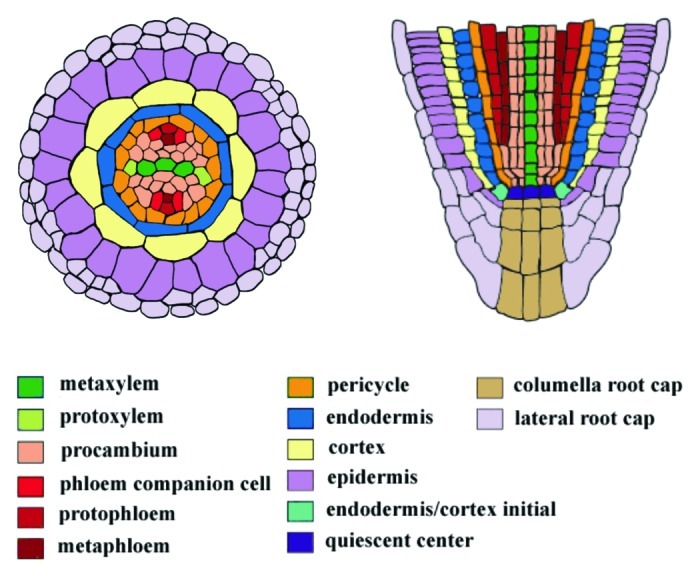
    1. Dolan L, Janmaat K, Willemsen V, Linstead P, Poethig S, Roberts K, et al. Cellular organisation of the Arabidopsis thaliana root. Development. 1993;119:71–84. - PubMed
    1. Benfey PN, Scheres B. Root development. Curr Biol. 2000;10:R813–5. doi: 10.1016/S0960-9822(00)00814-9. - DOI - PubMed
    1. van den Berg C, Willemsen V, Hendriks G, Weisbeek P, Scheres B. Short-range control of cell differentiation in the Arabidopsis root meristem. Nature. 1997;390:287–9. doi: 10.1038/36856. - DOI - PubMed
    1. Scheres B. Plant cell identity. The role of position and lineage. Plant Physiol. 2001;125:112–4. doi: 10.1104/pp.125.1.112. - DOI - PMC - PubMed
    1. van den Berg C, Willemsen V, Hage W, Weisbeek P, Scheres B. Cell fate in the Arabidopsis root meristem determined by directional signalling. Nature. 1995;378:62–5. doi: 10.1038/378062a0. - DOI - PubMed

Publication types

LinkOut - more resources