. 2012 Dec;41(6):2191-9.
doi: 10.3892/ijo.2012.1664. Epub 2012 Oct 16.

Wedelolactone, a medicinal plant-derived coumestan, induces caspase-dependent apoptosis in prostate cancer cells via downregulation of PKCε without inhibiting Akt

Affiliations

Wedelolactone, a medicinal plant-derived coumestan, induces caspase-dependent apoptosis in prostate cancer cells via downregulation of PKCε without inhibiting Akt

Sivalokanathan Sarveswaran et al. Int J Oncol. 2012 Dec.

Abstract

Emerging studies indicate that metabolism of arachidonic acid through the 5-lipoxygenase (5-Lox) pathway plays a critical role in the survival of prostate cancer cells raising the possibility that 5-Lox can be targeted for an effective therapy of prostate cancer. Wedelolactone (WDL), a medicinal plant-derived natural compound, is known to inhibit 5-Lox activity in neutrophils. However, its effect on apoptosis in prostate cancer cells has not been addressed. Thus, we tested the effects of WDL on human prostate cancer cells in vitro. We observed that WDL kills both androgen-sensitive as well as androgen-independent prostate cancer cells in a dose-dependent manner by dramatically inducing apoptosis. We also found that WDL-induced apoptosis in prostate cancer cells is dependent on c-Jun N-terminal Kinase (c-JNK) and caspase-3. Interestingly, WDL triggers apoptosis in prostate cancer cells via downregulation of protein kinase Cε (PKCε), but without inhibition of Akt. WDL does not affect the viability of normal prostate epithelial cells (PrEC) at doses that kill prostate cancer cells, and WDL-induced apoptosis is effectively prevented by 5-oxoETE, a metabolite of 5-Lox (but not by 15-oxoETE, a metabolite of 15-Lox), suggesting that the apoptosis-inducing effect of WDL in prostate cancer cells is mediated via inhibition of 5-Lox activity. These findings indicate that WDL selectivity induces caspase-dependent apoptosis in prostate cancer cells via a novel mechanism involving inhibition of PKCε without affecting Akt and suggest that WDL may emerge as a novel therapeutic agent against clinical prostate cancer in human.


Figures

Figure 1
Figure 1
Effect of WDL on the viability of prostate epithelial cells. Cancer and normal prostate epithelial cells (∼4×10³ per well) were plated in 96-well plates overnight in complete medium, and treated with varying doses of WDL. Plates were incubated further for 72 h at 37°C and cell viability was measured by Cell Titer assay as described in the Materials and methods section (20,33). Results are shown as mean value of each data point ± SE (n=6). WDL selectively affects the viability of prostate cancer cells sparing normal cells.
Figure 2
Figure 2
Morphological alteration of LNCaP cells by WDL. LNCaP prostate cancer cells (3×10⁵ per plate) were plated overnight in 60-mm diameter plates as described in the Materials and methods section (Microscopy) and treated either with doses of WDL or ibuprofen for 24 h at 37°C in the incubator. Control cells were treated with the vehicle only (0.2% DMSO). At the end of treatment period, cells were photographed at magnification, ×200. Data show a representative of three experiments with similar results.
Figure 3
Figure 3
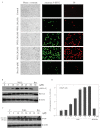
Induction of apoptosis by WDL LNCaP cells (3×10⁵ per plate) were plated as in Fig. 2 above and treated either with WDL or ibuprofen for 24 h. Control cells were treated with 0.2% DMSO. (A) At the end of incubation period, cells were stained with FITC-labeled Annexin V and propidium iodide, and observed under fluorescence microscope at ×200. A representative of two independent experiments is shown here with similar results. (B) At the end of incubation period, cells were lysed and phosphorylation of histone H2A.X at serine-139 was detected by western blot analysis. (C) Cleavage of PARP is shown as detected by western blot analysis. (D) Degradation of chromatin DNA to nucleosomal fragments was detected by Cell Death Detection ELISA. Results are shown as mean values of each data point ± SE (n=4).
Figure 4
Figure 4
Activation of c-Jun N-terminal Kinase (JNK) and mitochondrial permeability transition by WDL. LNCaP cells (3×10⁵ per plate) were plated as in Fig. 2 and treated with varying doses of WDL or ibuprofen (30 μM) at 37°C for 24 h. (A) Cell lysates were analyzed by western blot analysis with an antibody against phosphorylated-JNK. Antibodies against pan-specific JNK and β-actin were used as controls. A representative of three experiments with similar results is shown here. (B) Role of JNK in apoptosis was tested by pre-treating cells with specific inhibitor, SP600125. U0126, an inhibitor MAPK kinase, was used as negative control. Results are shown as mean values of each data point ± SE (n=4). (C) LNCaP cells were treated with WDL for 8 h and permeability transition of mitochondria was detected by treating cells with 40 nM Mitotracker red for 30 min at 37°C in the incubator. Hoechst dye 33342 was used to stain the nuclei. After washing, cells were photographed with a Nikon digital camera attached to a Leica fluorescence microscope at magnification, ×400.
Figure 5
Figure 5
Activation and role of caspase in WDL-induced apoptosis. (A) LNCaP cells (3×10⁵ per plate) were plated as in Fig. 2 above and treated with varying doses of WDL as indicated for 24 h. At the end of incubation period, enzymatic activities of caspase-3 in cell lysates were measured by colorimetric caspase-3 Cellular Activity Assay kit (no. AK-703) using DEVD-pNa as substrate (Biomol). (B) Cells were pretreated for 30 min with specific caspase-3 inhibitor (DEVD-FMK) before treatment with WDL (20 μM) for 24 h. Control cells were treated with the vehicle only (0.2% DMSO). Apoptosis was measured by detecting DNA degradation to nucleosomal fragments by Cell Death Detection ELISAplus (Roche). Data represent mean values of quadruplicate determination of each point ± SE.
Figure 6
Figure 6
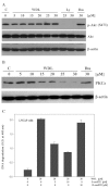
Effect of WDL on Akt and PKCε. (A) LNCaP cells (3×10⁵ per plate) in 60-mm diameter plates were treated with varying doses of WDL for 24 h. Control cells were treated with the solvent vehicle only (0.2% DMSO). Phosphorylation of Akt (at Serine 473) was detected by western blot analysis. LY294002 (LY) and ibuprofen (Ibu) were used as positive and negative controls, respectively. (B) Effect of WDL on the protein level of PKCε was detected by western blot analysis. β-actin was used as loading control. (C) Prevention of WDL-induced apoptosis by 5-oxoETE. LNCaP cells (3×10⁵ per plate) were plated in 60-mm diameter plates and treated with WDL (20 μM) with or without the addition of exogenous 5-oxoETE or 15-oxoETE. Plates were incubated at 37°C for 24 h in the CO2 incubator. Apoptosis was measured by Cell Death ELISA. Results show mean values of each data point ± SE (n= 4).

Similar articles

Cited by

References

    1. Govindachari T, Nagarajan K, Pai B. Wedelolactone from Eclipta alba. J Sci Indust Res. 1956;15B:664–665.
    1. Wagner H, Geyer B, Kiso Y, Hikino H, Rao GS. Coumestans as the main active principles of the liver drugs Eclipta alba and Wedelia calendulacea. Planta Med. 1986;52:370–374. - PubMed
    1. Singh B, Saxena AK, Chandan BK, Agarwal SG, Anand KK. In vivo hepatoprotective activity of active fraction from ethanolic extract of Eclipta alba leaves. Indian J Physiol Pharmacol. 2001;45:435–441. - PubMed
    1. Patel MB, Kadakia VM, Mishra SH. Simultaneous estimation of andrographolide and wedelolactone in herbal formulations. Indian J Pharma Sci. 2008;70:689–693. - PMC - PubMed
    1. Roy RK, Thakur M, Dixit VK. Hair growth promoting activity of Eclipta alba in male albino rats. Arch Dermatol Res. 2008;300:357–364. - PubMed

Publication types

MeSH terms