

Neural substrate of the late positive potential in emotional processing
- PMID: 23077042
- PMCID: PMC3516184
- DOI: 10.1523/JNEUROSCI.3109-12.2012
Neural substrate of the late positive potential in emotional processing
Abstract
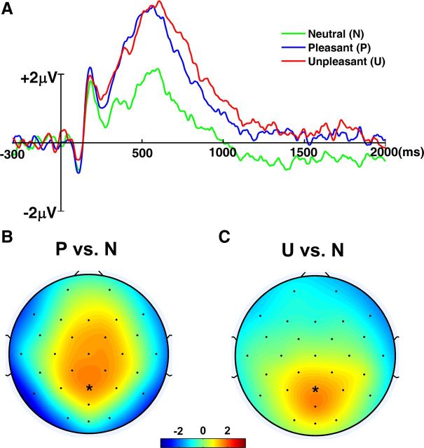
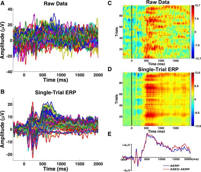
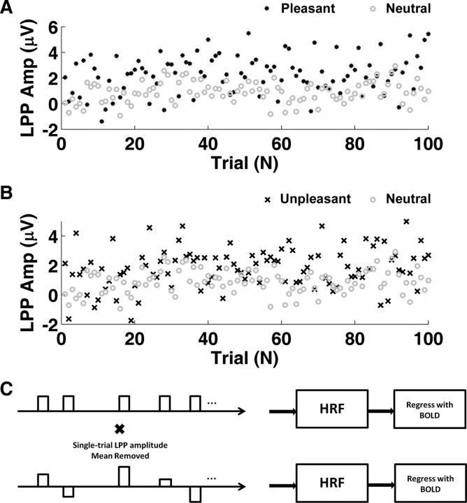
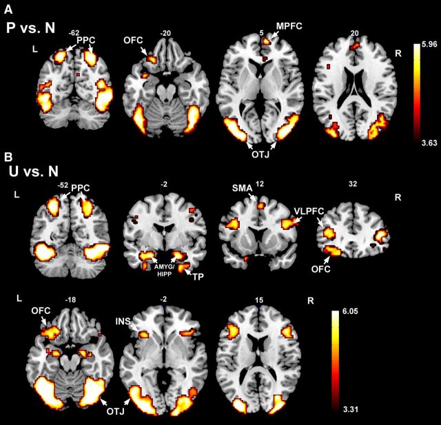
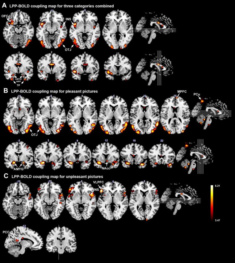
The late positive potential (LPP) is a reliable electrophysiological index of emotional perception in humans. Despite years of research, the brain structures that contribute to the generation and modulation of LPP are not well understood. Recording EEG and fMRI simultaneously, and applying a recently proposed single-trial ERP analysis method, we addressed the problem by correlating the single-trial LPP amplitude evoked by affective pictures with the blood oxygen level-dependent (BOLD) activity. Three results were found. First, relative to neutral pictures, pleasant and unpleasant pictures elicited enhanced LPP, as well as heightened BOLD activity in both visual cortices and emotion-processing structures such as amygdala and prefrontal cortex, consistent with previous findings. Second, the LPP amplitude across three picture categories was significantly correlated with BOLD activity in visual cortices, temporal cortices, amygdala, orbitofrontal cortex, and insula. Third, within each picture category, LPP-BOLD coupling revealed category-specific differences. For pleasant pictures, the LPP amplitude was coupled with BOLD in occipitotemporal junction, medial prefrontal cortex, amygdala, and precuneus, whereas for unpleasant pictures significant LPP-BOLD correlation was observed in ventrolateral prefrontal cortex, insula, and posterior cingulate cortex. These results suggest that LPP is generated and modulated by an extensive brain network composed of both cortical and subcortical structures associated with visual and emotional processing and the degree of contribution by each of these structures to the LPP modulation is valence specific.
Figures
References
-
- Adolphs R. Neural systems for recognizing emotion. Curr Opin Neurobiol. 2002;12:169–177. - PubMed
-
- Aharon I, Etcoff N, Ariely D, Chabris CF, O'Connor E, Breiter HC. Beautiful faces have variable reward value: fMRI and behavioral evidence. Neuron. 2001;32:537–551. - PubMed
-
- Allen PJ, Polizzi G, Krakow K, Fish DR, Lemieux L. Identification of EEG events in the MR scanner: the problem of pulse artifact and a method for its subtraction. Neuroimage. 1998;8:229–239. - PubMed
-
- Allen PJ, Josephs O, Turner R. A method for removing imaging artifact from continuous EEG recorded during functional MRI. Neuroimage. 2000;12:230–239. - PubMed
-
- Belouchrani A, Abed-Meraim K, Cardoso JF, Moulines E. Second-order blind separation of temporally correlated sources. Proc Int Conf on Digital Sig Proc; Cyprus. 1993. pp. 346–351.
Publication types
MeSH terms
Grants and funding
LinkOut - more resources
Full Text Sources
Other Literature Sources