. 2013 Jan;87(1):16-24.
doi: 10.1128/JVI.01853-12. Epub 2012 Oct 17.

Role of interferon regulatory factor 3-mediated apoptosis in the establishment and maintenance of persistent infection by Sendai virus

Affiliations

Role of interferon regulatory factor 3-mediated apoptosis in the establishment and maintenance of persistent infection by Sendai virus

Saurabh Chattopadhyay et al. J Virol. 2013 Jan.

Abstract

Infection of cultured cells by paramyxoviruses causes cell death, mediated by a newly discovered apoptotic pathway activated by virus infection. The key proapoptotic protein in this pathway is interferon regulatory factor 3 (IRF-3), which upon activation by virus infection binds BAX, translocates it to mitochondria, and triggers apoptosis. When IRF-3-knockdown cells were infected with Sendai virus (SeV), persistent infection (PI) was established. The PI cells produced infectious SeV continuously and constitutively expressed many innate immune genes. Interferon signaling was blocked in these cells. The elevated levels of IRF-3-driven genes in the PI cells indicated that the amount of residual IRF-3 activated by endogenous SeV was high enough to drive the transcriptional effects of IRF-3 but too low to trigger its apoptotic activity. We confirmed this IRF-3 threshold idea by generating a tetracycline (Tet)-inducible cell line for IRF-3 expression, which enabled us to express various levels of IRF-3. PI could be established in the Tet-off cell line, and as expected, when doxycycline was withdrawn, the cells underwent apoptosis. Finally, we tested for PI establishment in 12 mouse embryo fibroblasts by natural selection. Eleven lines became persistently infected; although seven out of them had low IRF-3 levels, four did not. When one of the latter four was further analyzed, we observed that it expressed a very low level of caspase 3, the final executor protease of the apoptotic pathway. These results demonstrated that SeV PI can arise from infection of normal wild-type cells, but only if they can find a way to impair the IRF-3-dependent apoptotic pathway.


Figures

Fig 1
Fig 1
SeV infection establishes persistence in the absence of IRF-3. (A) Cell lysates from mock-infected or SeV-infected (24 h postinfection) HT1080, 1K/SeV, or 1K cells were analyzed for IRF-3, SeV C protein, and actin by Western blotting. (B) 1K/SeV cells or their matched mock-infected control cells (1K) were immunostained with an antibody against SeV (red fluorescence). Nuclei were visualized by DAPI staining. (C) 1K cells were infected with virus produced by 1K/SeV cells (lane virus:1K/SeV), PI was established, and lysates from these cells, mock-infected 1K cells (lane −), or 1K/SeV cells (lane SeV) were analyzed for SeV C protein.
Fig 2
Fig 2
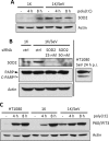
Elevated expression of SOD2 is not required to protect PI cells from apoptosis. (A) SOD2 protein expression in untreated 1K or 1K/SeV cells or cells transfected with poly(I·C) transfected for the indicated times was analyzed by Western blotting; (B) 1K or 1K/SeV cells were transfected with control (ctrl) or SOD2 siRNA (Dharmacon), and 96 h later the cells were analyzed by Western blotting for PARP cleavage (C-PARP, cleaved PARP), SOD2, and actin. A lysate from SeV-infected (MOI, 10; 24 h postinfection [p.i.]) HT1080 cells was used as a positive control for cleaved PARP (right); (C) induction of P60/IFIT3 protein following poly(I·C) transfection of HT1080, 1K, and 1K/SeV cells was analyzed by Western blotting.
Fig 3
Fig 3
Status of innate immune signaling in 1K/SeV cells. (A) HT1080, 1K, or 1K/SeV cells were left untreated or pretreated with IFN-β (1,000 U/ml) and infected with VSV (MOI, 0.1); culture fields were visualized for cell survival at 24 h postinfection; (B) antiviral states of 1K and 1K/SeV cells were compared by infecting them with VSV (MOI, 0.1) before or after IFN-β pretreatment for 8 h; infectious VSV production was determined by plaque assays (presented as numbers of PFU/ml); (C) STAT1 tyrosine 701 phosphorylation was analyzed in uninfected or SeV-infected (MOI, 10; 2 h postinfection) 1K cells or 1K/SeV cells in response to 30 min of IFN-β treatment; (D) IRF-3 serine 396 phosphorylation (p-IRF-3) and induction of A20 protein were analyzed at various times after poly(I·C) transfection of HT1080, 1K, and 1K/SeV cells. The panel for actin is identical to that in Fig. 2C, because the two experiments were done together. (E) IκB-α serine 32 phosphorylation (p-IκB-α) was analyzed before or 2 h after poly(I·C) (pIC) transfection of 1K and 1K/SeV cells. Total IκB-α and SeV C proteins were analyzed by Western blotting.
Fig 4
Fig 4
Inducible expression of IRF-3 and its functions in 1K cells. (A) 1K cells were transiently transfected with pTet-Off (Clontech) and V5-tagged human IRF-3 cloned in the pTRE2hyg vector (pTRE2hyg-V5-hIRF-3; Clontech), and 48 h later the cells were either left untreated or treated with Dox (1 μg/ml) and the cell lysates were analyzed for IRF-3 and actin by Western blotting; (B) a stable cell line (clone 10) was generated from the transiently transfected cells described in panel A and maintained in the presence of Dox (1 μg/ml); IRF-3 expression was analyzed in the presence of Dox or after removal of Dox; (C) clone 10 cells were maintained in the absence or the presence of various concentrations of Dox (100 ng/ml, 1 ng/ml, or none) and then infected with SeV (MOI, 10); IRF-3 levels gradually increased with decreasing Dox concentration (bottom), apoptosis was measured from the amount of cleaved PARP production (top), and gene induction was measured from the amount of P60/IFIT3 induction (middle); (D, E) clone 10 cells were maintained in the absence of Dox to induce the expression of IRF-3 and then transfected with siRNAs against BAX or a nontargeting sequence (NT) using DharmaFECT 4 reagent (Thermo Scientific); after 48 h, cells were infected with SeV (MOI, 10), and at 24 h postinfection, cell lysates were analyzed for cleaved PARP (D); BAX knockdown was confirmed by Western blotting (E).
Fig 5
Fig 5
Inducible expression of IRF-3 causes apoptosis in PI cells. (A) Clone 10/SeV cells were immunostained with an antibody against SeV (green fluorescence). Nuclei were visualized by DAPI staining; (B) persistently infected clone 10 cells (clone 10/SeV) were grown in the absence of Dox for the indicated times, when cell lysates were analyzed for cleaved PARP and actin by Western blotting; (C) clone 10/SeV cells were either grown in the presence of Dox (1 μg/ml) or washed extensively with PBS and grown in the absence of Dox for 72 h, when the culture fields were photographed.
Fig 6
Fig 6
Persistence in MEF cells. Cell lysates were prepared from PI MEF cells and their matched mock-infected controls and analyzed for IRF-3 and SeV C protein (A, B) and XIAP (C) levels by Western blotting. (A) PI MEFs which showed reduced levels of endogenous IRF-3; (B) PI MEFs with unchanged levels of endogenous IRF-3; (C) endogenous XIAP levels from the selected PI MEFs (as indicated).
Fig 7
Fig 7
PI in the presence of IRF-3 but absence of caspase 3. (A) Lysates from PI MEF1 cells or their matched mock-infected controls were analyzed for cleaved PARP, P54/Ifit2, or SeV C protein; (B) lysates from PI MEF1 cells or their matched mock-infected controls were analyzed for IRF-3, TRAF2, TRAF6, BAX, Bcl-XL, caspase 9, and caspase 3 by Western blotting; (C) persistently infected MCF-7 or 1K cells were immunostained with an antibody against SeV (green fluorescence). Nuclei were visualized by DAPI staining.

References

    1. Griffin DE, Hardwick JM. 1997. Regulators of apoptosis on the road to persistent alphavirus infection. Annu. Rev. Microbiol. 51:565–592 - PubMed
    1. Sarkar SN, Sen GC. 2004. Novel functions of proteins encoded by viral stress-inducible genes. Pharmacol. Ther. 103:245–259 - PubMed
    1. Jensen S, Thomsen AR. 2012. Sensing of RNA viruses: a review of innate immune receptors involved in recognizing RNA virus invasion. J. Virol. 86:2900–2910 - PMC - PubMed
    1. Yu M, Levine SJ. 2011. Toll-like receptor, RIG-I-like receptors and the NLRP3 inflammasome: key modulators of innate immune responses to double-stranded RNA viruses. Cytokine Growth Factor Rev. 22:63–72 - PMC - PubMed
    1. Colonna M. 2007. TLR pathways and IFN-regulatory factors: to each its own. Eur. J. Immunol. 37:306–309 - PubMed

Publication types

Substances

LinkOut - more resources