. 2013 Jan;87(1):94-109.
doi: 10.1128/JVI.01557-12. Epub 2012 Oct 17.

mRNA decay during herpes simplex virus (HSV) infections: mutations that affect translation of an mRNA influence the sites at which it is cleaved by the HSV virion host shutoff (Vhs) protein

Affiliations

mRNA decay during herpes simplex virus (HSV) infections: mutations that affect translation of an mRNA influence the sites at which it is cleaved by the HSV virion host shutoff (Vhs) protein

Lora A Shiflett et al. J Virol. 2013 Jan.

Abstract

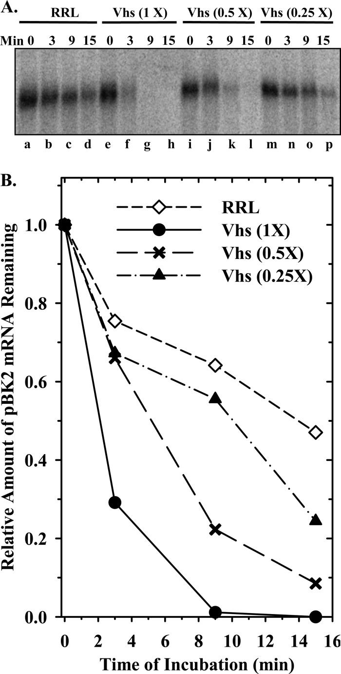
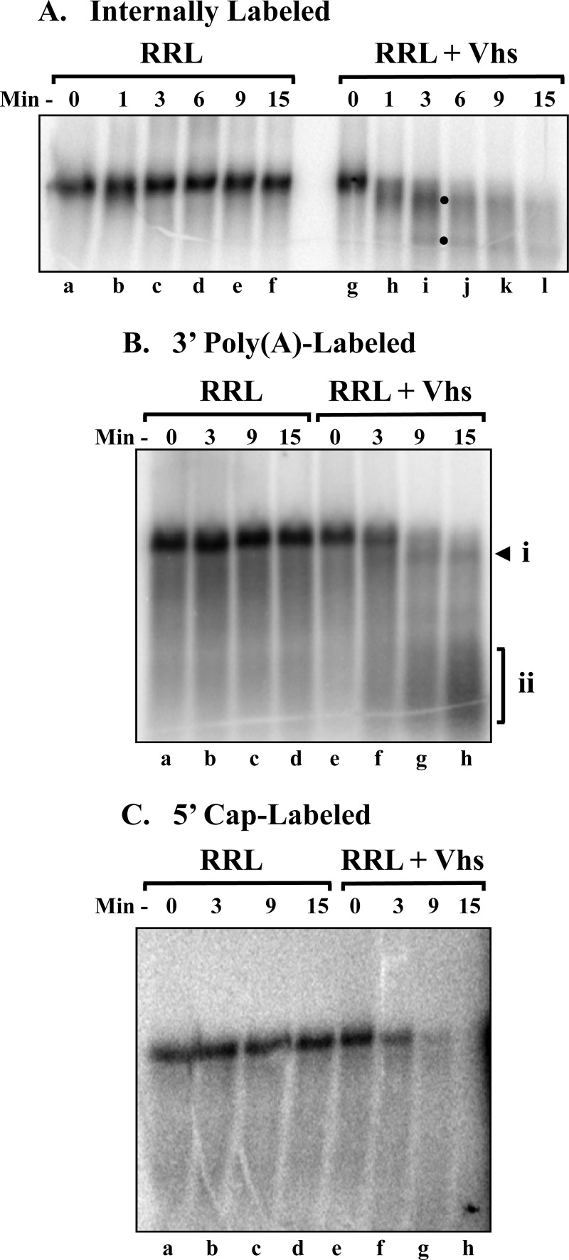
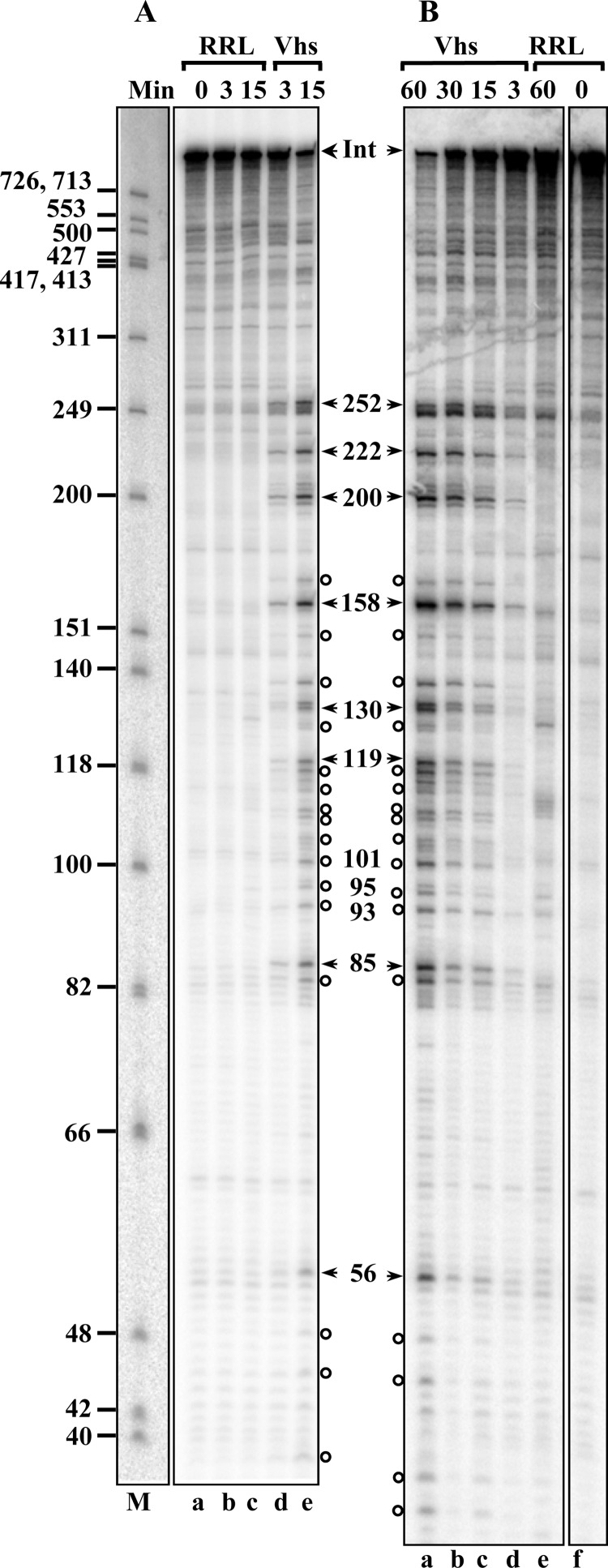
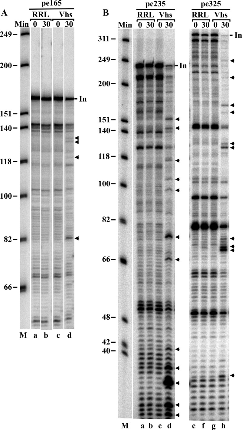
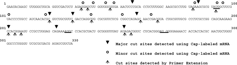
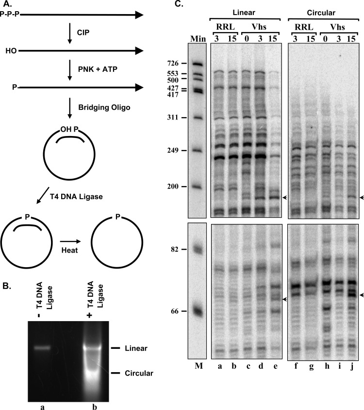
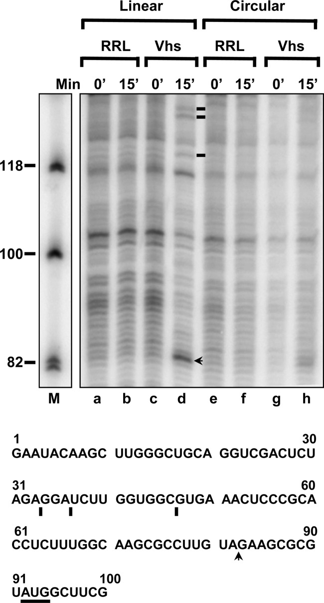
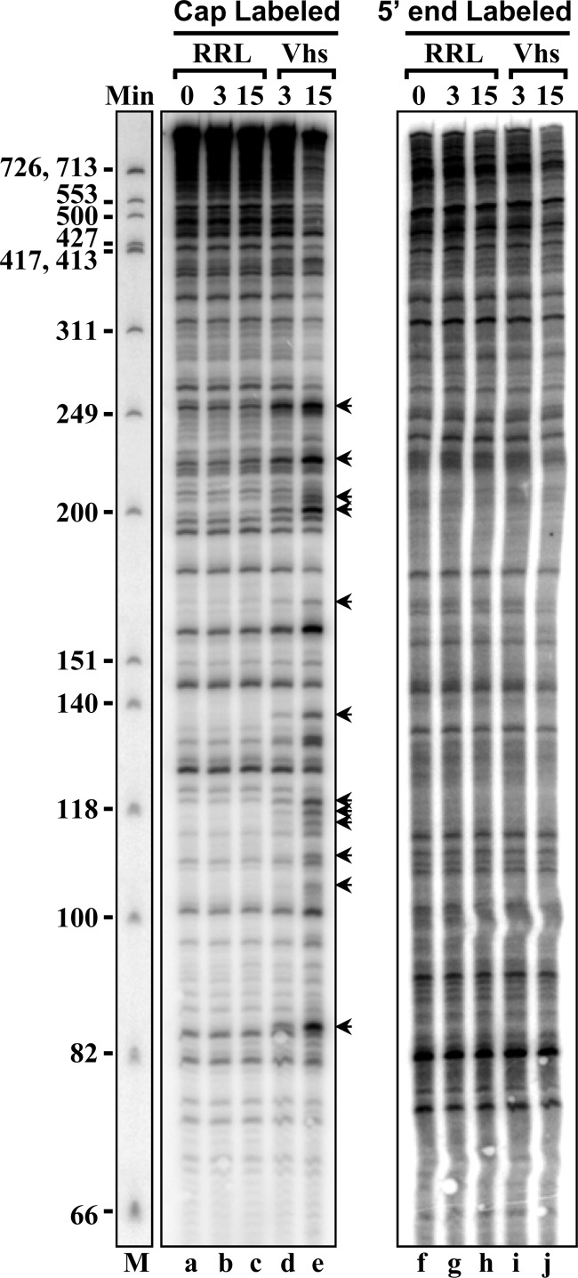
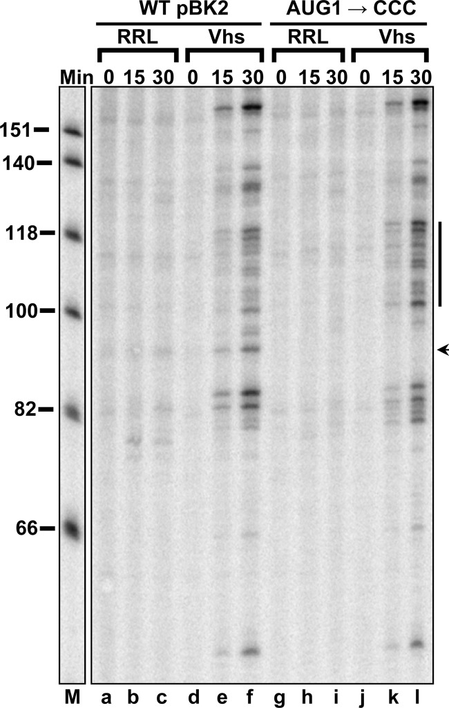
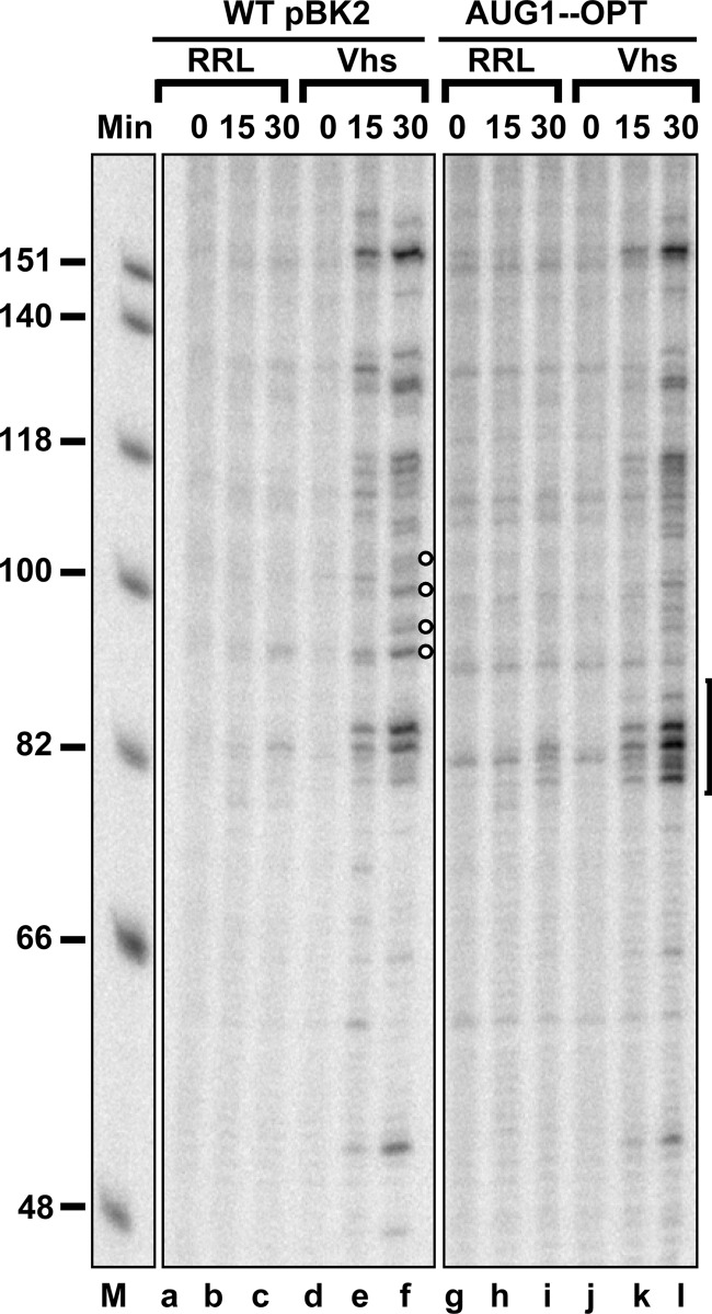
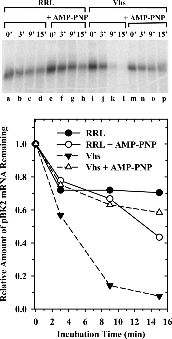
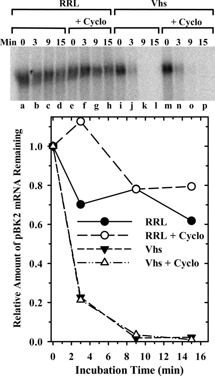
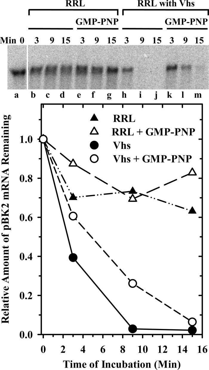
During lytic infections, the herpes simplex virus (HSV) virion host shutoff (Vhs) endoribonuclease degrades many host and viral mRNAs. Within infected cells it cuts mRNAs at preferred sites, including some in regions of translation initiation. Vhs binds the translation initiation factors eIF4H, eIF4AI, and eIF4AII, suggesting that its mRNA degradative function is somehow linked to translation. To explore how Vhs is targeted to preferred sites, we examined the in vitro degradation of a target mRNA in rabbit reticulocyte lysates containing in vitro-translated Vhs. Vhs caused rapid degradation of mRNAs beginning with cleavages at sites in the first 250 nucleotides, including a number near the start codon and in the 5' untranslated region. Ligation of the ends to form a circular mRNA inhibited Vhs cleavage at the same sites at which it cuts capped linear molecules. This was not due to an inability to cut any circular RNA, since Vhs cuts circular mRNAs containing an encephalomyocarditis virus (EMCV) internal ribosome entry site (IRES) at the same sites as linear molecules with the IRES. Cutting linear mRNAs at preferred sites was augmented by the presence of a 5' cap. Moreover, mutations that altered the 5' proximal AUG abolished Vhs cleavage at nearby sites, while mutations that changed sequences surrounding the AUG to improve their match to the Kozak consensus sequence enhanced Vhs cutting near the start codon. The results indicate that mutations in an mRNA that affect its translation affect the sites at which it is cut by Vhs and suggest that Vhs is directed to its preferred cut sites during translation initiation.


Figures

Fig 1
Fig 1
Dose-dependent degradation of mRNAs by Vhs. Vhs protein was produced by in vitro translation. Once translation was complete, the reaction [Vhs (1×)] was diluted using naïve rabbit reticulocyte lysate to yield lysates containing serial 2-fold dilutions of Vhs. mRNA degradation reactions were performed using cap-labeled pBK2 target mRNA and rabbit reticulocyte lysates containing 1×, 0.5×, or 0.25× amounts of Vhs or no Vhs (RRL). (A) Aliquots were removed after 0, 3, 9, or 15 min, and the RNAs were electrophoresed through a 1.3% (wt/vol) agarose-formaldehyde gel; (B) the relative amounts of full-length mRNAs were quantified using ImageQuant software and plotted to compare the relative mRNA decay rates.
Fig 2
Fig 2
Vhs degradation of pBK2 mRNA initiates near the 5′ end. Parallel in vitro mRNA degradation reactions were performed using rabbit reticulocyte lysates that contained (RRL + Vhs) or lacked (RRL) Vhs and target pBK2 mRNAs that were internally labeled (A), 3′ poly(A) tail labeled (B), or 5′ cap labeled (C). Aliquots were removed after 0, 1, 3, 6, 9, or 15 min, and the RNAs were electrophoresed through 1.3% (wt/vol) agarose-formaldehyde gels. Bands arising from shorter-than-full-length degradation products are indicated by the solid circles to the right of lane i in panel A and by the arrowhead labeled “i” and the bracket labeled “ii” to the right of lane h in panel B.
Fig 3
Fig 3
Mapping Vhs cut sites in cap-labeled pBK2 mRNA. Two identical in vitro mRNA degradation reactions were performed using rabbit reticulocyte lysates that contained (Vhs) or lacked (RRL) Vhs and cap-labeled pBK2 target mRNAs. One was carried out for 15 min (A), while the other was incubated for up to 60 min (B). Aliquots were removed after 0, 3, 15, 30, or 60 min, and the RNAs were electrophoresed through 8% polyacrylamide-8 M urea sequencing gels. The lane labeled “M” in panel A contains labeled marker fragments with the sizes (in nucleotides) of the fragments indicated on the left. The most prominent Vhs degradation products are indicated by the arrowheads to the right of panel A and to the left of panel B, along with the sizes of the products in nucleotides. Less prominent degradation products are designated by open circles. The band formed by intact pBK2 mRNA is indicated by the arrowheads labeled “Int.”
Fig 4
Fig 4
Mapping Vhs cut sites in pBK2 mRNA by primer extension. In vitro mRNA degradation reactions were performed using rabbit reticulocyte lysates that contained (Vhs) or lacked (RRL) Vhs and unlabeled pBK2 target mRNA. Aliquots were removed after 0 or 30 min, and the RNAs were analyzed by primer extension using the 5′-end-labeled primers pe165 (A), pe235 (B, lanes a through d), and pe325 (B, lanes e through h). The primer extension products were electrophoresed through 8% polyacrylamide-8 M urea sequencing gels. All of the samples were from the same experiment. Those analyzed using the primer pe165 were electrophoresed through the gel shown in panel A. Samples analyzed with the primers pe235 and pe325 were electrophoresed through a second gel shown in panel B. Bands due to prominent Vhs cleavage sites in pBK2 are indicated by arrowheads to the right of panel A, lane d, and to the right of panel B, lanes d and h. Bands due to extension of the primers to the end of intact pBK2 mRNA are indicated by the short lines labeled “In.”
Fig 5
Fig 5
Summary of Vhs cut sites identified by analysis of Vhs cutting of 5′ cap-labeled pBK2 mRNA and primer extension analysis of unlabeled pBK2 mRNA. The sequence of the first 330 nucleotides of pBK2 mRNA is shown. Downward-pointing arrowheads above the sequence indicate the most prominent Vhs cut sites identified by Vhs cutting of 5′ cap-labeled pBK2 mRNA. Open circles above the sequence indicate less prominent Vhs cut sites identified using cap-labeled pBK2 mRNA. Upward-pointing arrows below the sequence indicate the location of cut sites identified by primer extension analysis of Vhs degradation products. The first three AUG codons (beginning at nucleotides 92, 227, and 269) of pBK2 mRNA are underlined.
Fig 6
Fig 6
Vhs cuts circular mRNAs that have an EMCV IRES. (A) Diagram of the procedure for preparing circular mRNAs. (B) Linear and circular forms of the RNA were resolved by electrophoresis through a 1.6% (wt/vol) agarose gel. Lane b contains RNAs that were taken through the circularization procedure, and lane a contains an aliquot of RNA that was taken through the same procedure, except that T4 DNA ligase was omitted from the ligation reaction. (C) In vitro RNA degradation reactions were performed using rabbit reticulocyte lysates that contained (Vhs) or lacked (RRL) Vhs and capped linear or circular pCITE-RLuc RNA. Aliquots were removed after 0, 3, or 15 min, and the RNAs were analyzed by primer extension. Bands due to Vhs cleavage sites are indicated by arrows to the right of lanes e and j.
Fig 7
Fig 7
Vhs fails to cut circular pBK2 RNA at the same sites at which it cuts capped linear molecules. Circular (lanes e through h) and capped linear (lanes a through d) forms of pBK2 RNA were incubated in rabbit reticulocyte lysates that contained (Vhs) or lacked (RRL) in vitro-translated Vhs. Aliquots were removed after 0 or 15 min, and the RNAs were analyzed by primer extension using primer pe165. Bands due to prominent Vhs cleavage sites in capped linear pBK2 are indicated by the arrow and lines to the right of lane d. The sequence of the first 100 nucleotides of pBK2 mRNA is shown below the gel, with the 5′ proximal AUG underlined, and the prominent Vhs cut sites are indicated by the arrow and lines below the sequence.
Fig 8
Fig 8
Importance of a 5′ cap for Vhs cleavage of an mRNA at preferred sites. pBK2 RNA was labeled with a 32P-labeled 5′ cap (lanes a through e) or with a 32P-labeled 5′ monophosphate (lanes f through j) as described in the text. Cap-labeled or 5′ monophosphate-labeled RNAs were incubated in rabbit reticulocyte lysates that contained (Vhs) or lacked (RRL) in vitro-translated Vhs. Aliquots were removed after 0, 3, or 15 min, and the RNAs were analyzed by electrophoresis through an 8% polyacrylamide-8 M urea sequencing gel. The most prominent sites at which Vhs cuts cap-labeled pBK2 mRNA are indicated by arrows to the right of lane e.
Fig 9
Fig 9
Mutations that alter the 5′ proximal AUG of pBK2 mRNA abolish Vhs cleavage at nearby sites. In vitro mRNA degradation reactions were performed using wild-type (WT) pBK2 mRNA (lanes a through f) or the mutant AUG1→CCC (lanes g through l) in which the 5′ proximal AUG has been altered to CCC. Cap-labeled wild-type or mutant mRNAs were added to rabbit reticulocyte lysates that contained (Vhs) or lacked (RRL) in vitro-translated Vhs. Aliquots were removed after 0, 15, or 30 min, and the RNAs were analyzed by electrophoresis through an 8% polyacrylamide-8 M urea sequencing gel. The location of the prominent Vhs cleavage site at which cutting is abolished by the mutation is indicated by the arrow to the right of lane l, while sites at which cleavage was moderately enhanced are indicated by the line to the right of lane l.
Fig 10
Fig 10
Mutations that optimize the context of the 5′ proximal AUG of pBK2 mRNA enhance Vhs cleavage at nearby sites. In vitro mRNA degradation reactions were performed using wild-type pBK2 mRNA (lanes a through f) or the mutant AUG1-OPT (lanes g through l) in which nucleotides surrounding the 5′ proximal AUG have been altered to create a match to the Kozak consensus sequence. Cap-labeled wild-type or mutant mRNAs were added to rabbit reticulocyte lysates that contained (Vhs) or lacked (RRL) in vitro-translated Vhs. Aliquots were removed after 0, 15, or 30 min, and the RNAs were analyzed by electrophoresis through an 8% polyacrylamide-8 M urea sequencing gel. The bracket to the right of lane l denotes sites at which Vhs cleavage is enhanced by the mutations that optimize the context of the 5′ proximal AUG. Sites at which Vhs cleavage is reduced are indicated by the open circles to the right of lane f.
Fig 11
Fig 11
Vhs degradation of pBK2 mRNA in the presence of AMP-PNP. Rabbit reticulocyte lysates containing (lanes i through p) or lacking (lanes a through h) in vitro-translated Vhs were preincubated for 12 min in the presence (lanes e through h and m through p) or absence (lanes a through d and i through l) of 2 mM AMP-PNP, after which cap-labeled pBK2 mRNA was added and incubation continued. Aliquots were removed after 0, 3, 9, or 15 min, and the RNAs were electrophoresed through a 1.3% (wt/vol) agarose-formaldehyde gel. The relative amounts of full-length mRNAs were quantified using ImageQuant software and plotted to compare the relative decay rates of pBK2 mRNA.
Fig 12
Fig 12
Cycloheximide has no appreciable effect upon Vhs cleavage. Rabbit reticulocyte lysates containing (Vhs) or lacking (RRL) in vitro-translated Vhs were preincubated for 12 min in the presence (lanes e through h and m through p) or absence (lanes a through d and i through l) of 50 μg/ml of cycloheximide (Cyclo), after which cap-labeled pBK2 mRNA was added and incubation continued. Aliquots were removed after 0, 3, 9, or 15 min, and the RNAs were electrophoresed through a 1.3% (wt/vol) agarose-formaldehyde gel. The relative amounts of full-length mRNAs were quantified using ImageQuant software and plotted to compare the relative decay rates of pBK2 mRNA.
Fig 13
Fig 13
Vhs degradation of pBK2 mRNA in the presence of GMP-PNP. Rabbit reticulocyte lysates containing (lanes h through m) or lacking (lanes a through g) in vitro-translated Vhs were preincubated for 12 min in the presence (lanes e through g and k through m) or absence (lanes a through d and h through j) of 2 mM GMP-PNP, after which cap-labeled pBK2 mRNA was added and incubation continued. Aliquots were removed after 0, 3, 9, or 15 min, and the RNAs were electrophoresed through a 1.3% (wt/vol) agarose-formaldehyde gel. The relative amounts of full-length mRNAs were quantified using ImageQuant software and plotted to compare the relative decay rates of pBK2 mRNA.

References

    1. Roizman B, Knipe DM, Whitley RJ. 2007. Herpes simplex viruses, p 2501–2601 In Knipe DM, Howley PM, Griffin DE, Martin MA, Lamb RA, Roizman B, Straus SE. (ed), Fields virology, 5th ed Lippincott Williams & Wilkins, Philadelphia, PA
    1. Sandri-Goldin RM. 2007. Initiation of transcription and RNA synthesis, processing and transport in HSV and VZV infected cells. In Arvin A, Campadelli-Fiume G, Mocarski E, Moore PS, Roizman B, Whitley R, Yamanishi K. (ed), Human herpesviruses: biology, therapy, and immunoprophylaxis. Cambridge University Press, Cambridge, United Kingdom: http://www.ncbi.nlm.nih.gov/books/NBK47363/ - PubMed
    1. Ferenczy MW, Ranayhossaini DJ, DeLuca NA. 2011. Activities of ICP0 involved in the reversal of silencing of quiescent herpes simplex virus 1. J. Virol. 85:4993–5002 - PMC - PubMed
    1. Kristie TM. 2007. Early events pre-initiation of alphaherpes viral gene expression. In Arvin A, Campadelli-Fiume G, Mocarski E, Moore PS, Roizman B, Whitley R, Yamanishi K. (ed), Human herpesviruses: biology, therapy, and immunoprophylaxis. Cambridge University Press, Cambridge, United Kingdom: http://www.ncbi.nlm.nih.gov/books/NBK47386/ - PubMed
    1. Lester JT, DeLuca NA. 2011. Herpes simplex virus 1 ICP4 forms complexes with TFIID and mediator in virus-infected cells. J. Virol. 85:5733–5744 - PMC - PubMed

Publication types

LinkOut - more resources