. 2012 Jun;46(2):97-105.
doi: 10.2478/v10019-012-0016-0. Epub 2012 May 11.

Outcome of MRI-guided vacuum-assisted breast biopsy - initial experience at Institute of Oncology Ljubljana, Slovenia

Affiliations

Outcome of MRI-guided vacuum-assisted breast biopsy - initial experience at Institute of Oncology Ljubljana, Slovenia

Marta Zebic-Sinkovec et al. Radiol Oncol. 2012 Jun.

Abstract

Background: Like all breast imaging modalities MRI has limited specificity and the positive predictive value for lesions detected by MRI alone ranges between 15 and 50%. MRI guided procedures (needle biopsy, presurgical localisation) are mandatory for suspicious findings visible only at MRI, with potential influence on therapeutic decision. The aim of this retrospective study was to evaluate our initial clinical experience with MRI-guided vacuum-assisted breast biopsy as an alternative to surgical excision and to investigate the outcome of MRI-guided breast biopsy as a function of the MRI features of the lesions. PATIENTS AND METHODS.: In 14 women (median age 51 years) with 14 MRI-detected lesions, MRI-guided vacuum-assisted breast biopsy was performed. We evaluated the MRI findings that led to biopsy and we investigated the core and postoperative histology results and follow-up data.

Results: The biopsy was technically successful in 14 (93%) of 15 women. Of 14 biopsies in 14 women, core histology revealed 6 malignant (6/14, 43%), 6 benign (6/14, 43%) and 2 high-risk (2/14, 14%) lesions. Among the 6 cancer 3 were invasive and 3 were ductal carcinoma in situ (DCIS). The probability of malignancy in our experience was higher for non-mass lesion type and for washout and plateau kinetics.

Conclusions: Our initial experience confirms that MRI-guided vacuum-assisted biopsy is fast, safe and accurate alternative to surgical biopsy for breast lesions detected at MRI only.

Keywords: MRI; MRI guided vacuum assisted biopsy; breast cancer.


Figures
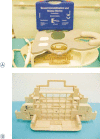
FIGURE 1
FIGURE 1
A,B. Biopsy coil device. Photographs show a four-channel breast biopsy coil with positioning device (A) and a grid-positioning device (B).
FIGURE 2
FIGURE 2
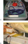
1.5 T Magnetom Avanto (Siemens, Erlangen, Germany). Patient lie face down on the exam table. Breast is moderately compressed in the mediolateral direction.
FIGURE 3A–D
FIGURE 3A–D
MRI-guided biopsy using axial, contrast-enhanced FLASH 3D T1W images. The pre-biopsy fat-suppressed image (A) shows an enhancing lesion in the right breast. The biopsy needle (B) is seen after localization, along with the lesion. Following the biopsy (D) the small magnetic susceptibility artefact due to the clip in situ is seen.
FIGURE 4
FIGURE 4
MRI-guided biopsy device (a) is inserted into breast to acquire tissue specimens (b)
FIGURE 5A–C
FIGURE 5A–C
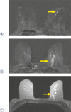
65 year old woman with a history of nipple-discharge in her left breast. Mammographic asimmetry. Ultrasound images showed dilated empty ducts, ductography was suspicious for papilloma. Axial T1-weighted subtracted image after Gadolinium injection (2nd minute) .(a) small round sharply circumscribed masses within a duct sistem–papillomas.(b) segmental, clumped, asymmetric enhancement, fast initial increase and postinitial wash out. (c) high signal intensity on T2–weighted image - BI-RADS 5. Pathologic diagnosis through MRI-guided vacuum-assisted biopsy was massive DCIS with foci of microinvasion.
FIGURE 6AB.
FIGURE 6AB.
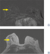
47 year old woman with history of invasive breast carcinoma 10 years ago in her left breast. Retraction of mammila on the right, mammograpic dense breast and benign calcifications. First look and second look ultrasound images showed a cyst. Axial T1-weighted subtracted image after Gadolinium injection (2nd minute).(A) 8×3 mm ductal - linear homogenous, asymmetric enhancement, fast initial increase with postinitial plateau (B) high signal intensity on T2-weighted images, categoriaztion BI-RADS 4. MRI-guided vacuum-assisted biopsy revealed high-grade DCIS withouth microcalcification.
FIGURE 7
FIGURE 7
64-year old woman with history of invasive breast carcinoma in her right breast 10 years ago. Mammography and ultrasound images showed discrete architectural distortion in the left breast. US-guided core biopsy was inconclusive. Axial T1-weighted subtracted image after Gadolinium injection (2nd minute) shows regional, heterogeneous, asymmetric, non-mass like enhancement, fast initial increase, post initial wash-out, intermediate signal intensity on T2-weighted images - BI-RADS 5. Pathologic diagnosis through MRI-guided vacuum-assisted biopsy was DCIS.
FIGURE 8
FIGURE 8
57-year old woman, US-guided fine needle biopsy performed at another institution was suspicious for pappilary carcinoma. Mammographically dense breast, first and second look ultrasound images at institute of oncology showed simple cyst. Axial T1-weighted subtracted image after. Gadolinium injection (2nd minute) shows ductal, reticular-dendritic, asymmetric, non-mass like enhancement, fast initial enhancement with post initial plateau. Intermediate signal intensity on T2-weighted images. BI-RADS 4. MRI-guided vacuum-assisted biopsy revealed invasive tubular carcinoma.
FIGURE 9
FIGURE 9
63-year old woman with history of invasive ductal carcinoma in her right breast 8 years ago. Clinical exam revealed enlarged lymph node in the left axilla. Fine needle aspiration biopsy showed metastatic lymph node. Mammographically discrete architectural distortion only in one projection in the left and postoperative changes in the right breast. Ultrasound was not performed at the discretion of the radiologist. Axial T1-weighted subtracted image after Gadolinium injection (2nd minute) shows focal, heterogeneous, asymmetric, non–mass like enhancement in the left breast, the kinetic curve shows fast initial enhancement with post-initial wash out, low signal intensity on T2-weighted images – BI-RADS 4. MRI–guided vacuum-assisted biopsy revealed invasive lobular carcinoma.
FIGURE 10
FIGURE 10
71-year old woman, MR mammography performed in the other institution. Axial T1-weighted subtracted image after Gadolinium injection (2nd minute) shows 3 round masses in her left breast, with spiculated margins, rim like enhancement, intermediate signal intensity on T2-weighted images. Kinetic curve shows fast initial enhancement with post-initial wash out. Lesions were categorized as BI-RADS 5. Pathologic diagnosis through MRI-guided vacuum-assisted biopsy was invasive lobular carcinoma.

References

    1. Podobnik J, Kocijancic I, Kovac V, Sersa I. 3T MRI in evaluation of asbestos-related thoracic diseases - preliminary results. Radiol Oncol. 2010;44:92–6. - PMC - PubMed
    1. Stanic K, Kovac V. Prophylactic cranial irradiation in patients with small-cell lung cancer: the experience at the Institute of Oncology Ljubljana. Radiol Oncol. 2010;44:180–6. - PMC - PubMed
    1. Kuhl CK, Schrading S, Bieling HB, Wardelmann E, Leutner CC, Koenig R, et al. MRI for diagnosis of pure ductal carcinoma in situ: a prospective observational study. Lancet. 2007;370:485–92. - PubMed
    1. Lehman CD, Deperi ER, Peacock S, McDonough MD, Demartini WB, Shook J. Clinical experience with MRI-guided vacuum-assisted breast biopsy. AJR Am J Roentgenol. 2005;184:1782–7. - PubMed
    1. Lehman CD, DeMartini W, Anderson BO, Edge SB. Indications for breast MRI in the patient with newly diagnosed breast cancer. Indications for breast MRI in the patient with newly diagnosed breast cancer. J Natl Compr Canc Netw. 2009;7:193–201. - PubMed

LinkOut - more resources