. 2012;7(10):e46287.
doi: 10.1371/journal.pone.0046287. Epub 2012 Oct 15.

Inhibition of BDNF in multiple myeloma blocks osteoclastogenesis via down-regulated stroma-derived RANKL expression both in vitro and in vivo

Affiliations

Inhibition of BDNF in multiple myeloma blocks osteoclastogenesis via down-regulated stroma-derived RANKL expression both in vitro and in vivo

Li-Sha Ai et al. PLoS One. 2012.

Abstract

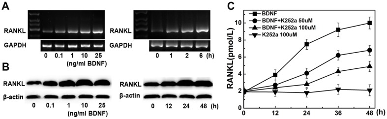
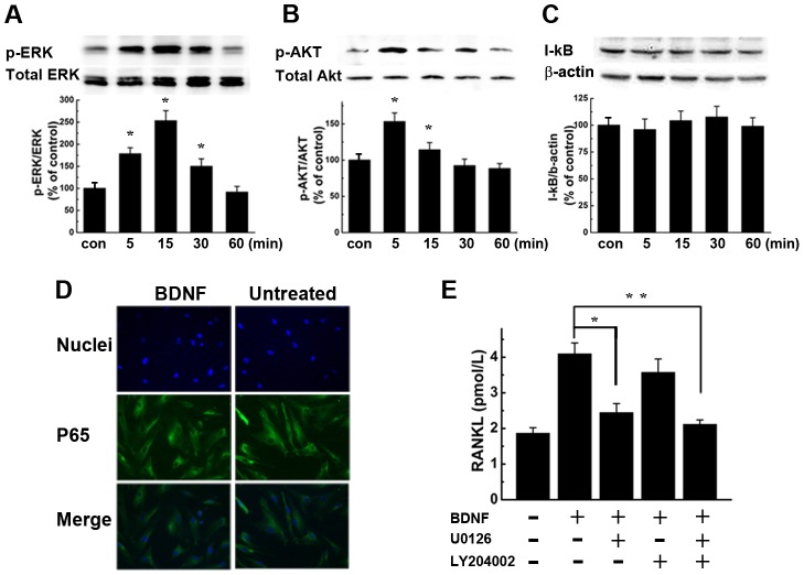
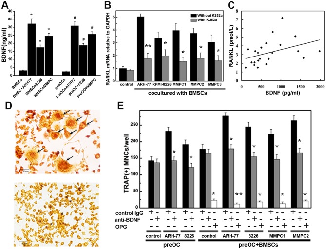
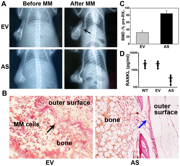
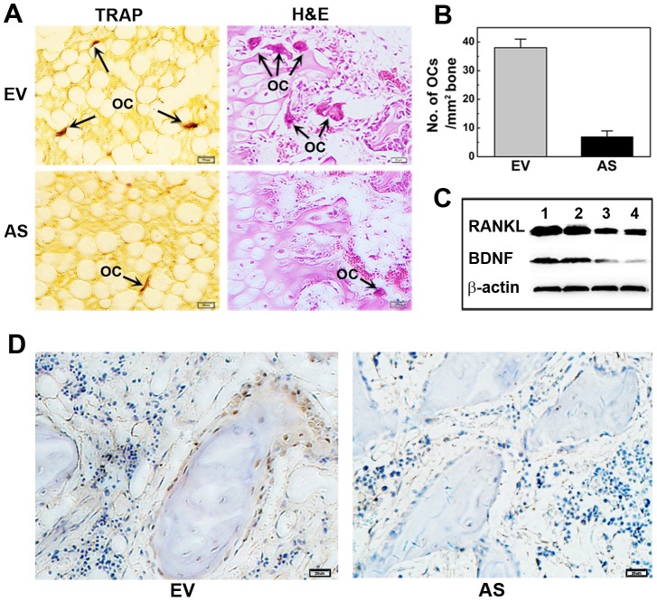
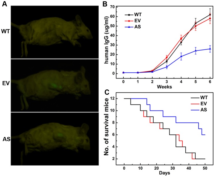
Brain-derived neurotrophic factor (BDNF) was recently identified as a factor produced by multiple myeloma (MM) cells, which may contribute to bone resorption and disease progression in MM, though the molecular mechanism of this process is not well understood. The purpose of this study was to test the effect of BDNF on bone disease and growth of MM cells both in vitro and in vivo. Co- and triple-culture systems were implemented. The in vitro results demonstrate that BDNF augmented receptor activator of nuclear factor kappa B ligand (RANKL) expression in human bone marrow stromal cells, thus contributing to osteoclast formation. To further clarify the effect of BDNF on myeloma bone disease in vivo, ARH-77 cells were stably transfected with an antisense construct to BDNF (AS-ARH) or empty vector (EV-ARH) to test their capacity to induce MM bone disease in SCID-rab mice. Mice treated with AS-ARH cells were preserved, exhibited no radiologically identifiable lytic lesions and, unlike the controls treated with EV-ARH cells, lived longer and showed reduced tumor burden. Consistently, bones harboring AS-ARH cells showed marked reductions of RANKL expression and osteoclast density compared to the controls harboring EV-ARH cells. These results provide further support for the potential osteoclastogenic effects of BDNF, which may mediate stromal-MM cell interactions to upregulate RANKL secretion, in myeloma bone diseases.


Conflict of interest statement

Competing Interests: The authors have declared that no competing interests exist.

Figures

Figure 1
Figure 1. Effects of BDNF on RANKL expression in human bone marrow stromal cells (BMSCs).
(A) The dose and time responses to BDNF of RANKL mRNA expression in BMSCs. PCR analysis indicates that BDNF markedly increases RANKL mRNA expression in human BMSCs (B) Western blot analysis shows that BDNF increased RANKL protein expression by human BMSCs in a dose- and time-dependent manner. (C) K252a diminishes BDNF-mediated RANKL secretion in human BMSCs. Stimulation with 25 ng/ml BDNF was performed in the absence or presence of K252a pretreatment (50, 100 nM; 60 min) in BMSCs. K252a without BDNF was used for control stimulation. RANKL levels in supernatants at different time points (0, 12, 24, 48 h) were detected by ELISA.
Figure 2
Figure 2. BDNF induces ERK1/2 and AKT phosphorylation in BMSCs, but has no effect on NF-κB activation.
(A, B, C) BMSCs were cultured in the presence of 25 ng/ml BDNF for 5, 15, 30, or 60 min. The expression of p-ERK1/2 (A) and p-Akt (B) began to increase at 5 min. However, no obvious change in the I-κB (C) level was detected by 60 min after BDNF stimulation. The results are representative of three independent experiments. Statistical analysis was conducted by ANOVA, * P<0.01 versus controls. (D) BDNF does not induce translocation of NF-κB p65 to the nucleus in BMSCs. BMSCs were treated with BDNF for 5, 15, 30 or 60 minutes, then fixed and probed with antibodies specific for NF-κB p65. Magnification ×200. Representative images from 15-min exposures to stimuli are exhibited. (E) MEK1/2 inhibitor U0126 and PI3K inhibitor LY204002 blunted the increase of RANKL protein secretion induced by BDNF. The results represent the means ± SEMs for all assays (performed in triplicate) (BDNF+U0126 vs. BDNF, * P<0.05; BDNF+LY204002 vs. BDNF, P = 0.069; BDNF+U0126+LY204002 vs. BDNF, ** P<0.05).
Figure 3
Figure 3. MM-derived BDNF promotes RANKL expression in co-culture systems and increases osteoclast formation in triple-culture systems.
(A) Soluble BDNF levels in culture medium measured by ELISA (ng/ml). BMSCs or preOCs were cultured alone or cocultured with MM cell (ARH-77, RPMI8226, MMPCs) using the transwell systems we described in methods before. After 48 hours, the supernatants were collected, and BDNF concentrations were analyzed with an ELISA kit. Each experiment was done in triplicate. The mean levels of BDNF secretion in cocultures of both MM-BMSC and MM-preOC were significantly higher than in BMSCs or preOCs alone (* P<0.01 compared to BMSCs cultured alone; # P<0.01 compared to preOCs cultured alone). (B) RANKL mRNA increased when BMSCs were cocultured with MM cells. These effects were abolished by K252a. The results represent the means ± SEMs for all assays (performed in triplicate). * P<0.05, ** P<0.01 compared to the control cultured without K252a. Control means the basal level of RANKL mRNA when BMSCs were cultured alone. (C) Correlation between BDNF levels in bone marrow plasma from 22 patients and marrow plasma-induced RANKL secretion by BMSCs (correlation coefficient = 0.35, P<0.01). (D) Representative images of TRAP-positive multinucleated osteoclast-like cells from human peripheral blood mononuclear cell cultures (labeled by black arrows) and the negative control. Magnification ×200. (E) Osteoclast precursors were cocultured with MM cells or triple-cultured with both BMSCs and MM cells. The enhancement of osteoclast formation in co- and triple-cultures was reversed by BDNF-neutralizing antibody (gray lanes) and was nearly completely abolished by OPG (white lanes). Goat IgG was used as a control (black lanes). * P<0.05, ** P<0.01 compared to the control.
Figure 4
Figure 4. Antisense inhibition of BDNF in ARH77 cells prevents tumor-induced osteolytic lesions in bone implants.
(A) Representative X-ray radiographs of the implanted myelomatous bones in each group, before cell engraftment (Before MM) and at the end of the experiment (After MM). Bones engrafted with EV-ARH cells were severely damaged, and tumors grew on the outer surface of the implanted bone (black arrow). However, no obvious osteolytic destruction was detected in bones harboring AS-ARH cells. (B) H&E staining of bone sections. Bones in the EV-ARH group were severely resorbed. Tumors damaged normal bone structures and infiltrated to the outer surface of the implanted bones (black arrow). In contrast, bones harboring AS-ARH cells had relatively normal structures and continuous cortexes (blue arrow). Magnification ×100. (C) Changes in the BMD of implanted bones. Error bars represent SEMs, P<0.01. (D) Changes in the level of soluble RANKL in rabbit bone implants. The mean levels of RANKL protein expression in WT-, EV-, and AS- group were 2205.9, 2130.5, and 286.3 pg/ml, respectively. RANKL levels in AS- group were significantly reduced when compared to EV- group (P<0.01).
Figure 5
Figure 5. Antisense inhibition of BDNF inhibits osteoclast formation and RANKL expression in myelomatous bones.
(A) TRAP and H&E staining of the myelomatous bones. Representative osteoclasts are labeled with arrows. Magnification ×200. (B) Quantitative analysis of osteoclasts on trabecular bone surface. Data represent the means ± SEMs of five separate high-power fields (HPFs). (C) Bone engrafts were fetched and cells in bones were obtained by flushing the bones repeatedly with 1 ml of PBS. Expression levels of RANKL and BDNF were measured by western blot Results indicate that RANKL and BDNF protein level in AS-ARH-infiltrated bones (lanes 3, 4) were much lower than those in EV-ARH group (lanes 1, 2). (D) Immunohistochemistry analysis indicates that the number of RANKL-positive cells in bone sections from the AS-ARH group was markedly decreased compared to the EV-ARH group .Magnification ×400.
Figure 6
Figure 6. Stable knockdown of BDNF activity inhibits tumor growth and prolongs overall survival in vivo.
(A) Representative fluorescence images of SCID-rab mice harboring the myeloma xenograft tumors. Mice harboring AS-ARH cells suffered lower tumor burden than animals harboring EV-ARH cells. (B) Circulating human IgG levels in SCID-rab mice were detected by ELISA. Human IgG levels began to show a difference 2 weeks after ARH inoculation, and the difference became increasingly marked. Circulating human IgG levels in the AS-ARH group was significantly lower than the EV-ARH and WT-ARH group at 6 weeks (P<0.05). (C) The effects of BDNF knockdown on overall survival were analyzed by Kaplan–Meier curves and long-rank tests.

References

    1. Roodman GD (2010) Pathogenesis of myeloma bone disease. J Cell Biochem 109: 283–291. - PubMed
    1. Basak GW, Srivastava AS, Malhotra R, Carrier E (2009) Multiple myeloma bone marrow niche. Curr Pharm Biotechnol 10: 345–346. - PubMed
    1. Tsubaki M, Kato C, Manno M, Ogaki M, Satou T, et al. (2007) Macrophage inflammatory protein-1alpha (MIP-1alpha) enhances a receptor activator of nuclear factor kappaB ligand (RANKL) expression in mouse bone marrow stromal cells and osteoblasts through MAPK and PI3K/Akt pathways. Mol Cell Biochem 304: 53–60. - PubMed
    1. Kwan Tat S, Padrines M, Theoleyre S, Heymann D, Fortun Y (2004) IL-6, RANKL, TNF-alpha/IL-1: interrelations in bone resorption pathophysiology. Cytokine Growth Factor Rev 15: 49–60. - PubMed
    1. Zannettino AC, Farrugia AN, Kortesidis A, Manavis J, To LB, et al. (2005) Elevated serum levels of stromal-derived factor-1alpha are associated with increased osteoclast activity and osteolytic bone disease in multiple myeloma patients. Cancer Res 65: 1700–1709. - PubMed

Publication types

MeSH terms