. 2012;7(10):e47447.
doi: 10.1371/journal.pone.0047447. Epub 2012 Oct 15.

A screen for selective killing of cells with chromosomal instability induced by a spindle checkpoint defect

Affiliations

A screen for selective killing of cells with chromosomal instability induced by a spindle checkpoint defect

Zeeshan Shaukat et al. PLoS One. 2012.

Abstract

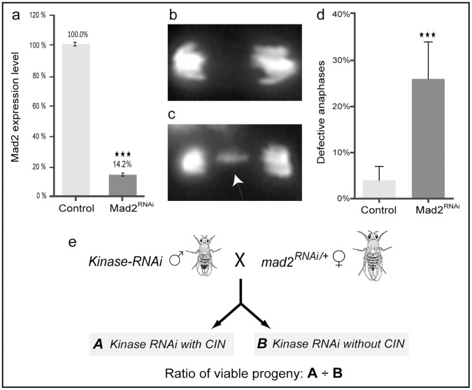
Background: The spindle assembly checkpoint is crucial for the maintenance of a stable chromosome number. Defects in the checkpoint lead to Chromosomal INstability (CIN), which is linked to the progression of tumors with poor clinical outcomes such as drug resistance and metastasis. As CIN is not found in normal cells, it offers a cancer-specific target for therapy, which may be particularly valuable because CIN is common in advanced tumours that are resistant to conventional therapy.

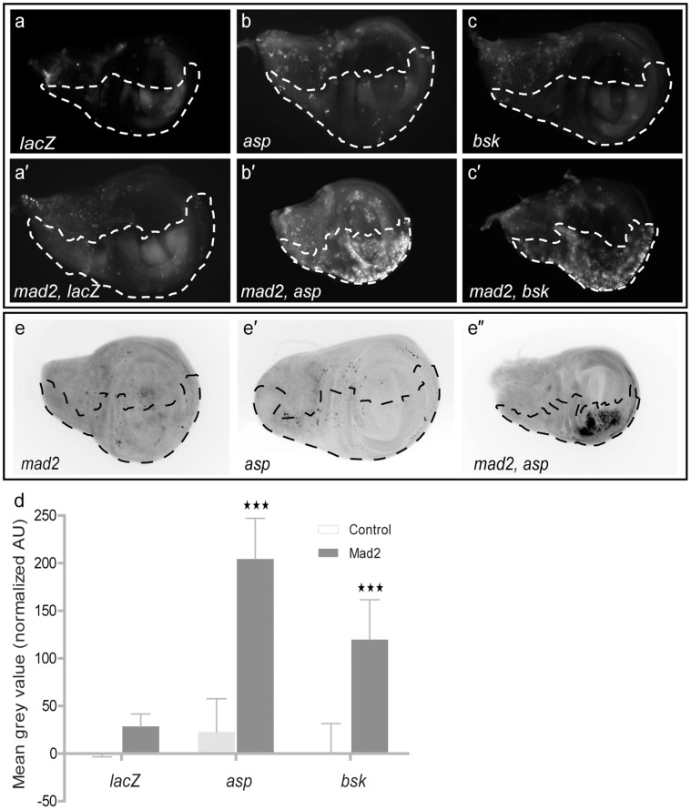
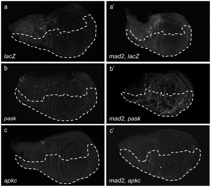
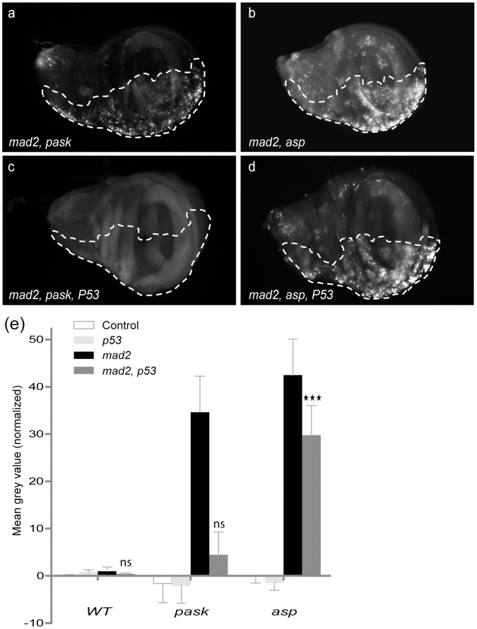
Principal findings: Here we identify genes that are required for the viability of cells with a CIN phenotype. We have used RNAi knockdown of the spindle assembly checkpoint to induce CIN in Drosophila and then screened the set of kinase and phosphatase genes by RNAi knockdown to identify those that induce apoptosis only in the CIN cells. Genes identified include those involved in JNK signaling pathways and mitotic cytoskeletal regulation.

Conclusions/significance: The screen demonstrates that it is feasible to selectively kill cells with CIN induced by spindle checkpoint defects. It has identified candidates that are currently being pursued as cancer therapy targets (e.g. Nek2: NIMA related kinase 2), confirming that the screen is able to identify promising drug targets of clinical significance. In addition, several other candidates were identified that have no previous connection with mitosis or apoptosis. Further screening and detailed characterization of the candidates could potentially lead to the therapies that specifically target advanced cancers that exhibit CIN.


Conflict of interest statement

Competing Interests: The authors have declared that no competing interests exist.

Figures

Figure 1
Figure 1. Establishment of a screening strategy using an induced-CIN model.
(a) Reverse transcriptase-qPCR shows that the ubiquitous expression of UAS-mad2 RNAi resulted in ∼85% knocked down of mad2 expression level (black bar) which is significantly less than the mad2 level in UAS-LacZ RNAi control (grey bar). Error bars represent SD. P-values are calculated by two-tailed Student’s t-test: p<0.001 = ★★★. (b–c) Third instar larval brain cells stained with Hoechst 33342 to label DNA. (b) Normal segregation in a wild type anaphase. (c) Defective anaphase in an induced-CIN brain cell (da>mad2) resulting in a lagging chromosome (arrowed). (d) The fraction of defective anaphases (lagging chromosomes or bridges) observed in mad2 knocked down (black bar) brain squashes and wild type controls (grey bar). Error bars represent 95% CIs. P-values are calculated by two-tailed Fisher’s exact test: p<0.001 = ★★★. (e) Diagrammatic representation of viability screen crosses. Males with Kinase-RNAi (UAS-kinasedsRNA) were crossed with females carrying the CIN background (UAS-mad2dsRNA; da-Gal4). Progeny were double knockdown (A: mad2 and kinase) or single knockdown (B: kinase only). The ratio of viable progeny A/B was used to rank candidates for further analysis.
Figure 2
Figure 2. Cell death assays on larval wing discs.
Dotted line shows the en-CD8GFP marked compartment or tester region in which genes were depleted. The other half of each wing disc expressed no transgenes and serves as an internal control. (a–c, a′–c′) Images of wing discs stained with Acridine Orange to show cell death. (a) Negative control (lacZ RNAi), (a′) LacZ and mad2 RNAi, (b & c) Candidate RNAi (asp and bsk), (b′ & c′) double knockdown of candidate and Mad2. (d) Graph shows quantitation of Acridine Orange staining (above wild type) in control and candidate imaginal wing disc halves with or without mad2 RNAi. Error bars represent 95% CIs, n≥8 in all cases. P-values were calculated by two-tailed t-tests with Welch’s correction: p<0.001 = ★★★. (e-e′) Cleaved caspase 3 staining showing apoptosis in e: mad2 RNAi, e′: asp RNAi and double knockdown (e″: asp RNAi and mad2 RNAi).
Figure 3
Figure 3. DNA damage (anti-P-H2AvD) staining of third instar larval wing discs.
(a–c′) Dotted line shows the en-CD8GFP marked test region in which genes were depleted. The other half of each wing disc expressed no transgenes. (a, a′) Negative control (LacZ RNAi) with and without Mad2 (b, b′) PASK depletion with and without Mad2. (c, c′) aPKC depletion with and without Mad2. Significant induction of DNA damage in the depleted area is seen in Pask, mad2 discs but not LacZ, mad2 or aPKC, mad2 discs.
Figure 4
Figure 4. P53-dependent and p53-independent apoptosis.
(a–d) Dotted line shows the en>CD8GFP marked test region and the other half expressed no transgenes. Acridine Orange staining on double (a–b: Candidate and mad2) and triple knockdown (c–d: Candidate, mad2 and p53) wing discs. (e) Graph shows quantitation of Acridine Orange staining (above wild type) in control and candidate knocked down imaginal wing disc halves. The first bar of each group represents candidate RNAi alone (control), the second bar represents candidate RNAi with p53RNAi (P53), the third or black bar represents Candidate and mad2 knocked down and the fourth bar represents triple knockdown (Candidate, mad2 and p53). Error bars represent 95%CIs, n≥8 in all cases. P-values are calculated by two-tailed t-tests with Welch’s correction: p<0.001 = ★★★ and p>0.05 = ns (not significant). Tests compare candidate mad2 p53 with candidate alone to test whether significant p53-independent cell death is seen when each candidate is co-depleted with Mad2. Significant levels of p53-independent cell death are seen for asp, mad2 but not Pask, mad2.

References

    1. Mertens F, Johansson B, Mitelman F (1994) Isochromosomes in neoplasia. Genes, Chromosomes and Cancer 10: 221–230. - PubMed
    1. Gao C, Furge K, Koeman J, Dykema K, Su Y, et al. (2007) Chromosome instability, chromosome transcriptome, and clonal evolution of tumor cell populations. Proceedings of the National Academy of Sciences 104: 8995–9000. - PMC - PubMed
    1. Heilig CE, Löffler H, Mahlknecht U, Janssen JWG, Ho AD, et al. (2009) Chromosomal instability correlates with poor outcome in patients with myelodysplastic syndromes irrespectively of the cytogenetic risk group. Journal of Cellular and Molecular Medicine 14: 895–902. - PMC - PubMed
    1. Swanton C, Nicke B, Schuett M, Eklund AC, Ng C, et al. (2009) Chromosomal instability determines taxane response. Proceedings of the National Academy of Sciences 106: 8671–8676. - PMC - PubMed
    1. Sotillo R, Schvartzman J-M, Socci ND, Benezra R (2010) Mad2-induced chromosome instability leads to lung tumour relapse after oncogene withdrawal. Nature 464: 436–440. - PMC - PubMed

Publication types

MeSH terms

LinkOut - more resources