

The viral transactivator HBx protein exhibits a high potential for regulation via phosphorylation through an evolutionarily conserved mechanism
- PMID: 23079056
- PMCID: PMC3533737
- DOI: 10.1186/1750-9378-7-27
The viral transactivator HBx protein exhibits a high potential for regulation via phosphorylation through an evolutionarily conserved mechanism
Abstract
Background: Hepatitis B virus (HBV) encodes an oncogenic factor, HBx, which is a multifunctional protein that can induce dysfunctional regulation of signaling pathways, transcription, and cell cycle progression, among other processes, through interactions with target host factors. The subcellular localization of HBx is both cytoplasmic and nuclear. This dynamic distribution of HBx could be essential to the multiple roles of the protein at different stages during HBV infection. Transactivational functions of HBx may be exerted both in the nucleus, via interaction with host DNA-binding proteins, and in the cytoplasm, via signaling pathways. Although there have been many studies describing different pathways altered by HBx, and its innumerable binding partners, the molecular mechanism that regulates its different roles has been difficult to elucidate.
Methods: In the current study, we took a bioinformatics approach to investigate whether the viral protein HBx might be regulated via phosphorylation by an evolutionarily conserved mechanism.
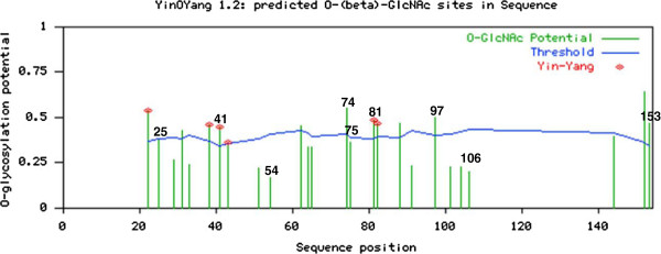
Results: We found that the phylogenetically conserved residues Ser25 and Ser41 (both within the negative regulatory domain), and Thr81 (in the transactivation domain) are predicted to be phosphorylated. By molecular 3D modeling of HBx, we further show these residues are all predicted to be exposed on the surface of the protein, making them easily accesible to these types of modifications. Furthermore, we have also identified Yin Yang sites that might have the potential to be phosphorylated and O-β-GlcNAc interplay at the same residues.
Conclusions: Thus, we propose that the different roles of HBx displayed in different subcellular locations might be regulated by an evolutionarily conserved mechanism of posttranslational modification, via phosphorylation.
Figures


Similar articles
-
Phosphorylation of Phylogenetically Conserved Amino Acid Residues Confines HBx within Different Cell Compartments of Human Hepatocarcinoma Cells.Molecules. 2021 Feb 26;26(5):1254. doi: 10.3390/molecules26051254. Molecules. 2021. PMID: 33652602 Free PMC article.
-
Smc5/6 Antagonism by HBx Is an Evolutionarily Conserved Function of Hepatitis B Virus Infection in Mammals.J Virol. 2018 Jul 31;92(16):e00769-18. doi: 10.1128/JVI.00769-18. Print 2018 Aug 15. J Virol. 2018. PMID: 29848586 Free PMC article.
-
Parvulin 14 and Parvulin 17 Bind to HBx and cccDNA and Upregulate Hepatitis B Virus Replication from cccDNA to Virion in an HBx-Dependent Manner.J Virol. 2019 Mar 5;93(6):e01840-18. doi: 10.1128/JVI.01840-18. Print 2019 Mar 15. J Virol. 2019. PMID: 30567987 Free PMC article.
-
The role of hepatitis B virus X protein is related to its differential intracellular localization.Acta Biochim Biophys Sin (Shanghai). 2011 Aug;43(8):583-8. doi: 10.1093/abbs/gmr048. Epub 2011 Jun 21. Acta Biochim Biophys Sin (Shanghai). 2011. PMID: 21693548 Review.
-
Hepatitis B virus, HBx mutants and their role in hepatocellular carcinoma.World J Gastroenterol. 2014 Aug 14;20(30):10238-48. doi: 10.3748/wjg.v20.i30.10238. World J Gastroenterol. 2014. PMID: 25132741 Free PMC article. Review.
Cited by
-
The Use of Intrinsic Disorder and Phosphorylation by Oncogenic Viral Proteins to Dysregulate the Host Cell Cycle Through Interaction with pRb.Viruses. 2025 Jun 10;17(6):835. doi: 10.3390/v17060835. Viruses. 2025. PMID: 40573426 Free PMC article. Review.
-
Mutations in Hepatitis-B X-Gene Region: Chronic Hepatitis-B versus Cirrhosis.J Clin Diagn Res. 2017 Mar;11(3):OC31-OC34. doi: 10.7860/JCDR/2017/22570.9498. Epub 2017 Mar 1. J Clin Diagn Res. 2017. PMID: 28511432 Free PMC article.
-
The Intrinsically Disordered Region of HBx and Virus-Host Interactions: Uncovering New Therapeutic Approaches for HBV and Cancer.Int J Mol Sci. 2025 Apr 10;26(8):3552. doi: 10.3390/ijms26083552. Int J Mol Sci. 2025. PMID: 40332052 Free PMC article. Review.
-
Sophisticated viral quasispecies with a genotype-related pattern of mutations in the hepatitis B X gene of HBeAg-ve chronically infected patients.Sci Rep. 2021 Feb 18;11(1):4215. doi: 10.1038/s41598-021-83762-4. Sci Rep. 2021. PMID: 33603102 Free PMC article.
-
Targeting Hepatitis B Virus Covalently Closed Circular DNA and Hepatitis B Virus X Protein: Recent Advances and New Approaches.ACS Infect Dis. 2019 Oct 11;5(10):1657-1667. doi: 10.1021/acsinfecdis.9b00249. Epub 2019 Sep 27. ACS Infect Dis. 2019. PMID: 31525994 Free PMC article.
References
-
- Murakami S, Cheong JH, Kaneko S. Human hepatitis B virus X gene encodes a regulatory domain which represses transactivation of X protein. J Biol Chem. 1994;269:15118–15123. - PubMed
-
- Gottlob K, Pagano S, Levrero M, Graessmann A. Hepatitis B virus X protein transcription activation domains are neither required nor sufficient for cell transformation. Cancer Res. 1998;58:3566–70. - PubMed
-
- Lin Y, Nomura T, Yamashita T, Dorjsuren D, Tang H, Murakami S. The transactivation and p53-interacting functions of hepatitis B virus X protein are mutually interfering but distinct. Cancer Res. 1997;57:5137–42. - PubMed
LinkOut - more resources
Full Text Sources
