. 2012 Oct 16;22(4):494-505.
doi: 10.1016/j.ccr.2012.08.017.

Elevated epidermal thymic stromal lymphopoietin levels establish an antitumor environment in the skin

Affiliations

Elevated epidermal thymic stromal lymphopoietin levels establish an antitumor environment in the skin

Shadmehr Demehri et al. Cancer Cell. .

Abstract

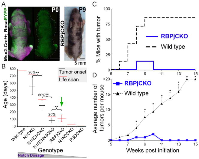
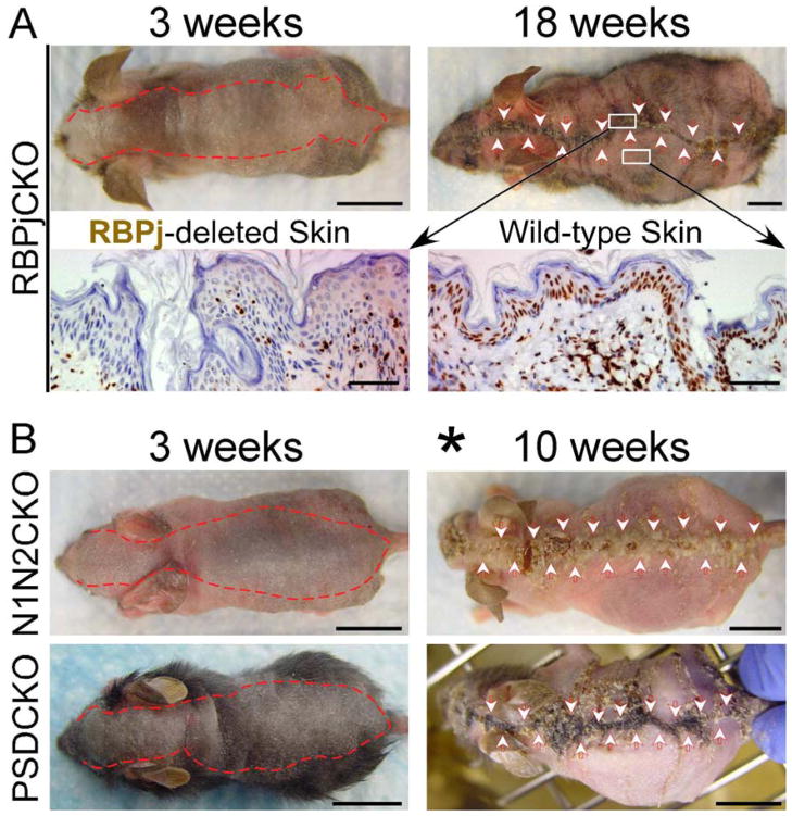
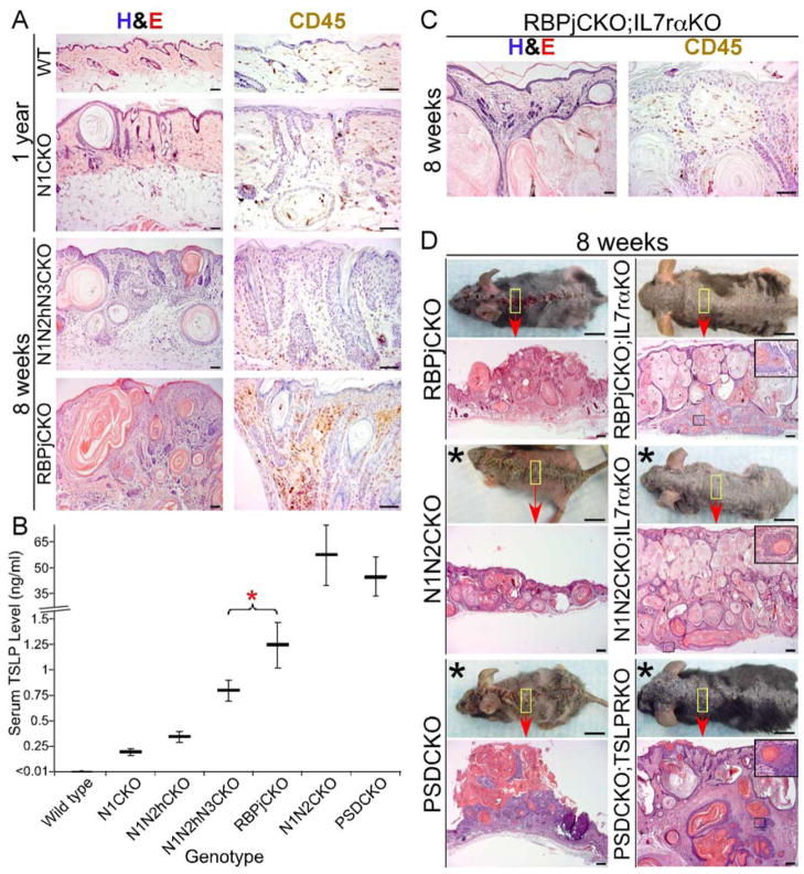
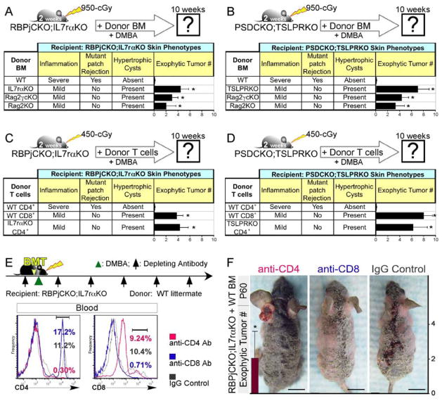
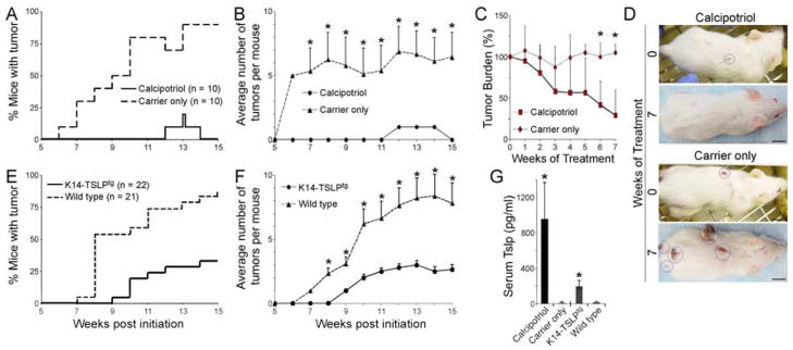
Thymic Stromal Lymphopoietin (TSLP), a cytokine implicated in induction of T helper 2 (Th2)-mediated allergic inflammation, has recently been shown to stimulate solid tumor growth and metastasis. Conversely, studying mice with clonal loss of Notch signaling in their skin revealed that high levels of TSLP released by barrier-defective skin caused a severe inflammation, resulting in gradual elimination of Notch-deficient epidermal clones and resistance to skin tumorigenesis. We found CD4(+) T cells to be both required and sufficient to mediate these effects of TSLP. Importantly, TSLP overexpression in wild-type skin also caused resistance to tumorigenesis, confirming that TSLP functions as a tumor suppressor in the skin.


Figures

Figure 1
Figure 1
Mice lacking RBPj in portions of their epidermis are resistant to skin tumorigenesis. (A) The calico pattern of EYFP expression (green) induced by Msx2-Cre-mediated gene deletion in a Msx2-Cretg/+; Rosa (LoxP_Stop_LoxP)-Eyfp (Msx2-Cre/+; RosaEYFP) newborn is shown. Image taken under tungsten illumination is shown in magenta. After birth, mutant clones become evident due to hair phenotypes. (B) Reduction in Notch signaling dosage in the skin correlates with shortening of life span and time to spontaneous tumor formation. This trend, however, does not extend to mice lacking RBPj (green arrow), which live ~100 days yet do not develop any skin tumors. n > 20 in each group; % indicates the percentage of mice that developed skin tumors; **: p <0.01, student’s t-test; error bars represent +/− SD. This figure is modified from (Demehri et al., 2009b). (C,D) Time to tumor onset (C; p <0.0001, log-rank test) and average tumor number (D) of RBPjCKO and wild-type littermates treated with the standard DMBA-TPA protocol from 3 to 18 weeks of age are shown. n = 7 for each group; *: p <0.05, student’s t-test; error bars represent +/− SEM. Genotypes: Msx2-Cretg/+; Notch1flox/flox (N1CKO), Msx2-Cretg/+; Notch1flox/flox; Notch2flox/+ (N1N2hCKO), Msx2-Cretg/+; Notch1flox/flox; Notch2flox/+; Notch3−/− (N1N2hN3CKO), Msx2-Cretg/+; Notch1flox/flox; Notch2flox/flox (N1N2CKO), Msx2-Cretg/+; Ps1flox/flox; Ps2−/− (PSDCKO), Msx2-Cretg/+; Rbpjflox/flox (RBPjCKO).
Figure 2
Figure 2
Notch signaling-deficient epidermal clones regress with age. (A) The red dotted line and arrowheads delineate the boundaries of the RBPj-deficient dorsal epidermis, as determined by hair/epidermal phenotype. Bottom panel shows α-RBPj antibody staining of RBPj-depleted midline skin (left) and wild-type skin in the periphery (right) of a 18-week-old RBPjCKO animal. (B) The red dotted line and arrowheads delineate the boundaries of the mutant dorsal epidermis in N1N2CKO and PSDCKO animals. Asterisk marks the recipients of a sublethal dose of irradiation in the second week of life; representative pictures are shown in all panels; scale bars: 1 cm (mice pictures); 50 μm (histology).
Figure 3
Figure 3
H-Ras-infected RBPjKO or PSDKO keratinocytes are highly tumorigenic in nude mice. (A) Schema showing the experimental procedure. Cells are infected with retrovirus containing oncogenic H-Ras and then with Adeno-Cre to delete the floxed alleles. H-Ras-infected RBPjKO or PSDKO and wild-type cells injected subcutaneously into the right and left flanks of the nude mice, respectively. (B) Nude mice with palpable tumors (red circles) are harvested 30 days after the injection. Hematoxylin and eosin (H&E) and α-RBPj antibody stainings on a tumor formed by RBPjKO keratinocytes are shown. Representative pictures are shown; scale bars: 1 cm (mice pictures), 50 μm (histology).
Figure 4
Figure 4
Blunting the skin inflammation by blocking TSLP signaling in Notch signaling-deficient animals results in mutant skin expansion and tumorigenesis. (A) H&E and CD45 antibody stainings show the level of skin inflammation in RBPjCKO animals compared to the tumor-prone N1CKO and N1N2hN3CKO mice. Scale bars: 50 μm. (B) The circulating TSLP levels during the second week of life are compared among the allelic series of Notch-deficient animals. *: p <0.05, student’s t-test; error bars represent +/− SD. This figure is modified from (Demehri et al., 2008). (C) H&E and CD45 antibody stainings highlight the level of dermal inflammation in RBPjCKO;IL7rαKO animals that lack TSLP signaling. Scale bars: 50 μm. (D) Gross and microscopic features of RBPjCKO;IL7rαKO, N1N2CKO;IL7rαKO and PSDCKO;TSLPRKO skin are compared to those of RBPjCKO, N1N2CKO and PSDCKO littermates, respectively. Asterisks mark the recipients of a sublethal dose of irradiation in the second week of life; insets: tumors penetrating the muscle layer; scale bars: 1 cm (mice pictures), 200 μm (histology); representative pictures are shown in all panels. See also Figure S1.
Figure 5
Figure 5
CD4+ T cells mediate the effects of TSLP on Notch-deficient skin rejection and tumor resistance. (A,B) The skin phenotypes of RBPjCKO;IL7rαKO (A) and PSDCKO;TSLPRKO (B) animals are analyzed 8 weeks after BMT from wild-type littermates, Rag2−/−,γc−/− (Rag2γcKO) or Rag2−/− (Rag2KO) animals. Note that Rag2γcKO and Rag2KO donors lack T and B cells. The level of dermal inflammation, the rejection of the mutant keratinocytes, the formation of hypertrophic cysts and the development of DMBA-dependent exophytic tumors are scored in the recipient animals at 10 weeks of age. BMT from TSLP receptor-deficient littermates are used as controls. *: p <0.05 compared to wild-type BM donor group. (C,D) 2-week-old RBPjCKO;IL7rαKO (C) and PSDCKO;TSLPRKO (D) pups were irradiated with 450-cGy and underwent adoptive T cell transfer using wild-type CD4+, wild-type CD8+ or TSLPR-deficient CD4+ T cells isolated from the spleens of their littermates as shown in the schematic diagrams. The level of dermal inflammation, the rejection of the mutant keratinocytes, the formation of hypertrophic cysts and the development of DMBA-dependent exophytic tumors are documented in the recipient animals at 10 weeks of age. *: p <0.05 compared to wild-type CD4+ T cell donor group. (E) 2-week-old RBPjCKO;IL7rαKO mice were treated with depleting antibodies (anti-CD4 or anti-CD8α; black arrows), lethally irradiated 2 days later and transplanted with T cell-depleted c-Kit+ BM progenitor cells from their wild-type littermates as shown in the schematic diagram. 2 days later the animals were treated with one dose DMBA while continuing to receive a weekly intraperitoneal injection of the indicated depleting antibody for 10 weeks. Flow cytometry shows the status of CD4+ and CD8+ T cells in the peripheral blood of RBPjCKO;IL7rαKO mice transplanted with c-Kit+ wild-type BM and treated with anti-CD4 or anti-CD8α antibody one week after the last antibody injection. Blood CD45+ leukocytes are traced in the figure. Note that PE-conjugated anti-CD8β antibody is used to detect CD8+ cells. (F) RBPjCKO;IL7rαKO mice transplanted with c-Kit+ BM cells and treated with anti-CD4, anti-CD8α or IgG control antibodies are compared. Bar graphs show the average number of DMBA-dependent exophytic tumors in each treatment group. *: p <0.05 compared to IgG control-treated group. For all experiments, mice were followed up to 90 days of age for exophytic tumor count; at least five mice were analyzed in each group; error bars on all bar graphs represent +/− SD; representative pictures are shown; scale bars: 1 cm. See also Figure S2.
Figure 6
Figure 6
TSLP creates a tumor-suppressing environment in the wild-type skin. (A,B) DMBA-TPA treated CD1 wild-type animals were topically treated with calcipotriol or carrier (EtOH), (A) time to tumor onset (p <0.0001, log-rank test) and (B) average number of tumors among tumor-bearing animals are shown. n = 10 for each group; error bars represent +/− SEM; *: p <0.01, student’s t-test. (C,D) The 8 tumor-bearing carrier-treated mice shown in “A&B” were randomly divided into two groups at the end of the 15-week DMBA-TPA treatment course. The “test” group was treated with 32 nmol calcipotriol while the “control” group continued to receive carrier alone 5 times per week. (C) After an additional 7-week treatment period, the tumor burdens of the calcipotriol-treated and carrier-treated mice are compared. n = 4 in each group; *: p <0.05, student’s t-test; error bars represent +/− SD. (D) The representative pictures show the size of the remaining tumors in calcipotriol-treated versus carrier-treated mice at the end of the 7-week follow-up period. Tumors are highlighted with blue circles; scale bars: 1 cm. (E,F) K14-TSLPtg female mice and their wild-type female littermates treated with DMBA-TPA are compared for (E) time to tumor onset (p <0.0001, log-rank test) and (F) average tumor number among tumor-bearing animals. n = 22 for K14-TSLPtg group and n = 21 for wild-type group; error bars represent +/− SEM; *: p <0.01, student’s t-test. (G) Circulating TSLP levels in calcipotriol-treated and K14-TSLPtg animals are compared to their controls. Error bars represent +/− SD; *: p <0.0001 compared to wild-type group, student’s t-test.

Comment in

References

    1. Al-Shami A, Spolski R, Kelly J, Fry T, Schwartzberg PL, Pandey A, Mackall CL, Leonard WJ. A role for thymic stromal lymphopoietin in CD4(+) T cell development. J Exp Med. 2004;200:159–168. - PMC - PubMed
    1. Al-Shami A, Spolski R, Kelly J, Keane-Myers A, Leonard WJ. A role for TSLP in the development of inflammation in an asthma model. J Exp Med. 2005;202:829–839. - PMC - PubMed
    1. Blanpain C, Lowry WE, Pasolli HA, Fuchs E. Canonical notch signaling functions as a commitment switch in the epidermal lineage. Genes Dev. 2006;20:3022–3035. - PMC - PubMed
    1. Campisi J. Senescent cells, tumor suppression, and organismal aging: good citizens, bad neighbors. Cell. 2005;120:513–522. - PubMed
    1. Cario G, Zimmermann M, Romey R, Gesk S, Vater I, Harbott J, Schrauder A, Moericke A, Izraeli S, Akasaka T, et al. Presence of the P2RY8-CRLF2 rearrangement is associated with a poor prognosis in non-high-risk precursor B-cell acute lymphoblastic leukemia in children treated according to the ALL-BFM 2000 protocol. Blood. 2010;115:5393–5397. - PubMed

Publication types