

Retinal haemorrhages and related findings in abusive and non-abusive head trauma: a systematic review
- PMID: 23079748
- PMCID: PMC3545381
- DOI: 10.1038/eye.2012.213
Retinal haemorrhages and related findings in abusive and non-abusive head trauma: a systematic review
Abstract
Aim: To report the retinal signs that distinguish abusive head trauma (AHT) from non-abusive head trauma (nAHT).
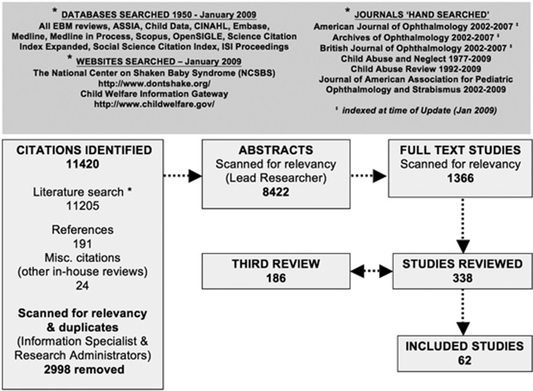
Methods: A systematic review of literature, 1950-2009, was conducted with standardised critical appraisal. Inclusion criteria were a strict confirmation of the aetiology, children aged <11 years and details of an examination conducted by an ophthalmologist. Post mortem data, organic disease of eye, and inadequate examinations were excluded. A multivariate logistic regression analysis was conducted to determine odds ratios (OR) and probabilities for AHT.
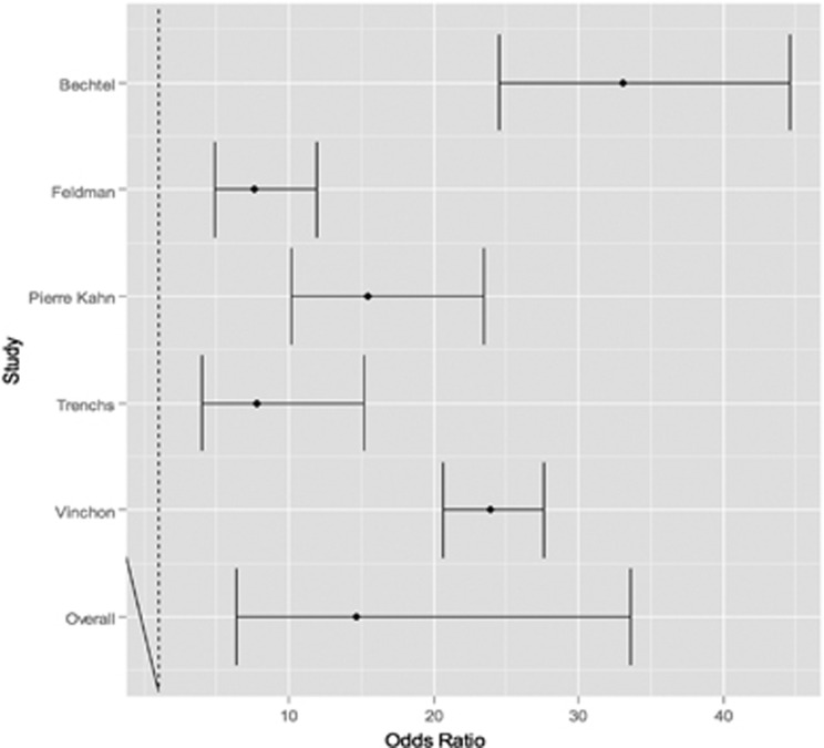
Results: Of the 62 included studies, 13 provided prevalence data (998 children, 504 AHT). Overall, retinal haemorrhages (RH) were found in 78% of AHT vs 5% of nAHT. In a child with head trauma and RH, the OR that this is AHT is 14.7 (95% confidence intervals 6.39, 33.62) and the probability of abuse is 91%. Where recorded, RH were bilateral in 83% of AHT compared with 8.3% in nAHT. RH were numerous in AHT, and few in nAHT located in the posterior pole, with only 10% extending to periphery. True prevalence of additional features, for example, retinal folds, could not be determined.
Conclusions: Our systematic review confirms that although certain patterns of RH were far commoner in AHT, namely large numbers of RH in both the eyes, present in all layers of the retina, and extension into the periphery, there was no retinal sign that was unique to abusive injury. RH are rare in accidental trauma and, when present, are predominantly unilateral, few in number and in the posterior pole.
Figures
References
-
- Levin AV. Ophthalmology of shaken baby syndrome. Neurosurg Clin N Am. 2002;13:201–211. - PubMed
-
- Maguire S, Pickerd N, Farewell D, Mann M, Tempest V, Kemp AM. Which clinical features distinguish inflicted from non-inflicted brain injury? a systematic review. Arch Dis Child. 2009;94:860–867. - PubMed
-
- Greenwald MJ, Weiss A, Oesterle CS, Friendly DS. Traumatic retinoschisis in battered babies. Ophthalmology. 1986;93:618–625. - PubMed
-
- Massicotte SJ, Folberg R, Torczynski E, Gilliland MG, Luckenbach MW. Vitreoretinal traction and perimacular retinal folds in the eyes of deliberately traumatized children. Ophthalmology. 1991;98:1124–1127. - PubMed
Publication types
MeSH terms
LinkOut - more resources
Full Text Sources
Medical
