Review
. 2012 Nov;11(11):860-72.
doi: 10.1038/nrd3864. Epub 2012 Oct 19.

MicroRNA therapeutics for cardiovascular disease: opportunities and obstacles

Affiliations
Review

MicroRNA therapeutics for cardiovascular disease: opportunities and obstacles

Eva van Rooij et al. Nat Rev Drug Discov. 2012 Nov.

Abstract

In recent years, prominent roles for microRNAs (miRNAs) have been uncovered in several cardiovascular disorders. The ability to therapeutically manipulate miRNA expression and function through systemic or local delivery of miRNA inhibitors, referred to as antimiRs, has triggered enthusiasm for miRNAs as novel therapeutic targets. Here, we focus on the pharmacokinetic and pharmacodynamic properties of current antimiR designs and their relevance to cardiovascular indications, and evaluate the opportunities and obstacles associated with this new therapeutic modality.


Conflict of interest statement

Competing interests statement

The authors declare competing financial interests: see Web version for details.

Figures

Figure 1 |
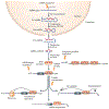
Figure 1 |. MicroRNA biogenesis and mechanism of action.
MicroRNA (miRNA) genes are usually transcribed by RNA polymerase II (Pol II) to form a capped and polyadenylated transcript. The miRNA precursor, termed primary miRNA (pri-miRNA), forms a hairpin-shaped loop structure that is cleaved by the RNase III Drosha and DiGeorge syndrome critical region 8 (DGCR8), yielding a hairpin-shaped precursor miRNA (pre-miRNA)that is ~70 nucleotides in length. The pre-miRNA is exported from the nucleus into the cytoplasm, where it is further cleaved by the RNase III enzyme Dicer, yielding an imperfect miRNA–miRNA* duplex that is about 22 nucleotides in length. Although either strand of the duplex may potentially act as a functional miRNA, only one strand is usually incorporated into the RNA-induced silencing complex (RISC). miRNAs incorporated in the RISC often recognize their targets — nucleotides 2–7 of miRNA (known as the ‘seed region’). Association of a miRNA with its mRNA target results in degradation of the mRNA as well astranslational inhibition. Recently, pseudogenes have also been implicated in regulating miRNA activity. Pseudogene transcripts are often conserved across species and many contain conserved miRNA binding sites, referred to as competing endogenous RNAs (ceRNAs), which act as decoys or sponges by seguestering miRNAs and preventing them from binding to their mRNA targets. Stress conditions can influence miRNA biogenesis at multiple levels (indicated on the figure by lightening bolts). IncRNA, long non-coding RNA; m7G, 7-methylguanosine (a modified form of guanosine attached to the 5′ ends of mRNAs); ORF, open reading frame.
Figure 2 |
Figure 2 |. AntimiR chemistries.
AntimiR (inhibitor of microRNA (miRNA)) chemistries currently use various high-affinity 2′ sugar modifications such as conformationally restricted nucleotides with 2′-O-methyl (2′-O-Me), 2′-O-methoxyethyl (2′-MOE), 2′-fluoro (2′-F) or locked nucleic acid (LNA) modifications. To increase nuclease resistance, most antimiR chemistriesto date harbour phosphorothioate backbone linkages, whereby a sulphur atom replaces one of the non-bridging oxygen atoms in the phosphate group. AntimiRs containing cholesterol, conjugated via a 2′-O-Me linkage, named antagomirs, are fully complementary to the mature miRNA sequence and contain several phosphorothioate moieties to increase their stability. Several unconjugated phosphorothioate antisense molecules with various high-affinity 2′sugar modifications (such as 2′-MOE, 2′-F or LNA) are currently also being used. Although all of these modifications improve nuclease resistance and increase duplex melting temperature, the high duplex melting temperature of LNA-modified oligonucleotides enables efficient miRNA inhibition with shorter antimiRs.
Figure 3 |
Figure 3 |. Multiple functions of miR-208 in the heart.
The microRNA miR-208a is encoded by an intron of the gene encoding myosin 6 (MYH6). miR-208a is reguired for the upregulation of myosin 7 (MYH7) and cardiac fibrosis in response to stress. miR-208a inhibits mediator of RNA polymerase II transcription subunit 13 (MED13), a component of the mediator complex, which regulates metabolic genes and additional targets. Inhibition of miR-208a with an antimiR (a miRNA inhibitor) inhibits cardiac remodelling and enhances systemic energy metabolism, resulting in beneficial effects in the settings of obesity and diabetes.
Figure 4 |
Figure 4 |. MicroRNAs often regulate related target genes.
Members of the miR-29 family exemplify the influence of microRNAs (miRNAs) on targets involved in common cellular processes. In this case, miR-29 coordinately inhibits the expression of numerous extracellular matrix proteins, such that the downregulation of this miRNA family under conditions of cardiovascular stress leads to vascular remodelling and fibrosis. ADAMTS, a disintegrin and metalloproteinase with thrombospondin motifs; COL1A1, collagen type 1 αl; ITGA11, integrin α11; MMP2, matrix metalloproteinase 2.

References

    1. van Rooij E et al. A signature pattern of stress-responsive microRNAs that can evoke cardiac hypertrophy and heart failure. Proc. Natl Acad. Sci. USA 103, 18255–18260 (2006). - PMC - PubMed
    2. This is the first description of changes in miRNA levels in murine and human heart disorders, and provides evidence for the relevance of miRNAs in heart disease.

    1. Ikeda S et al. Altered microRNA expression in human heart disease. Physiol. Genomics 31, 367–373 (2007). - PubMed
    2. This study describes unique miRNA signature patterns for different forms of human heart disease.

    1. Thum T et al. MicroRNAs in the human heart: a clue to fetal gene reprogramming in heart failure. Circulation 116, 258–267 (2007). - PubMed
    2. This study suggests a relationship between changes in miRNAs and the expression of fetal genes in human heart disease.

    1. Bonauer A, Boon RA & Dimmeler S Vascular microRNAs. Curr. Drug Targets 11, 943–949 (2010). - PubMed
    1. Zampetaki A & Mayr M MicroRNAs in vascular and metabolic disease. Circ. Res 110, 508–522 (2012). - PubMed

MeSH terms