

Cerebrovascular toxicity of PCB153 is enhanced by binding to silica nanoparticles
- PMID: 23081707
- PMCID: PMC3518694
- DOI: 10.1007/s11481-012-9403-y
Cerebrovascular toxicity of PCB153 is enhanced by binding to silica nanoparticles
Abstract
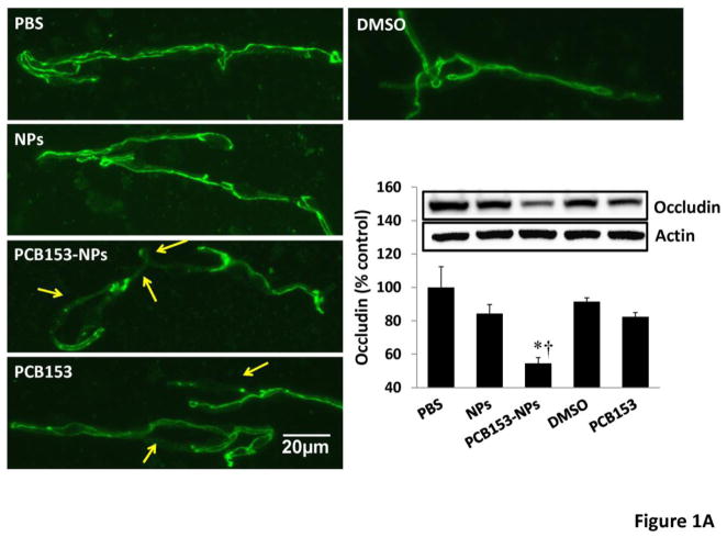
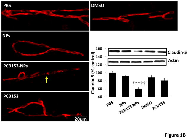
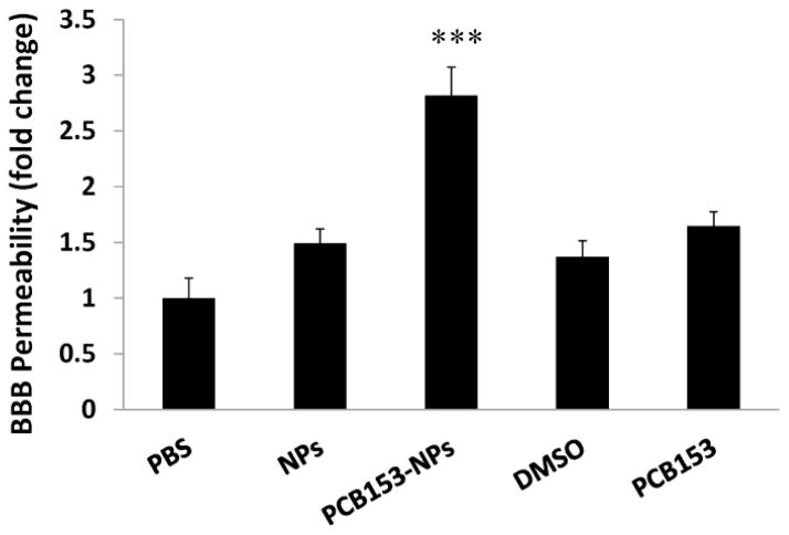
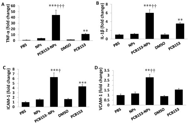
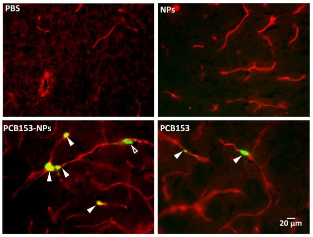
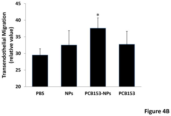
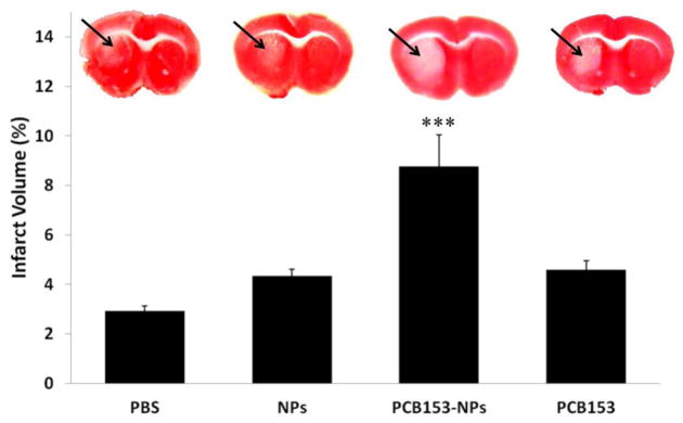
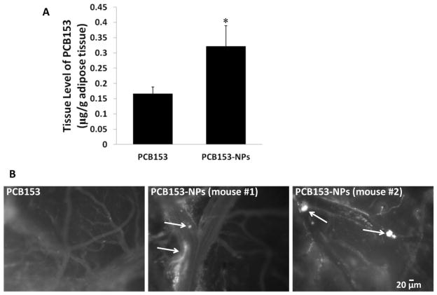
Environmental polychlorinated biphenyls (PCBs) are frequently bound onto nanoparticles (NPs). However, the toxicity and health effects of PCBs assembled onto nanoparticles are unknown. The aim of this study was to study the hypothesis that binding PCBs to silica NPs potentiates PCB-induced cerebrovascular toxicity and brain damage in an experimental stroke model. Mice (C57BL/6, males, 12-week-old) were exposed to PCB153 bound to NPs (PCB153-NPs), PCB153, or vehicle. PCB153 was administered in the amount of 5 ng/g body weight. A group of treated animals was subjected to a 40 min ischemia, followed by a 24 h reperfusion. The blood-brain barrier (BBB) permeability, brain infarct volume, expression of tight junction (TJ) proteins, and inflammatory mediators were assessed. As compared to controls, a 24 h exposure to PCB153-NPs injected into cerebral vasculature resulted in significant elevation of the BBB permeability, disruption of TJ protein expression, increased proinflammatory responses, and enhanced monocyte transmigration in mouse brain capillaries. Importantly, exposure to PCB153-NPs increased stroke volume and potentiated brain damage in mice subjected to ischemia/reperfusion. A long-term (30 days) oral exposure to PCB153-NPs resulted in a higher PCB153 content in the abdominal adipose tissue and amplified adhesion of leukocytes to the brain endothelium as compared to treatment with PCB153 alone. This study provides the first evidence that binding to NPs increases cerebrovascular toxicity of environmental toxicants, such as PCB153.
Conflict of interest statement
Figures
Similar articles
-
TLR4 signaling is involved in brain vascular toxicity of PCB153 bound to nanoparticles.PLoS One. 2013 May 14;8(5):e63159. doi: 10.1371/journal.pone.0063159. Print 2013. PLoS One. 2013. PMID: 23690990 Free PMC article.
-
Lipid rafts regulate PCB153-induced disruption of occludin and brain endothelial barrier function through protein phosphatase 2A and matrix metalloproteinase-2.Toxicol Appl Pharmacol. 2015 Sep 15;287(3):258-66. doi: 10.1016/j.taap.2015.06.011. Epub 2015 Jun 14. Toxicol Appl Pharmacol. 2015. PMID: 26080028 Free PMC article.
-
Lipopolysaccharide potentiates polychlorinated biphenyl-induced disruption of the blood-brain barrier via TLR4/IRF-3 signaling.Toxicology. 2012 Dec 16;302(2-3):212-20. doi: 10.1016/j.tox.2012.08.001. Epub 2012 Aug 13. Toxicology. 2012. PMID: 22906770 Free PMC article.
-
Effect of a two-week treatment with a low dose of 2,2'4,4',5,5'-hexachlorobiphenyl (PCB153) on tight junction protein expression in ovine choroid plexus during long and short photoperiods.Neurotoxicol Teratol. 2013 May-Jun;37:63-7. doi: 10.1016/j.ntt.2013.03.061. Epub 2013 Apr 6. Neurotoxicol Teratol. 2013. PMID: 23567221
-
Blood-brain barrier dysfunction in ischemic stroke: targeting tight junctions and transporters for vascular protection.Am J Physiol Cell Physiol. 2018 Sep 1;315(3):C343-C356. doi: 10.1152/ajpcell.00095.2018. Epub 2018 Jun 27. Am J Physiol Cell Physiol. 2018. PMID: 29949404 Free PMC article. Review.
Cited by
-
Protection of cultured brain endothelial cells from cytokine-induced damage by α-melanocyte stimulating hormone.PeerJ. 2018 May 15;6:e4774. doi: 10.7717/peerj.4774. eCollection 2018. PeerJ. 2018. PMID: 29780671 Free PMC article.
-
TLR4 signaling is involved in brain vascular toxicity of PCB153 bound to nanoparticles.PLoS One. 2013 May 14;8(5):e63159. doi: 10.1371/journal.pone.0063159. Print 2013. PLoS One. 2013. PMID: 23690990 Free PMC article.
-
Bridging Size and Charge Effects of Mesoporous Silica Nanoparticles for Crossing the Blood-Brain Barrier.Front Chem. 2022 Jun 27;10:931584. doi: 10.3389/fchem.2022.931584. eCollection 2022. Front Chem. 2022. PMID: 35880111 Free PMC article.
-
Lipid rafts regulate PCB153-induced disruption of occludin and brain endothelial barrier function through protein phosphatase 2A and matrix metalloproteinase-2.Toxicol Appl Pharmacol. 2015 Sep 15;287(3):258-66. doi: 10.1016/j.taap.2015.06.011. Epub 2015 Jun 14. Toxicol Appl Pharmacol. 2015. PMID: 26080028 Free PMC article.
-
Exposure to polychlorinated biphenyls selectively dysregulates endothelial circadian clock and endothelial toxicity.J Hazard Mater. 2023 Jul 15;454:131499. doi: 10.1016/j.jhazmat.2023.131499. Epub 2023 Apr 26. J Hazard Mater. 2023. PMID: 37126901 Free PMC article.
References
-
- Abbott NJ, Ronnback L, Hansson E. Astrocyte-endothelial interactions at the blood-brain barrier. Nat Rev Neurosci. 2006;7:41–53. - PubMed
-
- Albelda SM, Smith CW, Ward PA. Adhesion molecules and inflammatory injury. FASEB J. 1994;8:504–512. - PubMed
-
- ATSDR (Agency for Toxic Substances and Disease Registry) Health Consultation: Evaluation of Soil, Blood, and Air Data from Anniston, Alabama, Calhoun County, Alabama. Atlanta, GA: ATSDR; 2000. [Accessed 6 August 2012.]. http://www.atsdr.cdc.gov/hac/pha/pha.asp?docid=930&pg=0.
Publication types
MeSH terms
Substances
Grants and funding
LinkOut - more resources
Full Text Sources
Research Materials
