. 2013 Jan;31(1):59-70.
doi: 10.1002/stem.1254.

Functional dissection of Pax3 in paraxial mesoderm development and myogenesis

Affiliations

Functional dissection of Pax3 in paraxial mesoderm development and myogenesis

Alessandro Magli et al. Stem Cells. 2013 Jan.

Abstract

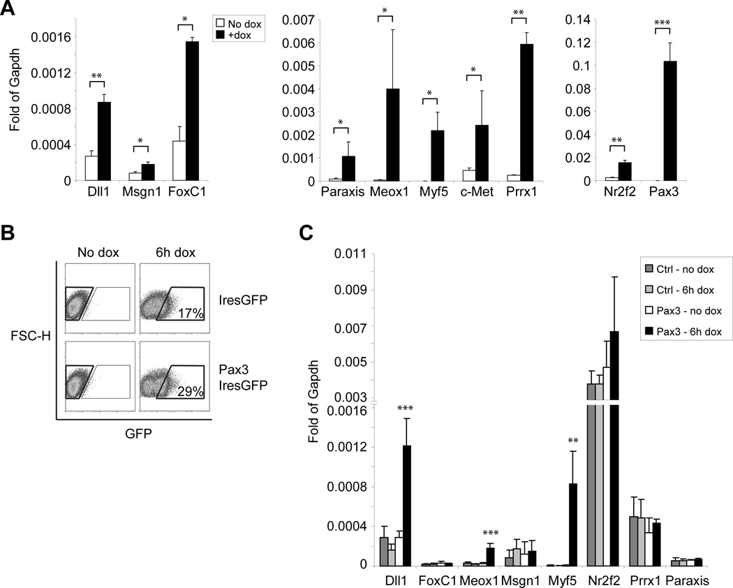
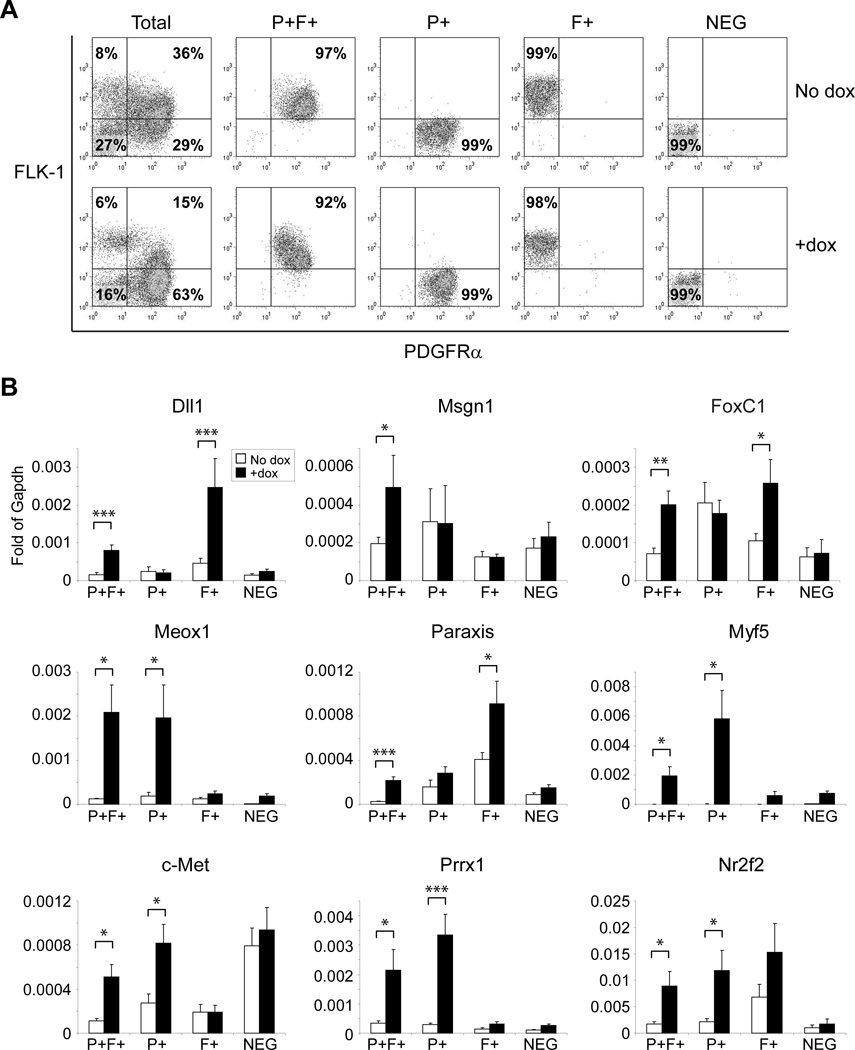
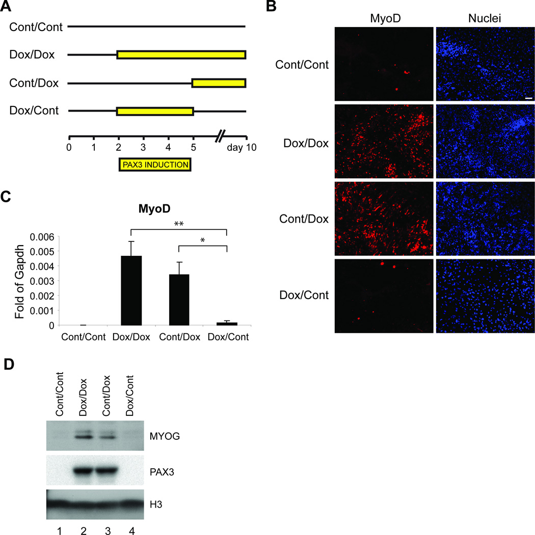
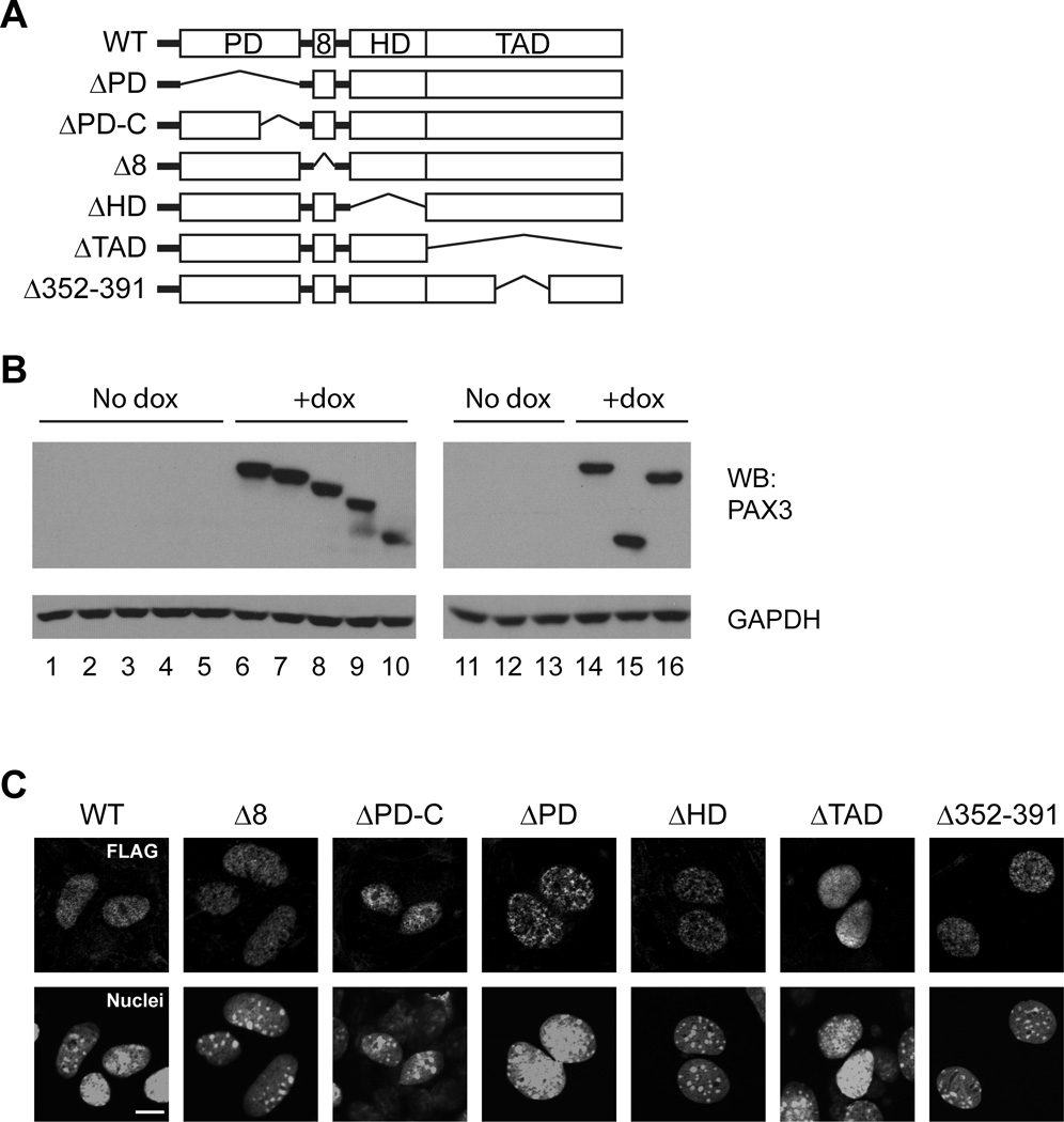
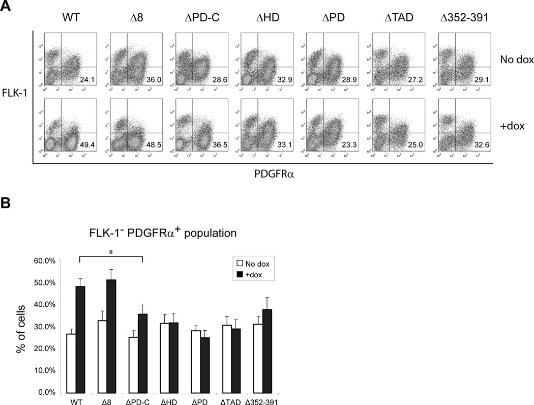
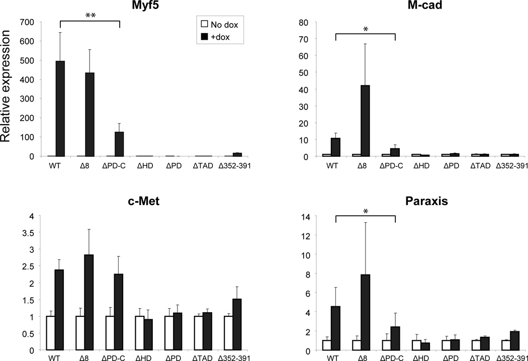
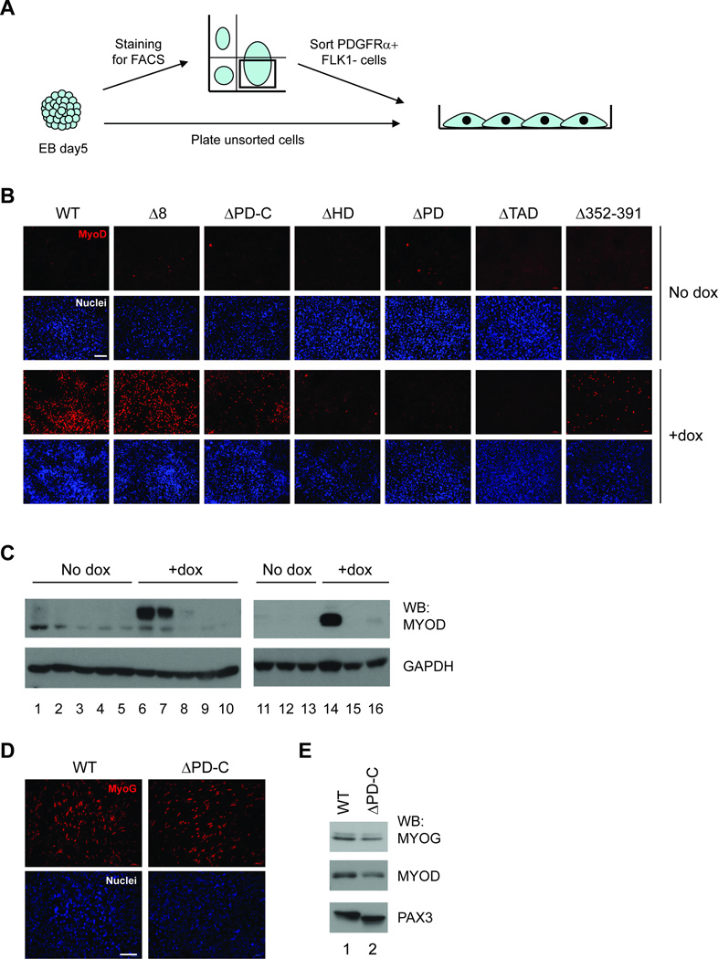
The paired box transcription factor Pax3 is well-known as a major regulator of embryonic myogenesis. Before Pax3 expression becomes restricted to the dermomyotome, this transcription factor is also expressed in the developing somites. The role of Pax3 at this early stage is unclear, in particular because of the scarce frequency of Pax3-positive cells in the early mouse embryo. Inducible gene expression in embryonic stem cells (ESCs) represents an excellent tool to overcome this limitation, since it can provide large quantities of otherwise rare embryonic populations expressing a factor of interest. Here we used engineered mouse ESCs to perform a functional analysis of Pax3 with the aim to identify the molecular determinants involved in the early functions of this transcription factor. We find that Pax3 induction during embryoid body differentiation results in the upregulation of genes expressed in the presomitic and somitic mesoderm. Moreover, we show that paraxial mesoderm induced by transient expression of Pax3 is not irreversibly committed to myogenesis rather requires sustained Pax3 expression. Using a series of deletion mutants of Pax3, which differentially affect its transcriptional activity, we map protein domains necessary for induction of paraxial mesoderm and induction of the myogenic program. The paired, homeo-, and transcriptional activation domains were each required for both processes, however, the paired-c-terminal RED domain showed a paraxial mesoderm-specific activity that was dispensable for myogenesis. These findings demonstrate and provide mechanistic insight into an early role for Pax3 in the generation of paraxial mesoderm.


Figures

Figure 1
Figure 1
Pax3 is necessary for the commitment of ES cells toward the paraxial mesoderm cell fate and skeletal myogenic lineage. A) Paraxial mesoderm and myogenic markers are up-regulated by Pax3 induction, as shown by quantitative RT-PCR analysis of total day 5 EBs. Results were normalized to Gapdh endogenous levels. Graphs represent the mean ±SD of at least 3 independent experiments. *p<0.05, **p<0.01, ***p<0.001. B) FACS plots show GFP expression following a short window of dox induction (6 hours) in induced and non-induced iPax3-Ires-GFP and Ires-GFP (empty vector control) day 5 EBs. C) Expression of Myf5, Meox1 and Dll1 is up-regulated within 6h of Pax3 induction, as shown by quantitative RT-PCR analysis performed on sorted cells (B). *p<0.05, **p<0.01, ***p<0.001.
Figure 2
Figure 2
A) FACS profile of total and sub-fractioned day 5 EBs from dox-induced and non-induced cultures. Cells were stained with anti-PDGFRα and anti-FLK-1 antibodies, sorted and re-analyzed to evaluate their purity. PDGFRα+FLK-1+ (P+F+), PDGFRα+FLK-1 (P+), PDGFRαFLK-1+ (F+), PDGFRαFLK-1 (NEG). The average frequency of cells in each sub-fraction is reported as percentage in the respective graph. B) Quantitative RT-PCR analysis of sorted day 5 EBs sub-fractions. Following Pax3 induction, paraxial mesoderm and myogenic genes were mainly up-regulated in P+F+ and P+ fractions. Graphs represent the mean ±SD of at least 3 independent experiments. *p<0.05, **p<0.01, ***p<0.001.
Figure 3
Figure 3
Temporal windows of Pax3-responsiveness during EB differentiation. A) Outline of time windows of Pax3 induction during EB differentiation (D2–D5) and monolayer stage (D5–D10). The following 4 experimental groups were assessed: i) no dox added in any differentiation stage (cont/cont), ii) dox added in both stages, EB and monolayer, from day 2 to day 10 (dox/dox), iii) dox added only from day 5 at the monolayer stage (cont/dox), and iv) dox added only at the EB stage, from day 2 to day 5 (dox/cont). B–D) Pax3 is necessary to drive the myogenic commitment of mesodermal precursors. In all experimental arms, day 5 EBs were sorted for PDGFRα+FLK-1cells, and then cultured as monolayer, on gelatin-coated glass coverslips for additional 5 days (D5–D10): the first 4 days in the presence of proliferation medium, and in the last 24 hours in differentiation myogenic medium to induce myotubes formation, at which point samples were analyzed by immunofluorescence staining (B), real time PCR analyses (C) or western blot (D). Pax3 was induced as outlined in (A). (B) Immunofluorescence staining for MYOD. Cells were fixed and stained with anti-MYOD (red) antibody and Dapi (nuclei – blue). Pax3 induction at the monolayer stage results in expression levels of MYOD that are comparable to continuous induction. Bar: 50µm. C) MyoD expression by RT-qPCR analysis. Results were normalized to Gapdh. Graphs represent the mean ±SD of at least 3 independent experiments. *p<0.05, **p<0.01. D) Western blot analyses for MYOG and PAX3. Samples were lysed with RIPA buffer and analyzed with the indicated antibodies. Histone H3 antibody was used for loading control. Cont/Cont (lane 1), Dox/Dox (lane 2), Cont/Dox (lane 3) and Dox/Cont (lane 4).
Figure 4
Figure 4
Generation of Pax3 deletion mutants. A) Schematic representation of Pax3 mutant constructs used to generate inducible ES cell lines (PD: paired domain, 8: octapeptide, HD: homeodomain, TAD: transactivation domain). B) Expression of the Pax3 mutant genes upon Dox induction during EB differentiation of inducible ES cell lines. Day 5 EBs were lysed with RIPA buffer and protein extracts were analyzed by western blot using anti-GAPDH and anti-PAX3 antibodies directed toward the amino- (lanes 11–16) and carboxy-terminals (lanes 1–10). Pax3 (lanes 1, 6, 11 and 14); Pax3Δ8 (lanes 2 and 7); Pax3 ΔPD-C (lanes 3 and 8); Pax3 ΔHD-C (lanes 4 and 9); Pax3 ΔPD (lanes 5 and 10); Pax3 ΔTAD (lanes 12 and 15); Pax3 Δ352-391 (lanes 13 and 16). C) Day 5 EBs were trypsinized, plated on glass coverslips in the presence of dox, and after 2 days, cells were fixed and subjected to immunofluorescence staining with anti-PAX3 antibody. Pictures were acquired with a confocal microscope. Bar: 10µm.
Figure 5
Figure 5
Effects of Pax3 mutants on mesoderm patterning. A) A representative FACS profile for PDGFRα and FLK-1 in induced and non-induced ES cells encoding wild-type Pax3 or Pax3 mutated on the selected domains at EB day 5. Transgene expression was induced by adding dox to the culture medium from day 2. Fluorescence intensity for FLK-1 is indicated on the y axis and PDGFRα on the x axis. B) Frequency of paraxial mesoderm progenitors, measured by the percentage of PDGFRα+FLK-1 cells. Data represent mean ±SD of 3 independent experiments. * p<0.05.
Figure 6
Figure 6
Gene expression analysis of Pax3 mutants. Relative levels of Myf5, M-cadherin, c-Met, and Paraxis in day 5 EBs. Transcripts are normalized to Gapdh, and reported as relative expression to the non-induced sample. The graphs report the mean ±SD from at least 3 independent experiments. *p<0.05, **p<0.01.
Figure 7
Figure 7
Identification of critical domains for Pax3 activity on the myogenic program. A) Schematic representation of the experiment layout. To investigate the ability of Pax3 to induce the paraxial mesoderm and the myogenic lineage, unsorted (panel B and C) or sorted PDGFRα+FLK-1 cells (panel D and E) were plated as monolayer on gelatin-coated glass coverslips and cultured for 4 days in proliferation medium and 1 day in differentiation medium. B) Cell were fixed and stained with anti-MYOD (red) antibody and Dapi (nuclei – blue). Samples were analyzed by epifluorescence microscopy. Bar: 100µm. C) Similarly, cells from differentiated cultures were analyzed by western blot for the expression of MYOD. Cells were lysed with RIPA buffer and protein extracts were analyzed using anti-MYOD, anti-GAPDH antibodies. Pax3 (lanes 1, 6, 11 and 14); Pax3Δ8 (lanes 2 and 7); Pax3 ΔPD-C (lanes 3 and 8); Pax3 ΔHD-C (lanes 4 and 9); Pax3 ΔPD (lanes 5 and 10); Pax3 ΔTAD (lanes 12 and 15); Pax3 Δ352-391 (lanes 13 and 16). D) Immunostaining with anti-MYOG antibody (red) and Dapi (nuclei – blue) of differentiated sorted PDGFRα+FLK-1 cells from dox-treated Pax3 and Pax3 ΔPD-C cell lines. Bar: 100µm. E) Similarly to panel D, differentiated cultures were analyzed by western blot with anti-MYOD, anti-MYOG and anti-PAX3 antibodies. Pax3 (lane 1) and Pax3 ΔPD-C (lane 2).

Similar articles

Cited by

References

    1. Tam PP, Loebel DA. Gene function in mouse embryogenesis: get set for gastrulation. Nat Rev Genet. 2007;8:368–381. - PubMed
    1. Pourquie O. Vertebrate segmentation: from cyclic gene networks to scoliosis. Cell. 2011;145:650–663. - PMC - PubMed
    1. Brand-Saberi B, Wilting J, Ebensperger C, et al. The formation of somite compartments in the avian embryo. Int J Dev Biol. 1996;40:411–420. - PubMed
    1. Buckingham M, Relaix F. The role of Pax genes in the development of tissues and organs: Pax3 and Pax7 regulate muscle progenitor cell functions. Annu Rev Cell Dev Biol. 2007;23:645–673. - PubMed
    1. Epstein DJ, Vekemans M, Gros P. Splotch (Sp2H), a mutation affecting development of the mouse neural tube, shows a deletion within the paired homeodomain of Pax-3. Cell. 1991;67:767–774. - PubMed

Publication types

MeSH terms

Substances