. 2013 Mar;57(3):995-1004.
doi: 10.1002/hep.26099. Epub 2013 Feb 12.

Aging promotes the development of diet-induced murine steatohepatitis but not steatosis

Affiliations

Aging promotes the development of diet-induced murine steatohepatitis but not steatosis

Luis Fontana et al. Hepatology. 2013 Mar.

Abstract

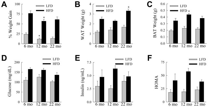
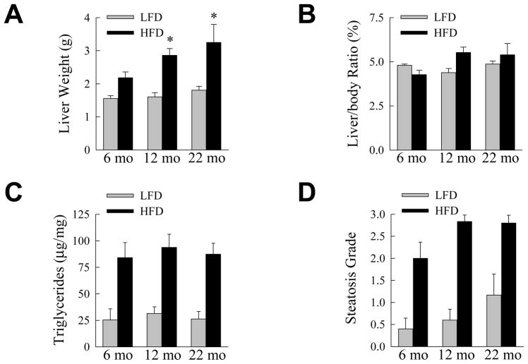
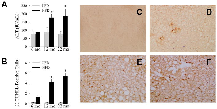
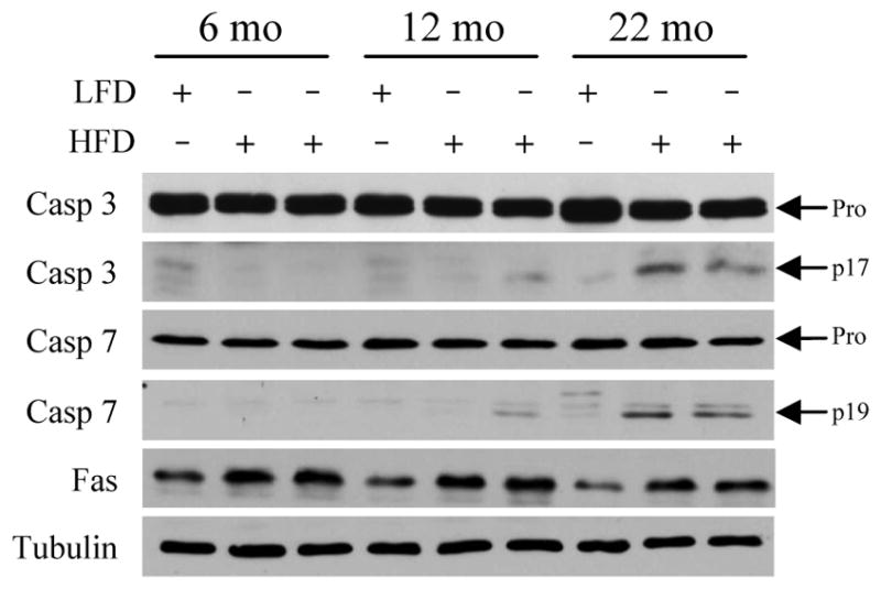
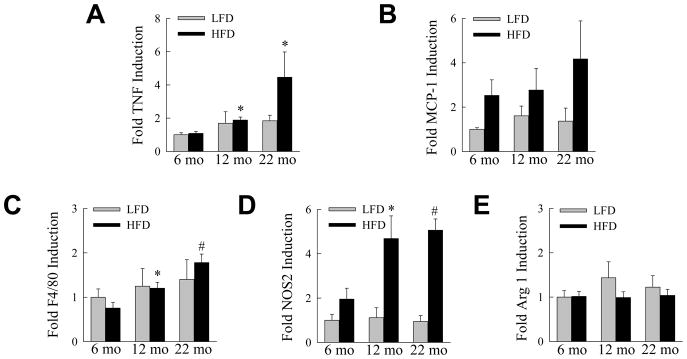
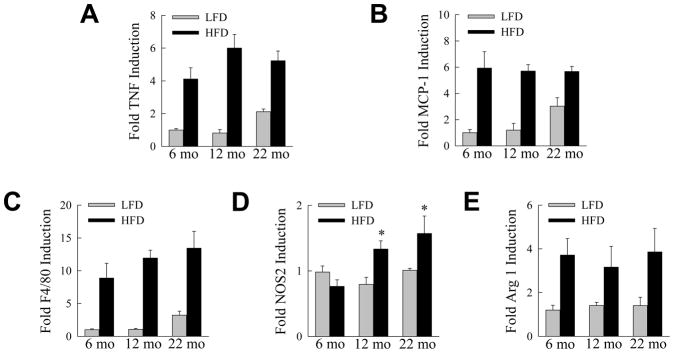
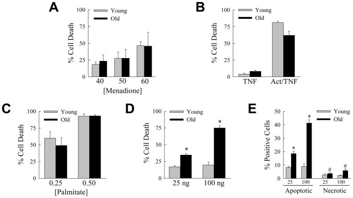
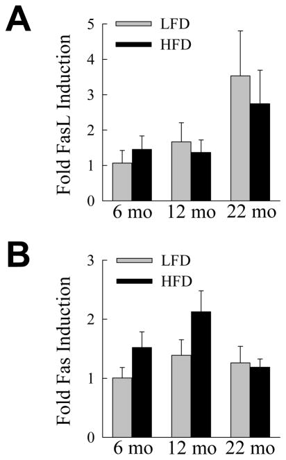
The prevalence of the metabolic syndrome and nonalcoholic fatty liver disease (NAFLD) in humans increases with age. It is unknown whether this association is secondary to the increased incidence of risk factors for NAFLD that occurs with aging, reflects the culmination of years of exposure to lifestyle factors such as a high-fat diet (HFD), or results from physiological changes that characterize aging. To examine this question, the development of NAFLD in response to a fixed period of HFD feeding was examined in mice of different ages. Mice aged 2, 8, and 18 months were fed 16 weeks of a low-fat diet or HFD. Increased body mass and insulin insensitivity occurred in response to HFD feeding irrespective of the age of the mice. The amount of HFD-induced hepatic steatosis as determined biochemically and histologically was also equivalent among the three ages. Liver injury occurred exclusively in the two older ages as reflected by increased serum alanine aminotransferase levels, positive terminal deoxynucleotide transferase-mediated deoxyuridine triphosphate nick end-labeling, and caspase activation. Older mice also had an elevated innate immune response with a more pronounced polarization of liver and adipose tissue macrophages into an M1 phenotype. Studies of cultured hepatocytes from young and old mice revealed that aged cells were selectively sensitized to the Fas death pathway.

Conclusion: Aging does not promote the development of hepatic steatosis but leads to increased hepatocellular injury and inflammation that may be due in part to sensitization to the Fas death pathway and increased M1 macrophage polarization.


Figures

Fig. 1
Fig. 1
Aging does not affect HFD-induced increases in body and adipose mass and insulin resistance. (A) Percentage weight gain from 16 weeks of LFD or HFD feeding in mice sacrificed at the indicated ages (*P<0.02 as compared to 6-month old mice fed the same diet; n=5–10). (B) WAT weights from the same animals (*P<0.01 as compared to HFD-fed, 6-month old mice; n=5–10). (C) BAT weights (*P<0.03 as compared to HFD-fed, 6-month old mice; n=5–10). (D) Serum glucose levels (n=5–10). (E) Serum insulin levels (n=5). (F) HOMA values (n=5).
Fig. 2
Fig. 2
The development of HFD-induced steatosis is not affected by aging. (A) Liver weights in LFD- and HFD-fed mice sacrificed at the indicated ages (*P<0.05 as compared to HFD-fed, 6-month old mice; n=5–10). (B) Liver weight to body weight ratios in the same mice (n=5–10). (C) Hepatic triglyceride content normalized to liver weight (n=5–10). (D) Histological grade of steatosis (n=5–6).
Fig. 3
Fig. 3
Aging amplifies HFD-induced liver injury. (A) Serum ALTs in LFD- and HFD-fed mice sacrificed at the ages shown (*P<0.03 as compared to HFD-fed, 6-month old mice; n=5–10). (B) Percentage of TUNEL-positive hepatocytes in the livers of LFD- and HFD-fed mice (*P<0.003 as compared to HFD-fed, 6-month old mice; n=4). (C–F) Images of TUNEL-stained cells from a LFD-fed 6-month old mouse (C), a HFD-fed 6-month old mouse (D), a HFD-fed 12-month old mouse (E) and a HFD-fed 22-month old mouse (F).
Fig. 4
Fig. 4
Effector caspases are activated in the livers of aged HFD-fed mice. Immunoblots of total hepatic protein from LFD- and HFD-fed mice for caspase 3 (Casp 3), caspase 7 (Casp 7), Fas and tubulin as a loading control. Arrows indicate the procaspase (Pro) and the cleaved caspase 3 (p17) and 7 (p19) forms.
Fig. 5
Fig. 5
Aging increases hepatic inflammation in response to a HFD. LFD- and HFD-fed mice were sacrificed at the ages shown and mRNA levels in whole liver determined by real time PCR for (A) TNF, (B) MCP-1, (C) F4/80, (D) NOS2 and (E) arginase 1 (*P<0.03, #P<0.002 as compared to HFD-fed, 6-month old mice; n=5–6).
Fig. 6
Fig. 6
Aging promotes an M1 macrophage phenotype in adipose tissue. mRNA levels in white adipose tissue from LFD- and HFD-fed mice of the indicated ages for (A) TNF, (B) MCP-1, (C) F4/80, (D) NOS2 and (E) arginase 1 (*P<0.01 as compared to HFD-fed, 6-month old mice; n=5–6).
Fig. 7
Fig. 7
Aged hepatocytes are selectively sensitized to death from the Fas death pathway. (A) Primary hepatocytes were isolated from young (2 month) and old (18 month) mice, cultured and the percentage cell death determined by MTT assay 24 h after treatment with the indicated μM concentrations of menadione (n=7–11). (B) Percentage cell death by MTT assay 24 h after treatment with TNF alone, or cotreatment with actinomycin D and TNF (Act/TNF) (n=4–8). (C) Percentage cell death after 24 h treatment with 0.25 or 0.5 mM palmitate (n=7–8). (D) Percentage cell death by MTT assay 24 h after treatment with the indicated amounts (ng/ml) of Jo2 antibody (*P<0.0001; n=4–8). (E) Percentage of acridine orange/ethidium bromide costained cells that were apoptotic or necrotic by immunofluorescence 12 h after treatment with 25 or 100 ng/ml of Jo2 antibody (*P<0.005, #P<0.03 as compared to young mice; n=3).
Fig 8
Fig 8
Hepatic FasL and Fas mRNA levels are unaffected by aging. (A) Relative FasL mRNA levels in whole liver from LFD- and HFD-fed mice (n=5–6). (B) Fas mRNA levels in the same livers (n=5–6).

Comment in

  • Reply: To PMID 23081825.
    Czaja MJ. Czaja MJ. Hepatology. 2013 Aug;58(2):831. doi: 10.1002/hep.26214. Epub 2013 Jun 24. Hepatology. 2013. PMID: 23280403 No abstract available.
  • Old age and steatohepatitis: a dangerous liaison?
    Vinciguerra M. Vinciguerra M. Hepatology. 2013 Aug;58(2):830-1. doi: 10.1002/hep.26212. Epub 2013 Jun 12. Hepatology. 2013. PMID: 23281079 No abstract available.

References

    1. Tiniakos DG, Vos MB, Brunt EM. Nonalcoholic fatty liver disease: pathology and pathogenesis. Annu Rev Pathol. 2010;5:145–171. - PubMed
    1. Eckel RH, Grundy SM, Zimmet PZ. The metabolic syndrome. Lancet. 2005;365:1415–1428. - PubMed
    1. Ford ES, Giles WH, Dietz WH. Prevalence of the metabolic syndrome among US adults: findings from the third National Health and Nutrition Examination Survey. JAMA. 2002;287:356–359. - PubMed
    1. Lee JY, Kim KM, Lee SG, Yu E, Lim YS, Lee HC, et al. Prevalence and risk factors of non-alcoholic fatty liver disease in potential living liver donors in Korea: a review of 589 consecutive liver biopsies in a single center. J Hepatol. 2007;47:239–244. - PubMed
    1. Amarapurkar D, Kamani P, Patel N, Gupte P, Kumar P, Agal S, et al. Prevalence of non-alcoholic fatty liver disease: population based study. Ann Hepatol. 2007;6:161–163. - PubMed

Publication types

MeSH terms