Comparative Study
. 2012 Nov 27;53(12):7847-54.
doi: 10.1167/iovs.12-10793.

Protective effect of carnosic acid, a pro-electrophilic compound, in models of oxidative stress and light-induced retinal degeneration

Affiliations
Comparative Study

Protective effect of carnosic acid, a pro-electrophilic compound, in models of oxidative stress and light-induced retinal degeneration

Tayebeh Rezaie et al. Invest Ophthalmol Vis Sci. .

Abstract

Purpose: The herb rosemary has been reported to have antioxidant and anti-inflammatory activity. We have previously shown that carnosic acid (CA), present in rosemary extract, crosses the blood-brain barrier to exert neuroprotective effects by upregulating endogenous antioxidant enzymes via the Nrf2 transcriptional pathway. Here we investigated the antioxidant and neuroprotective activity of CA in retinal cell lines exposed to oxidative stress and in a rat model of light-induced retinal degeneration (LIRD).

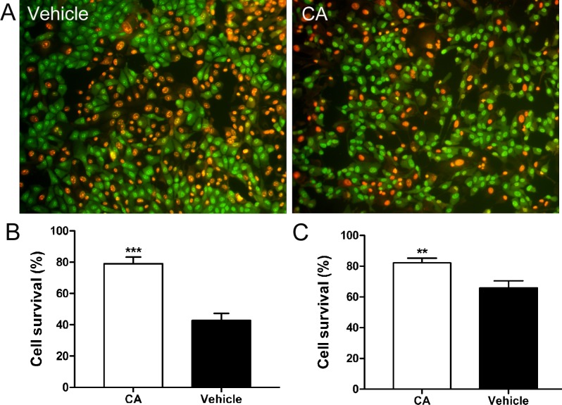
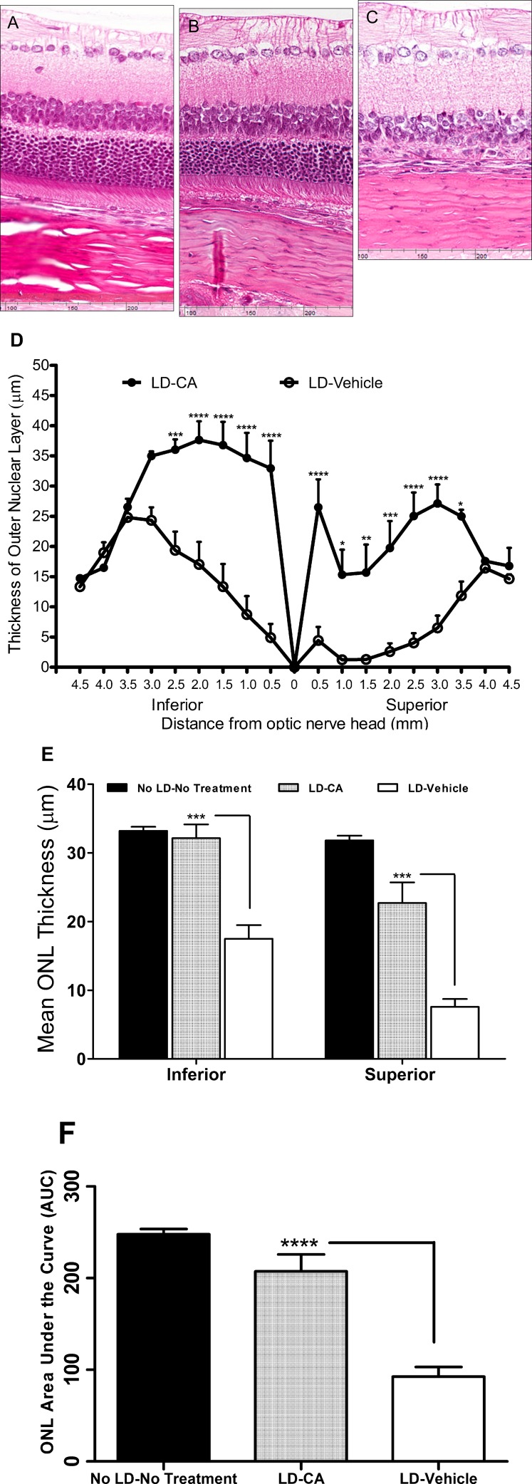
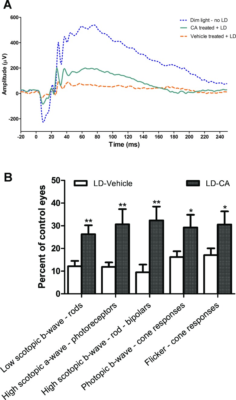
Methods: Retina-derived cell lines ARPE-19 and 661W treated with hydrogen peroxide were used as in vitro models for testing the protective activity of CA. For in vivo testing, dark-adapted rats were given intraperitoneal injections of CA prior to exposure to white light to assess protection of the photoreceptor cells. Retinal damage was assessed by measuring outer nuclear layer thickness and by electroretinogram (ERG).

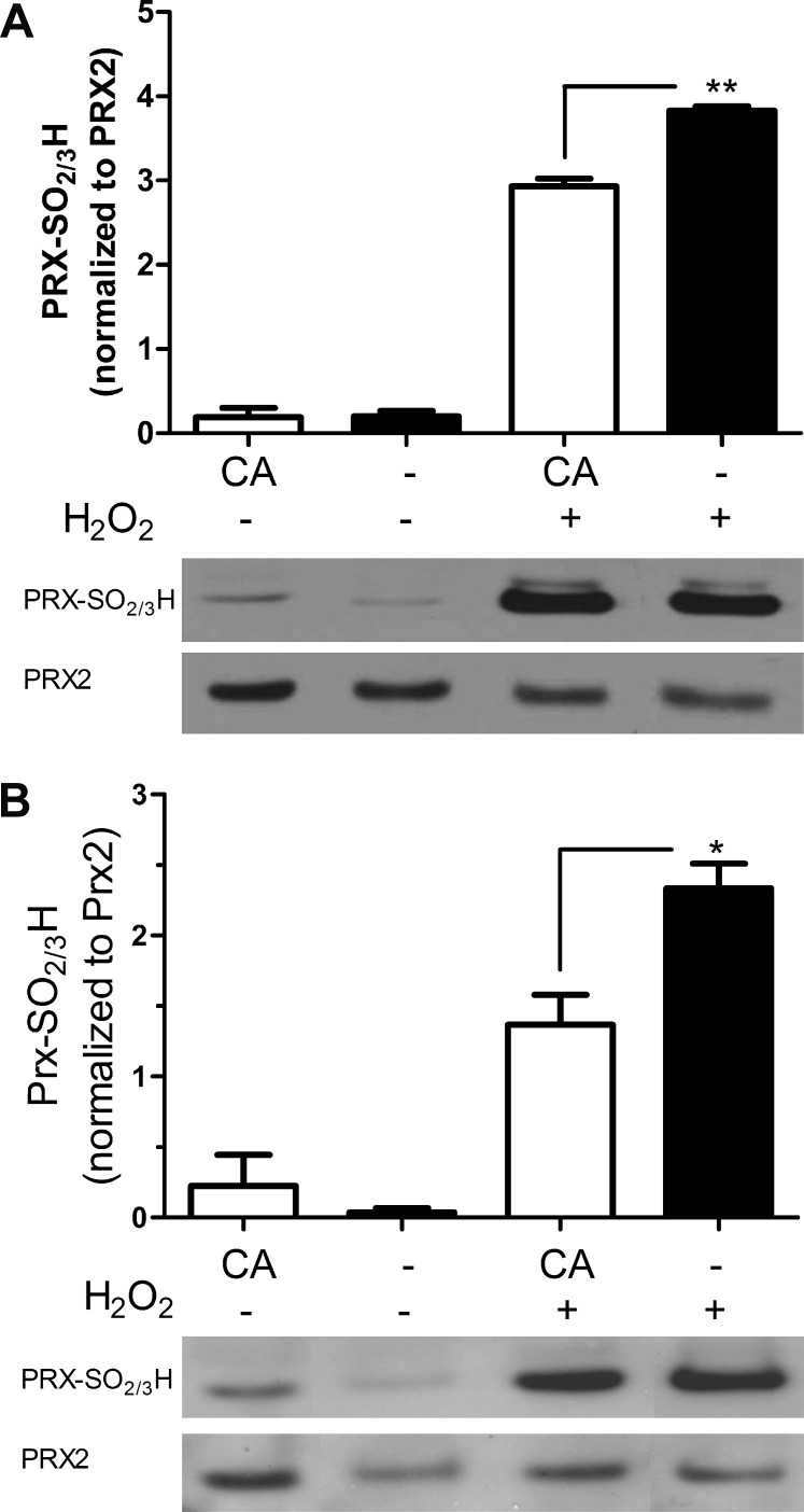
Results: In vitro, CA significantly protected retina-derived cell lines (ARPE-19 and 661W) against H(2)O(2)-induced toxicity. CA induced antioxidant phase 2 enzymes and reduced formation of hyperoxidized peroxiredoxin (Prx)2. Similarly, we found that CA protected retinas in vivo from LIRD, producing significant improvement in outer nuclear layer thickness and ERG activity.

Conclusions: These findings suggest that CA may potentially have clinical application to diseases affecting the outer retina, including age-related macular degeneration and retinitis pigmentosa, in which oxidative stress is thought to contribute to disease progression.


Conflict of interest statement

Disclosure: T. Rezaie, None; S.R. McKercher, None; K. Kosaka, None; M. Seki, None; L. Wheeler, None; V. Viswanath, None; T. Chun, None; R. Joshi, None; M. Valencia, None; S. Sasaki, None; T. Tozawa, None; T. Satoh, P; S.A. Lipton, P

Figures

Figure 1.
Figure 1.
Protective effect of CA from H2O2-induced cytotoxicity. (A) Representative images of ARPE-19 cells stained with acridine orange and ethidium bromide after exposure to H2O2 and treatment with CA or vehicle control. Live cells stained green, while dead cell nuclei stained red. (B, C) Histograms of surviving ARPE-19 cells (B) and 661W photoreceptor cells (C) treated with vehicle or 10 μM of CA in serum-free medium for 21 hours followed by a 4-hour exposure to H2O2. Values are means ± SEM. Pretreatment with CA resulted in significantly enhanced survival of both ARPE-19 cells and 661W cells (n ≥ 3 experiments, each performed in triplicate). ***P < 0.0001, **P < 0.005, Student's t-test.
Figure 2.
Figure 2.
CA induces expression of antioxidant genes and proteins in the ARPE-19 retinal cell line. (A) CA-induced activation of ARE by luciferase assay (P < 0.001 by t-test). Vaues are mean + SEM. (B) CA-induced upregulation of ARE-dependent expression of HO-1, NQO1, GCLM, and xCT genes, as shown by RT-PCR. (C) Immunoblot demonstrating upregulation of HO-1, NQO1, and SRXN1 antioxidant proteins 24 hours after 10 μM CA treatment (n ≥ 4 for each panel).
Figure 3.
Figure 3.
CA reduces hyperoxidation of Prx2. CA treatment of ARPE-19 retinal cells (A) and 661W photoreceptor cells (B) significantly protected against H2O2-induced hyperoxidation of Prx2 (n = 4; *P < 0.05; **P < 0.006 by t-test). Values are means ± SEM.
Figure 4.
Figure 4.
CA protects photoreceptors from light damage in the intact retina. (AC) Eye sections from normal rats stained with H&E stain in the superior retinal area, 500 to 1000 μm from the ONH (A), after light insult and treatment with CA (25 mg/kg) (B), and after light insult with vehicle treatment (C). (D) Graph shows retinal ONL thickness in rats treated with CA or vehicle and exposed to damaging light. Values are means ± SEM. (E) ONL thickness in the inferior and the superior hemispheres of control retinas or after light damage (LD) with and without CA treatment. (F) AUC analysis for the three different treatment groups. Significantly thicker ONLs were observed in rats treated with CA than vehicle controls (n = 7 for No LD, 15 for LD + vehicle, 14 for LD + CA; *P < 0.05, **P < 0.01; ***P < 0.001; ****P < 0.0001 by ANOVA). Values are means ± SEM.
Figure 5.
Figure 5.
Beneficial effect of CA treatment on retinal electrophysiology by ERG analysis. (A) Representative ERG traces from rat retinas of dim light-exposed control, CA-treated after light damage (LD), and vehicle-treated after LD. LD resulted in significant loss of ERG responses in vehicle-treated animals. Administration of CA enhanced ERG responses compared to vehicle (n = 5 for dim light/no LD, n = 7 for CA-treated/LD, n = 8 for vehicle-treated/LD groups). The initial downward deflection in the voltage response represents the a-wave, while the subsequent positive voltage response is the b-wave, as described in text (see Results section). (B) In rats treated with CA compared to vehicle, there was significant protection from light damage on both the ERG a-wave and b-wave (n = 8 for LD-vehicle, 7 for LD-CA; values normalized to the “no treatment” group, where n = 5; *P < 0.05; **P < 0.005). Values are means ± SEM.

References

    1. Beatty S, Koh H, Phil M, Henson D, Boulton M. The role of oxidative stress in the pathogenesis of age-related macular degeneration. Surv Ophthalmol. 2000;45:115–134 - PubMed
    1. Winkler BS, Boulton ME, Gottsch JD, Sternberg P. Oxidative damage and age-related macular degeneration. Mol Vis. 1999;5:32. - PMC - PubMed
    1. Ambati J, Ambati BK, Yoo SH, Ianchulev S, Adamis AP. Age-related macular degeneration: etiology, pathogenesis, and therapeutic strategies. Surv Ophthalmol. 2003;48:257–293 - PubMed
    1. Mata NL, Weng J, Travis GH. Biosynthesis of a major lipofuscin fluorophore in mice and humans with ABCR-mediated retinal and macular degeneration. Proc Natl Acad Sci U S A. 2000;97:7154–7159 - PMC - PubMed
    1. Radu RA, Mata NL, Bagla A, Travis GH. Light exposure stimulates formation of A2E oxiranes in a mouse model of Stargardt's macular degeneration. Proc Natl Acad Sci U S A. 2004;101(16):5928–5933 - PMC - PubMed

Publication types

MeSH terms