

Expression and role of VEGF--a in the ciliary body
- PMID: 23081980
- PMCID: PMC3493183
- DOI: 10.1167/iovs.12-10098
Expression and role of VEGF--a in the ciliary body
Abstract
Purpose: The role of VEGF-A in the normal ciliary body is largely unexplored. The ciliary body is similar in many respects to the choroid plexus of the brain, and we demonstrated previously the importance of VEGF-A in maintenance of choroid plexus vasculature and ependymal cells. Therefore, the role of VEGF-A in ciliary body homeostasis was explored.
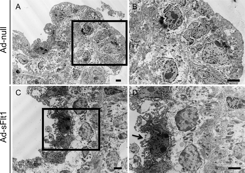
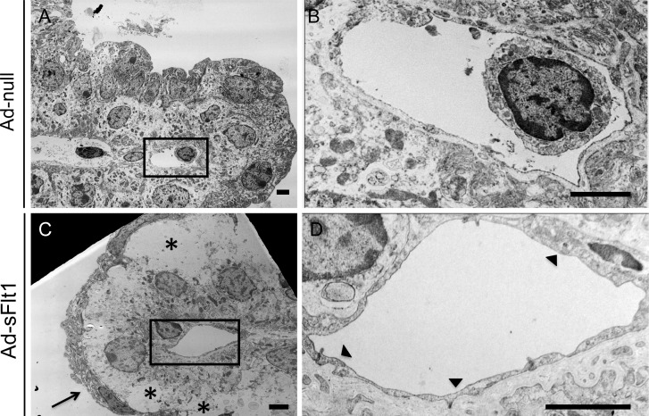
Methods: Swiss-Webster mice (VEGF-LacZ) were used to determine VEGF-A expression during ciliary body development and in the adult. VEGFR2 expression was determined in adult wild type C56BL/6J mice. Systemic VEGF-A neutralization in vivo was achieved with adenovirus-mediated overexpression of soluble VEGFR1 (sFlt1). Following VEGF-A neutralization, the ciliary epithelium was analyzed by light microscopy and transmission electron microscopy (TEM). The effect of VEGF-A blockade on ciliary body function also was assessed by measuring intraocular pressure.
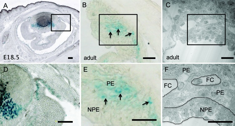
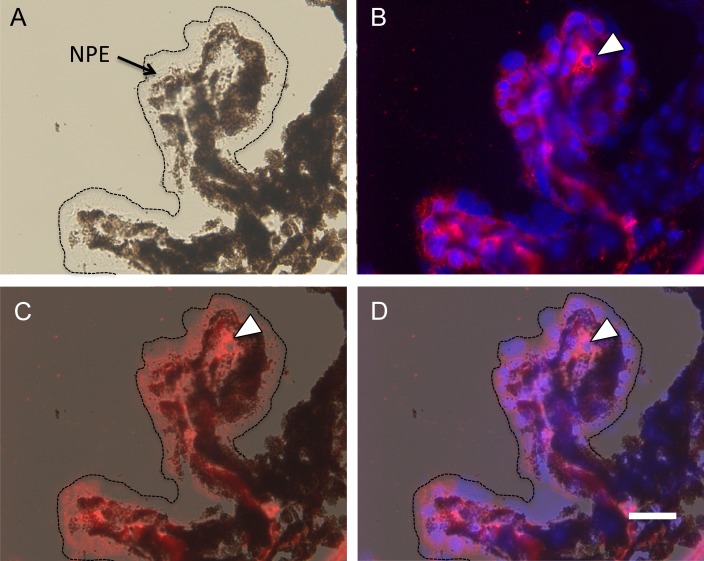
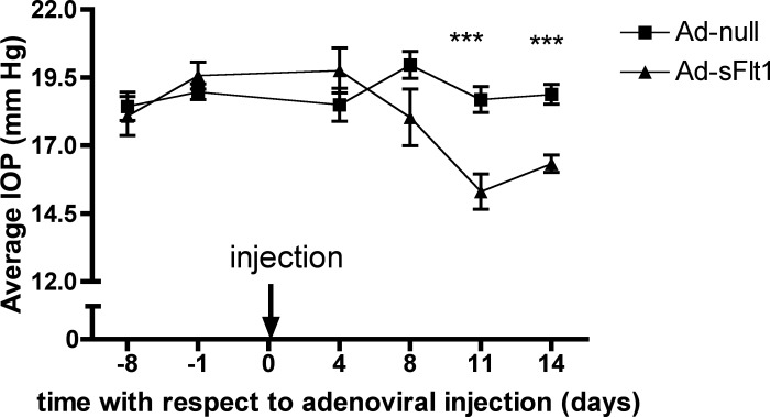
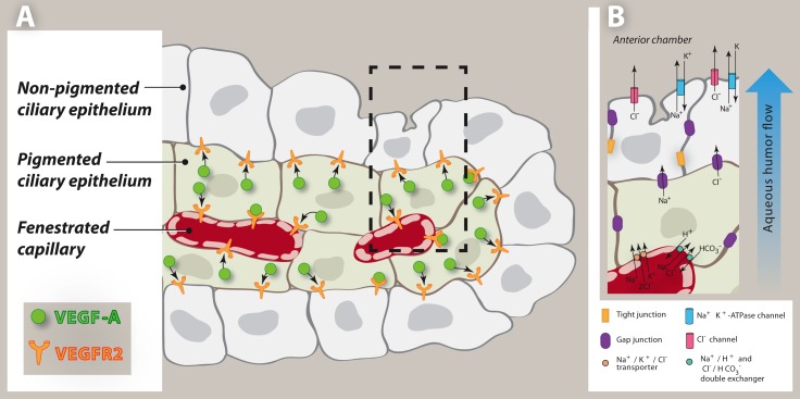
Results: VEGF-A expression was detected at embryonic day 18.5 (E18.5), the onset of ciliary process formation. In the adult ciliary body, VEGF-A was expressed by the pigmented epithelium, whereas VEGFR2 was localized primarily to the capillary endothelium and nonpigmented epithelium. Systemic VEGF-A neutralization led to a thinning of the nonpigmented epithelium, vacuolization of the pigmented epithelium, loss of capillary fenestrations, and thrombosis. These changes were associated with impaired ciliary body function, as evidenced by decreased intraocular pressure in sFlt1-overexpressing animals (15.31 ± 2.06 mm Hg) relative to controls (18.69 ± 1.49 mm Hg).
Conclusions: VEGF-A has an important role in ciliary body homeostasis. Potential for undesired off-target effects should be considered with the chronic use of anti-VEGF-A therapies.
Conflict of interest statement
Disclosure:
Figures

Similar articles
-
Expression and role of VEGF in the adult retinal pigment epithelium.Invest Ophthalmol Vis Sci. 2011 Dec 9;52(13):9478-87. doi: 10.1167/iovs.11-8353. Invest Ophthalmol Vis Sci. 2011. PMID: 22058334 Free PMC article.
-
Depression of intraocular pressure following inactivation of connexin43 in the nonpigmented epithelium of the ciliary body.Invest Ophthalmol Vis Sci. 2009 May;50(5):2185-93. doi: 10.1167/iovs.08-2962. Epub 2009 Jan 24. Invest Ophthalmol Vis Sci. 2009. PMID: 19168903 Free PMC article.
-
The effects of VEGF-A-inhibitors aflibercept and ranibizumab on the ciliary body and iris of monkeys.Graefes Arch Clin Exp Ophthalmol. 2016 Jun;254(6):1117-25. doi: 10.1007/s00417-016-3344-8. Epub 2016 Apr 22. Graefes Arch Clin Exp Ophthalmol. 2016. PMID: 27106625
-
[Variation of inflammatory reaction of ciliary body--harmony between clinic and basic science].Nippon Ganka Gakkai Zasshi. 2004 Dec;108(12):717-48; discussion 749. Nippon Ganka Gakkai Zasshi. 2004. PMID: 15656085 Review. Japanese.
-
Development of the ciliary body: a brief review.Trans Ophthalmol Soc U K (1962). 1986;105 ( Pt 2):123-30. Trans Ophthalmol Soc U K (1962). 1986. PMID: 3541302 Review.
Cited by
-
Low wnt/β-catenin signaling determines leaky vessels in the subfornical organ and affects water homeostasis in mice.Elife. 2019 Apr 1;8:e43818. doi: 10.7554/eLife.43818. Elife. 2019. PMID: 30932814 Free PMC article.
-
Recent perspectives on the delivery of biologics to back of the eye.Expert Opin Drug Deliv. 2017 May;14(5):631-645. doi: 10.1080/17425247.2016.1227783. Epub 2016 Sep 6. Expert Opin Drug Deliv. 2017. PMID: 27573097 Free PMC article. Review.
-
Cellular models and therapies for age-related macular degeneration.Dis Model Mech. 2015 May;8(5):421-7. doi: 10.1242/dmm.017236. Dis Model Mech. 2015. PMID: 26035859 Free PMC article. Review.
-
Loss of caveolin-1 causes blood-retinal barrier breakdown, venous enlargement, and mural cell alteration.Am J Pathol. 2014 Feb;184(2):541-55. doi: 10.1016/j.ajpath.2013.10.022. Epub 2013 Dec 8. Am J Pathol. 2014. PMID: 24326256 Free PMC article.
-
Femtosecond laser-assisted selective reduction of neovascularization in rat cornea.Lasers Med Sci. 2014 Jul;29(4):1417-27. doi: 10.1007/s10103-014-1545-0. Epub 2014 Feb 26. Lasers Med Sci. 2014. PMID: 24570086 Free PMC article.
References
-
- Raviola G, Raviola E. Intercellular junctions in the ciliary epithelium. Invest Ophthalmol Vis Sci. 1978;17:958–981 - PubMed
-
- Kamba T, Tam BY, Hashizume H, et al. VEGF-dependent plasticity of fenestrated capillaries in the normal adult microvasculature. Am J Physiol Heart Circ Physiol. 2006;290:H560–H576 - PubMed
Publication types
MeSH terms
Substances
Grants and funding
LinkOut - more resources
Full Text Sources
Other Literature Sources
Molecular Biology Databases
