. 2012 Nov 20;126(21):2491-501.
doi: 10.1161/CIRCULATIONAHA.111.076950. Epub 2012 Oct 18.

The angiogenic factor secretoneurin induces coronary angiogenesis in a model of myocardial infarction by stimulation of vascular endothelial growth factor signaling in endothelial cells

Affiliations

The angiogenic factor secretoneurin induces coronary angiogenesis in a model of myocardial infarction by stimulation of vascular endothelial growth factor signaling in endothelial cells

Karin Albrecht-Schgoer et al. Circulation. .

Abstract

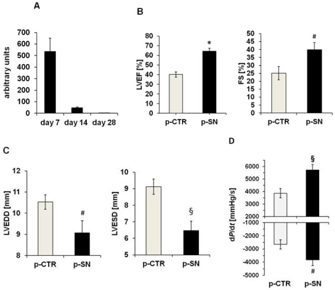
Background: Secretoneurin is a neuropeptide located in nerve fibers along blood vessels, is upregulated by hypoxia, and induces angiogenesis. We tested the hypothesis that secretoneurin gene therapy exerts beneficial effects in a rat model of myocardial infarction and evaluated the mechanism of action on coronary endothelial cells.

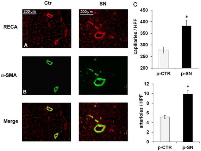
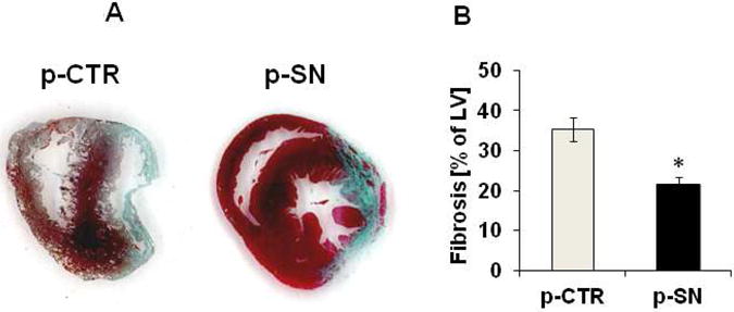
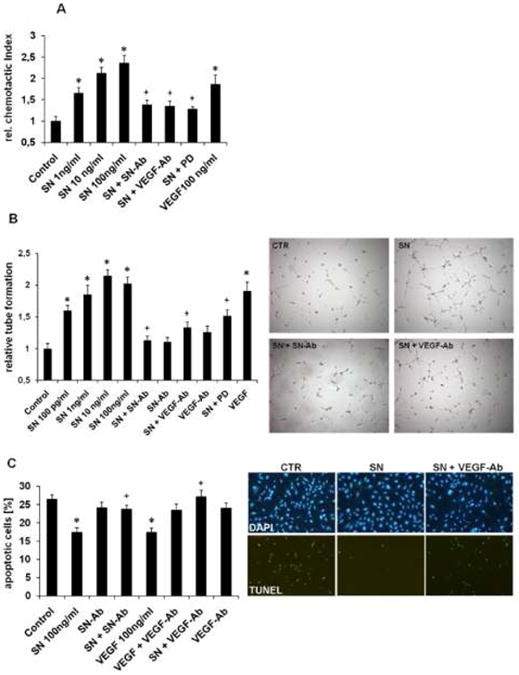
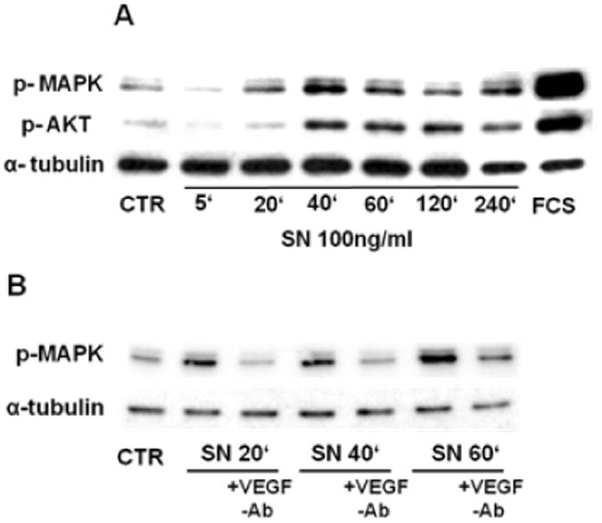
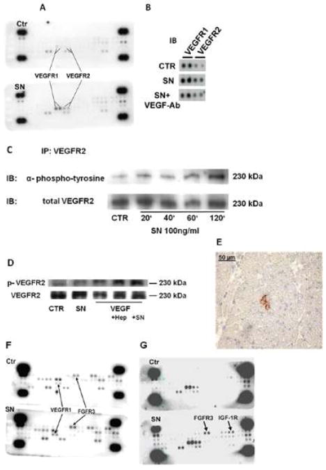
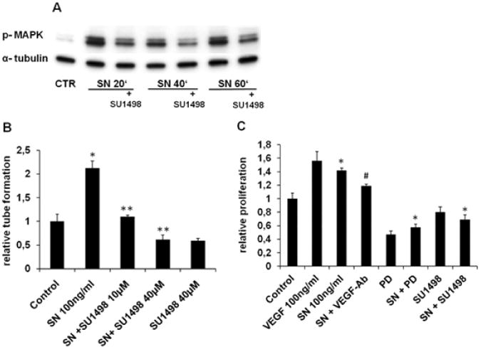
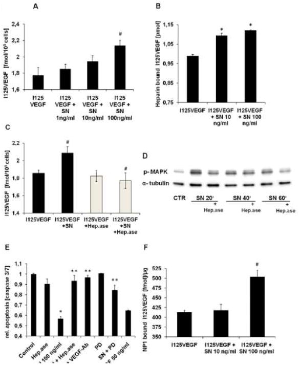
Methods and results: In vivo secretoneurin improved left ventricular function, inhibited remodeling, and reduced scar formation. In the infarct border zone, secretoneurin induced coronary angiogenesis, as shown by increased density of capillaries and arteries. In vitro secretoneurin induced capillary tubes, stimulated proliferation, inhibited apoptosis, and activated Akt and extracellular signal-regulated kinase in coronary endothelial cells. Effects were abrogated by a vascular endothelial growth factor (VEGF) antibody, and secretoneurin stimulated VEGF receptors in these cells. Secretoneurin furthermore increased binding of VEGF to endothelial cells, and binding was blocked by heparinase, indicating that secretoneurin stimulates binding of VEGF to heparan sulfate proteoglycan binding sites. Additionally, secretoneurin increased binding of VEGF to its coreceptor neuropilin-1. In endothelial cells, secretoneurin also stimulated fibroblast growth factor receptor-3 and insulin-like growth factor-1 receptor, and in coronary vascular smooth muscle cells, we observed stimulation of VEGF receptor-1 and fibroblast growth factor receptor-3. Exposure of cardiac myocytes to hypoxia and ischemic heart after myocardial infarction revealed increased secretoneurin messenger RNA and protein.

Conclusions: Our data show that secretoneurin acts as an endogenous stimulator of VEGF signaling in coronary endothelial cells by enhancing binding of VEGF to low-affinity binding sites and neuropilin-1 and stimulates further growth factor receptors like fibroblast growth factor receptor-3. Our in vivo findings indicate that secretoneurin may be a promising therapeutic tool in ischemic heart disease.


Figures

Figure 1
Figure 1
SN gene therapy improves myocardial function after MI. A Plasmid derived SN expression in rat myocardium after MI. Real time PCR analysis for plasmid derived SN in myocardium after 7, 14 and 28 days revealed high SN expression after 7 and 14 days. Values are arbitrary units (n=4). B Effect of SN gene therapy on cardiac function. Echocardiographic assessment of myocardial function showed improved left ventricular ejection fraction and fractional shortening (LVEF and LVFS) 4 weeks after MI and treatment with SN-plasmid (p-SN) compared to control plasmid (p-CTR). Results are shown as mean ± SEM (n=12 per group; *p<0.001; #p<0.05 p-SN vs. p-CTR). C Effects of SN gene therapy on left ventricular remodeling. Echocardiographic analysis of left ventricular end diastolic and systolic diameter (LVEDD and LVESD) revealed inhibition of left ventricular dilatation 4 weeks after MI by SN gene therapy compared to treatment with control vector. Results are shown as mean ± SEM (n=12 per group; #p<0.05; §p<0.005 p-SN vs. p-CTR). D Cardiac Hemodynamic data reveal improved ventricular function after SN gene therapy. 4 weeks after MI, left ventricular maximum positive (+dP/dt) and negative (−dP/dt) pressure development were improved in the SN-treated animal group. Results are shown as mean ± SEM (n=16; §p<0.005 and #p<0.05 p-SN vs. p-CTR).
Figure 2
Figure 2
Effects of SN gene therapy on angiogenesis and arteriogenesis in-vivo. Immunofluorescence staining for RECA (A) and for α-SMA (B) in rat myocardium of the infarct border zone treated with SN-plasmid or control plasmid 4 weeks after MI. (C) Values are expressed as RECA positive cells (capillaries) or as α-SMA positive cells (arteries/arterioles) per high power field (HPF, n=15; *p<0.001 p-SN vs. p-CTR).
Figure 3
Figure 3
Effects of SN gene therapy on ventricular fibrosis after MI. A SN prevents anterior wall fibrosis after MI. Representative Masson's trichrome-stained sections in the area of infarction, 4 weeks after MI (blue=collagen). B Quantification of ventricular fibrosis. Quantification of Masson's trichrome-stained sections, 4 weeks after MI showed significantly reduced fibrosis in animals treated with SN-plasmid compared to control animals (n=13; * p<0.001 p-SN vs. p-CTR).
Figure 4
Figure 4
Effects of SN on HCAECs in cell culture. A SN induces HCAEC migration. HCAEC migration is expressed as chemotactic index relative to control. SN induced cell migration with a maximum effect at 100 ng/ml could be blocked with neutralizing SN-antibody, VEGF-antibody and the MAPK inhibitor PD98.059 (PD). VEGF served as positive control (n=4; *p<0.001 vs. Control; +p<0.01 vs. SN 100 ng/ml). B SN induces capillary tube formation in-vitro. HCAECs were seeded on matrigel for 6 hours and capillary tubes were counted. SN induced capillary tube formation with a maximum effect at 10 ng/ml. Neutralizing SN-antibody, VEGF-antibody and PD blocked SN-induced tube formation. VEGF served as positive control. (n=6; *p<0.001 vs. Control; +p<0.01 vs. SN 100 ng/ml). Representative images of findings with control medium, SN 100ng/ml, SN and neutralizing SN-antibody, SN and VEGF-antibody are shown. C SN inhibits HCAEC apoptosis. HCAECs were starved and stained for TUNEL and DAPI (apoptotic cells are given as % of TUNEL positive cells of all DAPI positive cells). SN reduced HCAEC apoptosis with a maximum at 100 ng/ml concentration whereas co-incubation with SN-antibody and VEGF-antibody inhibited SN-induced protective effects. VEGF served as positive control. (n=4; *p<0.001 vs. Control; +p<0.01 vs. SN 100 ng/ml). Representative images of TUNEL and DAPI stains with control medium, SN 100ng/ml, SN and VEGF-antibody are shown.
Figure 5
Figure 5
SN stimulates intracellular signaling pathways in HCAECs. A SN stimulates MAPK (ERK) and Akt activation in HCAECs. HCAECs were incubated with SN 100 ng/ml for different time points, cell lysates were collected and further treated for Western blotting. SN activated MAPK after 20 minutes and Akt after 40 minutes, lasting until 4 hours. B SN induced MAPK (ERK) phosphorylation is mediated by VEGF. HCAECs were incubated with SN 100 ng/ml ± VEGF-antibody and extracts were analyzed for MAPK phosphorylation by Western blotting. Co-incubation with a VEGF-antibody inhibits SN-induced MAKP activation.
Figure 6
Figure 6
SN stimulates VEGF- and FGF-receptors. A Treatment of HCAECs with SN induces phosphorylation of VEGFR1 and 2. Profiler assays were used to investigate the role of receptor tyrosine kinases in SN stimulated cells. HCAECs were stimulated with SN 100 ng/ml for 40 min and further treated as recommended by the manufacturer. In comparison to untreated cells, SN induced phosphorylation of VEGFR1 and 2. B SN-induced VEGF receptor activation is mediated by endogenous VEGF. For receptor tyrosine kinase Profiler assays, HCAECs were treated with control medium or SN 100 ng/ml with or without VEGF-antibody. SN-mediated VEGF receptor activation was blocked by the VEGF-antibody. C SN induces phosphorylation of VEGFR2 (Immunoprecipitaton). HCAECs were stimulated with SN 100 ng/ml for different time periods, cell lysates were collected and immunoprecipitated for VEGFR2. After blotting, membranes were probed with anti-phospho-tyrosine antibody. SN induced activation of VEGFR2 after 120 minutes. D SN effects on VEGF-mediated VEGFR2 activation are comparable to those of heparin. HCAECs were treated with control medium, SN 100 ng/ml and VEGF 50 ng/ml in combination with either heparin 1 μg/ml or SN 100 ng/ml. Levels of VEGFR2 phosphorylation were determined by Western blot analysis using a specific phospho-VEGFR2 antibody. SN, like heparin stimulated VEGF mediated VEGFR2 stimulation. E VEGFR2 phosphorylation in vivo. Frozen Sections of rat heart 3 days after MI and SN gene therapy showed positive staining for phospho-VEGFR2. F Treatment of HCASMCs with SN induced phosphorylation of VEGFR1 and FGFR3. HCASMCs were stimulated with SN 100 ng/ml for 40 min and cell lysates were used for profiler assays. In comparison to untreated cells, SN induced phosphorylation of VEGFR1 and FGFR3. G SN also activates FGFR3 and IGF-1R in HCAECs. In unstarved HCAECs, profiler assays revealed stimulation of FGFR3 and IGF-1 receptor with SN 100 ng/ml after 40 minutes, beside the already observed phosphorylation of VEGFR1 and 2.
Figure 7
Figure 7
VEGFR2 mediates SN-induced effects on HCAECs. A SN-induced MAPK activation is blocked by SU1498. HCAECs were preincubated with SU1498 (40 μM) for 60 min. Thereafter cells were stimulated with SN 100 ng/ml and further processed for Western blotting. The specific VEGFR2 inhibitor SU1498 abrogated SN-induced ERK activation at different time-points. B SN mediated tube formation is impaired with SU1498. HCAECs were seeded on matrigel and after 6 hours, capillary tubes were counted. SN 100 ng/ml induced capillary tube formation was blocked with SU1498 (10 and 40 μM). (n=8; *p<0.001 SN vs. Control, **p<0.001 SN vs. SN + SU1498). C Proliferation of HCAECs is modulated by ERK and VEGFR2. HCAECs were cultured and stimulated with SN 100 ng/ml with or without VEGF-antibody, PD and SU1498 (10μM). Both PD and SU1498 abolished SN-induced cell proliferation (n=6; **p<0.001 SN vs. SN + SU1498, SN + PD; SN vs. Control *p<0.001; SN vs. SN + VEGF-Ab +p<0.01).
Figure 8
Figure 8
SN stimulates binding of VEGF to heparan sulfate proteoglycans and neuropilin 1. A SN increases I125VEGF-binding to HCAECs. For binding assays 10 ng/ml I125VEGF with or without increasing amounts of SN (1, 10 and 100 ng/ml), was added to HCAECs for 2 hours at 4°C. Binding of I125VEGF was significantly increased in the presence of SN 100ng/ml (I125VEGF vs. SN 100 ng/ml + I125VEGF, # p<0.05 SN; n=4). B SN enhances binding of I125VEGF to heparin. Maxisorb tubes were coated with heparin-BSA complex and incubated with 0.5 ng/ml I125 VEGF ± 10 or 100 ng/ml SN for 2 hours at room temperature. In the presence of SN, binding of I125VEGF to heparin is significantly increased compared to I125VEGF alone (I125VEGF vs. SN + I125VEGF, *p<0.001; n=5). C Heparinase pre-treatment blocks SN mediated I125VEGF-binding to HCAECs. To investigate the influence of heparan sulfate proteoglycans on I125VEGF-binding, HCAECs were pre-incubated with heparinase for 4 hours at 37°C. Thereafter binding assays were performed with I125VEGF ± SN 100ng/ml. Pre-incubation with heparinase diminished SN-induced increase of I125VEGF-binding to HCAECs (I125VEGF vs. SN+I125VEGF and I125VEGF + SN vs. I125VEGF + SN + Heparinase, #p<0.05; n=3). D Heparinase inhibits SN mediated MAPK activation. After preincubation of HCAECs with heparinase, cells were stimulated with SN 100 ng/ml for different time periods and lysates were processed for Western blotting. E VEGF-Ab, PD and heparinases abrogate SN-induced anti-apoptotic effects. HCAECs were starved over night in medium without supplements ± SN 100 ng/ml, PD (10 μM), VEGF-antibody and heparinase. VEGF 50 ng/ml served as positive control (SN vs. Control, *p<0.001; SN + Heparinase, SN + VEGF-Ab and SN + PD vs. SN 100 ng/ml, ** p<0.001; n=4). F SN increases binding of I125VEGF to neuropilin 1. Maxisorb tubes were coated with recombinant, human neuropilin 1 and binding assays in the presence or absence of SN 10 and 100 ng/ml were performed. SN 100 ng/ml significantly increased I125VEGF-binding to its co-receptor neuropilin 1 (I125VEGF vs. I125VEGF + SN, #p<0.05; n=3).

Similar articles

Cited by

References

    1. Gheorghiade M, Bonow RO. Chronic heart failure in the united states: A manifestation of coronary artery disease. Circulation. 1998;97:282–289. - PubMed
    1. Kawamoto A, Murayama T, Kusano K, Ii M, Tkebuchava T, Shintani S, Iwakura A, Johnson I, von Samson P, Hanley A, Gavin M, Curry C, Silver M, Ma H, Kearney M, Losordo DW. Synergistic effect of bone marrow mobilization and vascular endothelial growth factor-2 gene therapy in myocardial ischemia. Circulation. 2004;110:1398–1405. - PubMed
    1. Kondo I, Ohmori K, Oshita A, Takeuchi H, Fuke S, Shinomiya K, Noma T, Namba T, Kohno M. Treatment of acute myocardial infarction by hepatocyte growth factor gene transfer: The first demonstration of myocardial transfer of a “functional” gene using ultrasonic microbubble destruction. J Am Coll Cardiol. 2004;44:644–653. - PubMed
    1. Kusano KF, Pola R, Murayama T, Curry C, Kawamoto A, Iwakura A, Shintani S, Ii M, Asai J, Tkebuchava T, Thorne T, Takenaka H, Aikawa R, Goukassian D, von Samson P, Hamada H, Yoon YS, Silver M, Eaton E, Ma H, Heyd L, Kearney M, Munger W, Porter JA, Kishore R, Losordo DW. Sonic hedgehog myocardial gene therapy: Tissue repair through transient reconstitution of embryonic signaling. Nat Med. 2005;11:1197–1204. - PubMed
    1. Yanagisawa-Miwa A, Uchida Y, Nakamura F, Tomaru T, Kido H, Kamijo T, Sugimoto T, Kaji K, Utsuyama M, Kurashima C, Ito H. Salvage of infarcted myocardium by angiogenic action of basic fibroblast growth factor. Science. 1992;257:1401–1403. - PubMed

Publication types

MeSH terms