. 2012 Dec 15;18(24):6690-701.
doi: 10.1158/1078-0432.CCR-12-0770. Epub 2012 Oct 18.

Ethacrynic acid and a derivative enhance apoptosis in arsenic trioxide-treated myeloid leukemia and lymphoma cells: the role of glutathione S-transferase p1-1

Affiliations

Ethacrynic acid and a derivative enhance apoptosis in arsenic trioxide-treated myeloid leukemia and lymphoma cells: the role of glutathione S-transferase p1-1

Rui Wang et al. Clin Cancer Res. .

Abstract

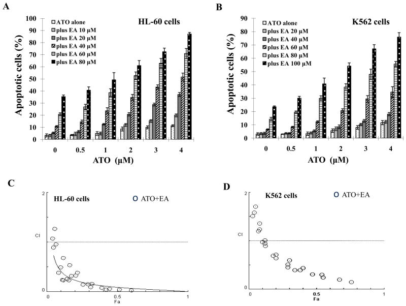
Purpose: Arsenic trioxide (ATO) as a single agent is used for treatment of acute promyelocytic leukemia (APL) with minimal toxicity, but therapeutic effect of ATO in other types of malignancies has not been achieved. We tested whether a combination with ethacrynic acid (EA), a glutathione S-transferase P1-1 (GSTP1-1) inhibitor, and a reactive oxygen species (ROS) inducer will extend the therapeutic effect of ATO beyond APL.

Experimental design: The combined apoptotic effects of ATO plus ethacrynic acid were tested in non-APL leukemia and lymphoma cell lines. The role of ROS, GSTP1-1, glutathione (GSH), and Mcl-1 in apoptosis was determined. The selective response to this combination of cells with and without GSTP1-1 expression was compared.

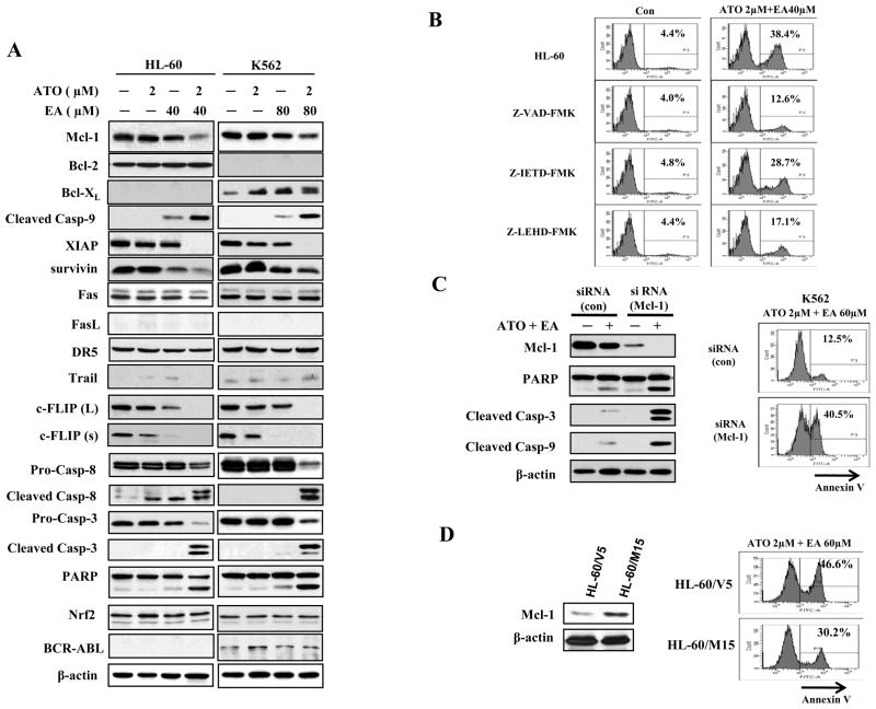
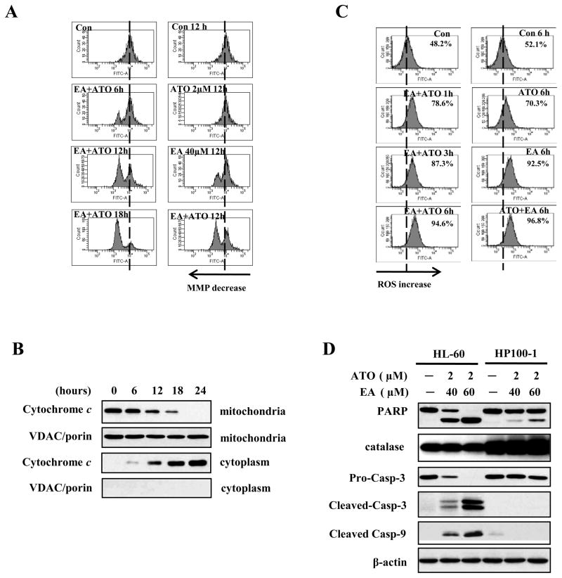
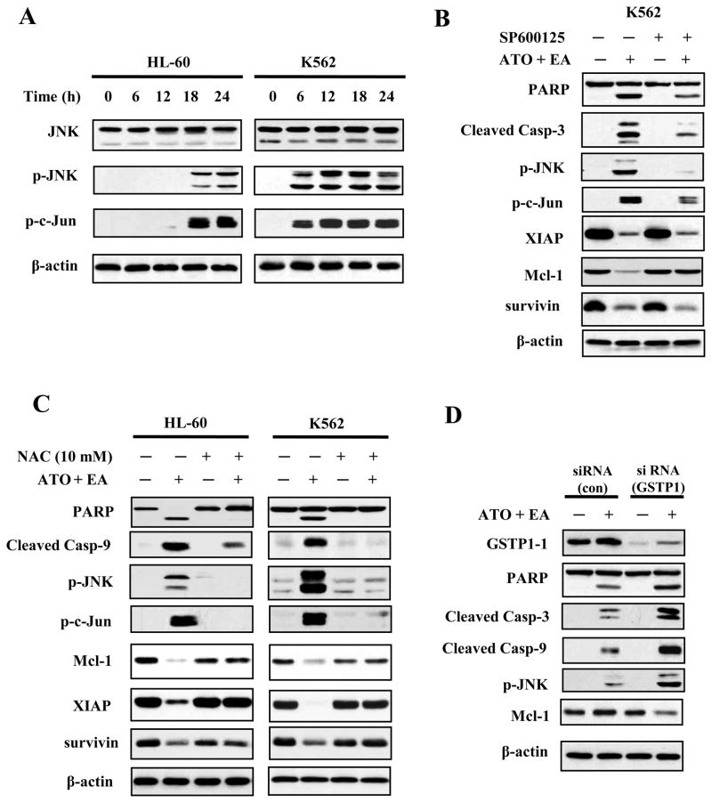
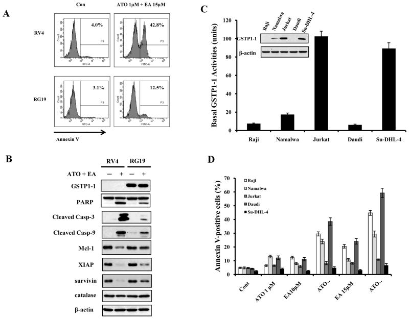
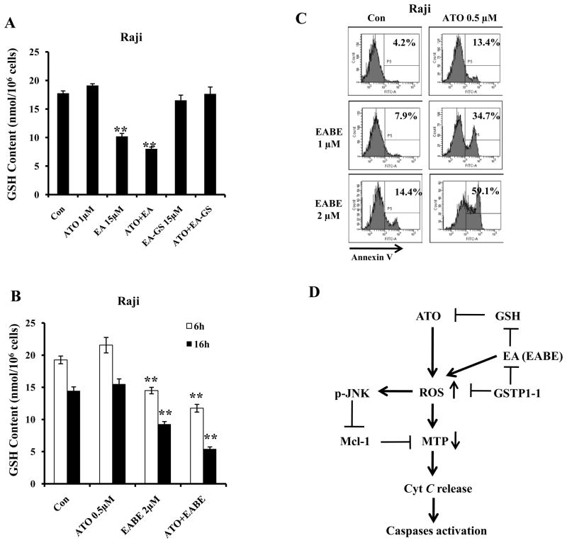
Results: ATO/EA combination synergistically induced apoptosis in myeloid leukemia and lymphoma cells. This treatment produced high ROS levels, activated c-jun-NH(2)-kinase (JNK), and reduced Mcl-1 protein. This led to the decrease of mitochondrial transmembrane potential, release of cytochrome c, and subsequently, to activation of caspase-3 and -9. Induction of apoptosis in leukemia and lymphoma cells expressing GSTP1-1 required high ethacrynic acid concentrations to be combined with ATO. Silencing of GSTP1 in leukemia cells sensitized them to ATO/EA-induced apoptosis. In a subgroup of B-cell lymphoma, which does not express GSTP1-1, lower concentrations of ethacrynic acid and its more potent derivative, ethacrynic acid butyl-ester (EABE), decreased intracellular GSH levels and synergistically induced apoptosis when combined with ATO.

Conclusion: B-cell lymphoma cells lacking GSTP1-1 are more sensitive than myeloid leukemia cells to ATO/EA-induced apoptosis.


Conflict of interest statement

Conflict of interest disclosure:JG is one of named inventors for the formulation of arsenic trioxide and receives royalty payment for the use of arsenic trioxide in patients with acute promyelocytic leukemia.

Figures

Figure 1
Figure 1. ATO/EA has synergistic effect on apoptosis in HL-60 and K562 cells
(A & B) Quantification of apoptosis. HL-60 (A) and K562 (B) cells were treated for 24 h with a fixed ratio of ATO (0.5–4 μM) and EA (10–100 μM). Apoptotic cells were quantified by microscopic detection and counting of AO and EB-stained cells. (C & D) Combination index (CI) of ATO with EA on apoptosis induction in HL-60 (C) and K562 (D) cells. The nature of interaction between ATO and EA was characterized by median dose effect analysis using CompuSyn software. CI values less than 1.0 (horizontal line) corresponds to a synergistic interaction. Fa on the x-axis denotes the fraction affected (e.g. Fa of 0.5 is equivalent to a 50% apoptosis induction.)
Figure 2
Figure 2. ATO and EA combination induces activation of caspase-3, caspase-8, and caspase-9 and dowregulates antiapoptotic proteins
(A) Modulation of apoptotic proteins by ATO/EA. HL-60 and K562 cells were treated with EA and ATO at the indicated concentrations for 24 h. The relative levels of indicated proteins were determined by Western blotting using specific antibodies. β-actin served as loading control. (B) Caspase 9 (but not caspase 8) is the major effector of ATO/E-induced apoptosis. HL-60 cells were pretreated for 4 h with 50 μM Z-VAD-FMK (a general-caspase inhibitor), 50 μM Z-IETD-FMK (a caspase-8 inhibitor), 50 μM Z-LEHD-FMK (a caspase-9 inhibitor), and then incubated with 2 μM ATO and 40 μM EA for another 24 h. Apoptotic cells were quantified using annexin V-FITC staining and FACS analysis. Con.-control. (C) Silencing of Mcl-1 enhances ATO/EA-induced apoptosis in K562 cells. K562 cells were transfected with Mcl-1siRNAor negative control siRNA and after 18 h, treated with 2 μM ATO and 60 μM EA for additional 24 h. The protein levels were determined by Western blotting and the apoptotic cells were determined by FACS after staining with annexin V-FITC. (D) Increased Mcl-1 level attenuated ATO/EA-induced apoptosis in HL-60 cells. HL-60 cells transfected with an empty vector (HL-60/V5) and Mcl-1 expression plasmid (HL-60/M15) were treated with 2 μM ATO and 60 μM EA for additional 24 h. The Mcl-1 level was determined by Western blotting and the apoptotic cells were determined by FACS after staining with annexin V-FITC.
Figure 3
Figure 3. ROS production is involved in the mitochondria-mediated apoptotic pathway activated by ATO/EA treatment
(A) Combination treatment reduces mitochondrial transmembrane potential (MTP). MTP was measured using changes in fluorescence density upon DiOC6(3) loading (see Material and Methods) of untreated HL-60 cells, or cells treated with 2 μM ATO alone, 40 μM EA alone, or the combination, for the indicated times. The peak shift to the left indicates lower MTP. (B) Time-course of cytochrome c release from mitochondria to cytoplasm. HL-60 cells were treated with the combination of 2 μM ATO/40 μM EA for the indicated times, cytosolic and mitochondrial fractions were obtained and analyzed by Western blotting with anti-cytochrome c antibody. VDAC/porin was used as control for purity of the mitochondrial fraction. (C) EA alone and ATO/EA combination increase the H2O2 (ROS) content. HL-60 cells untreated, or treated for the indicated times with 2 μM ATO, or 40 μM EA, or the combination were used to determine the intracellular H2O2 content by DCFH-DA. The peak shift to the right indicates increased levels of H2O2 content. (D) High catalase level blocks ATO/EA-0induced mitochondrial apoptotic pathway. HL-60 and its derivative, catalase over-expressing HP100-1 cells, untreated or treated with 2 μM ATO/40 μM or 60 μM EA for 24 h. The relative levels of PARP, catalase, caspase-3, -9, and β-actin were determined with Western blotting using specific antibodies.
Figure 4
Figure 4. JNK signaling is activated by ATO/EA treatment
(A) Activation of JNK. HL-60 and K562 cells treated with ATO/EA combinations for the indicated times, were lysed and tested for levels of total JNK protein, p-JNK and p-c-Jun, a target of JNK, using specific antibodies in a Western blot analysis. β-actin was used as a loading control. The combinations of ATO/EA are: 2μM ATO and 40 μM EA for HL-60 cells, and 2 μM ATO and 80 μl EA for K562 cells. (B) JNK activation is crucial for ATO/EA induced apoptotic pathway. K562 cells pretreated with 40 μM JNK inhibitor (SP600125) followed by treatment with ATO/EA for 24 h were used to determine the levels of PARP, caspase-3, p-JNK, p-c-Jun, XIAP, Mcl-1, survivin, and β-actin using Western blotting. (C) Treatment with an anti-oxidant NAC inhibits the apoptotic pathway. HL-60 and K562 cells were pretreated with 10 mM NAC for 4 h, followed by ATO/EA treatment for 24 h. The levels of PARP, caspase-3, -9, p-JNK, p-c-Jun, XIAP, Mcl-1, surviving and β-actin were determined using Western blotting. (D) Knock-down of GSTP1-1 enhances the activation of caspases and JNK. K562 cells were transfected with GSTP1siRNA or control siRNA and after 18 h treated with 2 μM ATO/60 μM EA for additional 24 h. Protein levels of GSTP1-1, PARP, caspase-3, -9, p-JNK, Mcl-1 and β-actin were determined with Western blotting using specific antibodies.
Figure 5
Figure 5. GSTP1-1 levels determine the sensitivity of lymphoma cells to ATO/EA treatment-induced apoptosis
(A) RV4 lymphoma cells which lack GSTP1-1 expression are sensitive to ATO/EA induced apoptosis at low EA concentration. RV4 cells and RG19 (high GSTP1-1 expression) cells were treated with 1 μM ATO/15 μM EA for 24 h and the apoptotic cells were detected by FACS after staining with annexin V-FITC. (B) ATO/EA induced mitochondrial apoptosis pathway is muted in RG19 cells. RV4 and RG19 cells were treated with 1 μM ATO/15 μM EA for 24 h and the protein levels of the mitochondrial apoptotic pathways were analyzed by Western blot analysis. (C) Lymphoma cell lines differ widely in GSTP1-1protein and activity. The protein levels of GSTP1-1 were determined by Western blotting and the activity was determined biochemically. (D) Low GSTP1-1 activity lymphoma cells, Raji, Namalwaand Daudi, are highly sensitive to apoptosis induction by low concentrations of ATO/EA; Jurkat and Su-DHL-4 cells with high GSTP1-1activity are resistant. Raji, Namalwa, Daudi, Su-DHL-4 and Jurkat cells were treated with 1 μM ATO together with EA at the indicated concentrations for 24 h. Apoptotic cells were detected by FACS after staining with annexin V-FITC.
Figure 6
Figure 6. GSH level is crucial for synergistic apoptosis induction by a combination of ATO with EA and its derivatives in lymphoma cells without GSTP1-1 expression. (A) EA, but not EA-GS, decreases the levels of intracellular GSH
Raji cell were treated with 1 μM ATO, 15 μM EA, 15 μM EA-GS for 6 h. (B) EABE decreases GSH levels in Raji cells. Raji cells were treated with 0.5 μM ATO, 2 μM EABE or their combination for 6 h and 16 h. The intracellular GSH levels were determined as described in Materials and Methods. (C) EABE enhances ATO apoptosis in Raji cells. Raji cells were treated with 0.5 μM ATO, 1 μM or 2 μM EABE or their combination for 3 days with; medium with fresh drug supplements was changed daily. The apoptotic cells were detected by FACS after staining with annexin V-FITC. (D) A working model of ATO/EA-induced apoptosis in lymphoma cells with or without GSTP1-1 expression. ATO or EA/EABE, can individually increase ROS through separate pathways. When combined, the two drugs strongly increase ROS causing reduction in MTP as well as activation of JNK. Active JNK participates in down-regulation of Mcl-1, leading to maximal cytochrome c release, activation of caspase-9 and induction of apoptosis. EA enhances ATO ability to produce ROS by decreasing intracellular GSH levels. GSTP1-1 detoxifies EA/EABE, ROS and ATO and blocks apoptosis induction. ATO combined with low concentrations of EA/EABE selectively induces apoptosis in B-cell lymphoma lacking GSTP1-1 expression.

Similar articles

Cited by

References

    1. Niu C, Yan H, Yu T, Sun HP, Liu JX, Li XS, et al. Studies on treatment of acute promyelocytic leukemia with arsenic trioxide: remission induction, follow-up, and molecular monitoring in 11 newly diagnosed and 47 relapsed acute promyelocytic leukemia patients. Blood. 1999;94:3315–24. - PubMed
    1. Soignet SL, Frankel SR, Douer D, Tallman MS, Kantarjian H, Calleja E, et al. United States multicenter study of arsenic trioxide in relapsed acute promyelocytic leukemia. J Clin Oncol. 2001;19:3852–60. - PubMed
    1. Jing Y, Wang L, Xia L, Chen GQ, Chen Z, Miller WH, et al. Combined effect of all-trans retinoic acid and arsenic trioxide in acute promyelocytic leukemiacells in vitro and in vivo. Blood. 2001;97:264–9. - PubMed
    1. Chen GQ, Zhou L, Styblo M, Walton F, Jing Y, Weinberg R, et al. Methylated metabolites of arsenic trioxide are more potent than arsenic trioxide as apoptotic but not differentiation inducers in leukemia and lymphoma cells. Cancer Res. 2003;63:1853–9. - PubMed
    1. Dai J, Weinberg RS, Waxman S, Jing Y. Malignant cells can be sensitized to undergo growth inhibition and apoptosis by arsenic trioxide through modulation of the glutathione redox system. Blood. 1999;93:268–77. - PubMed

Publication types

MeSH terms