

Duodenal obstruction following acute pancreatitis caused by a large duodenal diverticular bezoar
- PMID: 23082068
- PMCID: PMC3471120
- DOI: 10.3748/wjg.v18.i38.5485
Duodenal obstruction following acute pancreatitis caused by a large duodenal diverticular bezoar
Abstract
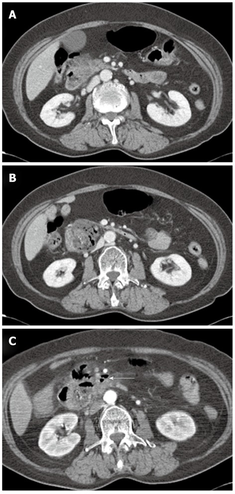
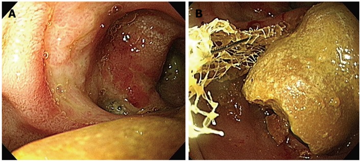
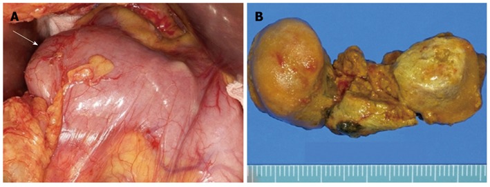
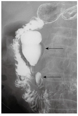
Bezoars are concretions of indigestible materials in the gastrointestinal tract. It generally develops in patients with previous gastric surgery or patients with delayed gastric emptying. Cases of periampullary duodenal divericular bezoar are rare. Clinical manifestations by a bezoar vary from no symptom to acute abdominal syndrome depending on the location of the bezoar. Biliary obstruction or acute pancreatitis caused by a bezoar has been rarely reported. Small bowel obstruction by a bezoar is also rare, but it is a complication that requires surgery. This is a case of acute pancreatitis and subsequent duodenal obstruction caused by a large duodenal bezoar migrating from a periampullary diverticulum to the duodenal lumen, which mimicked pancreatic abscess or microperforation on abdominal computerized tomography. The patient underwent surgical removal of the bezoar and recovered completely.
Keywords: Bezoar; Diverticulum; Duodenal obstruction; Pancreatitis.
Figures
References
-
- Andrus CH, Ponsky JL. Bezoars: classification, pathophysiology, and treatment. Am J Gastroenterol. 1988;83:476–478. - PubMed
-
- Robles R, Parrilla P, Escamilla C, Lujan JA, Torralba JA, Liron R, Moreno A. Gastrointestinal bezoars. Br J Surg. 1994;81:1000–1001. - PubMed
-
- Katapadi M, Kostandy G, Wang A, Gutierrez R, Malik A, Pachter BR. Can a bezoar cause acute pancreatitis? J Clin Gastroenterol. 1997;24:120–121. - PubMed
Publication types
MeSH terms
LinkOut - more resources
Full Text Sources
Medical
