. 2012;2(9):905-15.
doi: 10.7150/thno.4908. Epub 2012 Sep 30.

Porphyrins as theranostic agents from prehistoric to modern times

Affiliations

Porphyrins as theranostic agents from prehistoric to modern times

Yumiao Zhang et al. Theranostics. 2012.

Abstract

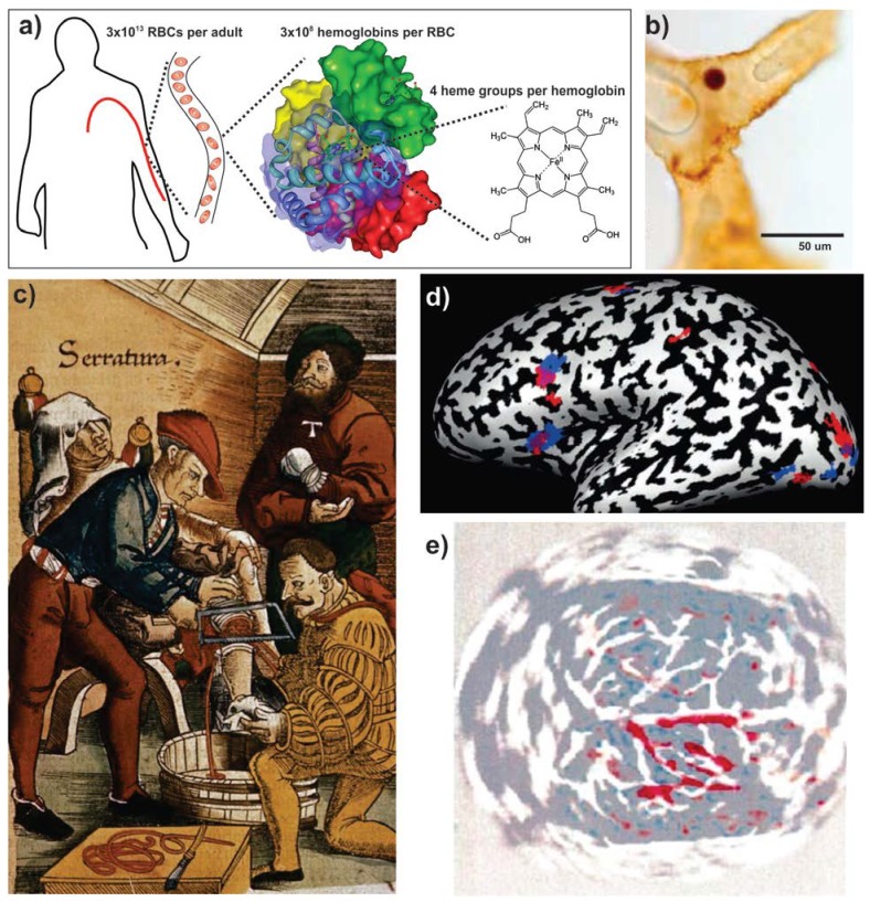
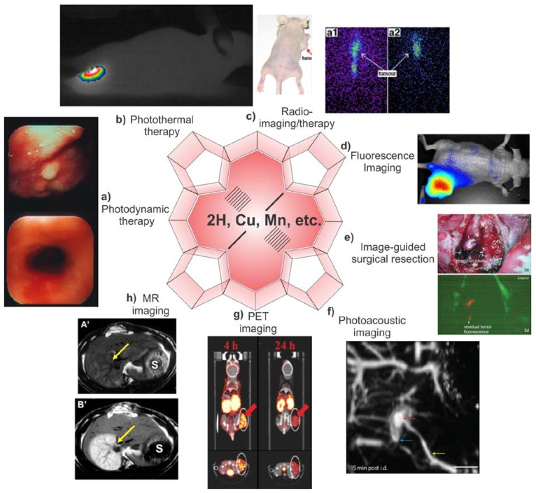
Long before humans roamed the planet, porphyrins in blood were serving not only as indispensable oxygen carriers, but also as the bright red contrast agent that unmistakably indicates injury sites. They have proven valuable as whole body imaging modalities have emerged, with endogenous hemoglobin porphyrins being used for new approaches such as functional magnetic resonance imaging and photoacoustic imaging. With the capability for both near infrared fluorescence imaging and phototherapy, porphyrins were the first exogenous agents that were employed with intrinsic multimodal theranostic character. Porphyrins have been used as tumor-specific diagnostic fluorescence imaging agents since 1924, as positron emission agents since 1951, and as magnetic resonance (MR) contrast agents since 1987. Exogenous porphyrins remain in clinical use for photodynamic therapy. Because they can chelate a wide range of metals, exogenous porphyrins have demonstrated potential for use in radiotherapy and multimodal imaging modalities. Going forward, intrinsic porphyrin biocompatibility and multimodality will keep new applications of this class of molecules at the forefront of theranostic research.

Keywords: Porphyrins; theranostics.


Conflict of interest statement

Competing Interests: The authors have declared that no competing interest exists.

Figures

Figure 1
Figure 1
Endogenous porphyrins. a) Schematic organization of heme in the body ,. b) A possible heme-containing red blood cell identified in 65 million year old dinosaur tissue . c) Surgical illustration of an amputation from circa 1500 AD. Excessive bleeding can readily be observed due to the bright red heme to guide tourniquet application (Archives & Special Collections, Columbia University Health Sciences Library). d) BOLD MR imaging using heme oxygenation. In this case neural differences between English and Hebrew speech patterns are shown. Blue and red regions are involved in morphological processing in Hebrew and English, respectively and regions of overlap are shown in purple . e) Non-invasive, rat brain transcranial photoacoustic imaging following right-side whisker stimulation . Reproduced with permission from the publishers of corresponding references.
Figure 2
Figure 2
Multimodal theranostic capabilities of exogenous porphyrins. a) Human esophageal cancer successfully treated with PDT . b) Photothermal image of a xenograft bearing mouse injected with porphysomes and then irradiated by laser for 1 min showing tumor temperature rapidly rising above 60 ºC . c) Radiotherapy: Melanoma imaging of 188Re-T3,4 CPP in tumor bearing mice. Scintigraphic images were collected at 8h (a1) and 24 h (a2), showing porphyrin potential in radiotherapy and imaging . d) Near infrared fluorescence imaging of porphysome activation in a KB xenograft bearing mouse . e) Fluorescence Guided Resection (FGR): the surgical cavity after white light resection of brain tumor in rabbit (top), fluorescence imaging of PpIX showing tumor margins. Additional FGR can improve the accuracy of resection . f) Photoacoustic image of rat lymphatics mapped following intradermal injection of porphysomes in rats . g) PET imaging showing clear delineation between the tumor and other tissues by PET was obtained at 4, 24 h after intravenous injection of a targeted 64Cu porphyrin . h) MR imaging: In the top precontrast T1 weighted image, the infarcted right liver lobe (arrow) was barely detected whereas 24 h after injection of Gadophrin-2 at 0.05 mmol/kg the infarcted liver lobe was strongly enhanced (bottom) . Reproduced with permission from the publishers of corresponding references.

References

    1. Meinschein WG, Barghoorn ES, Schopf JW. Biological remnants in a precambrian sediment. Science. 1964;145:262–3. - PubMed
    1. Hodgson GW, Peterson E, Kvenvold KA, Bunnenbe E, Halpern B, Ponnampe C. Search for prophyrins in lunar dust. Science. 1970;167:763–5. - PubMed
    1. Barghoorn ES. The oldest fossils. Sci Amer. 1971;224:30–42. - PubMed
    1. Canham GWR. Porphyrins and evolutions. Canadian Chemical Education. 1972;8:5–6.
    1. Zuckerkandl E. The evolution of hemoglobin. Sci Amer. 1965;212:110–8. - PubMed