. 2012;7(10):e47869.
doi: 10.1371/journal.pone.0047869. Epub 2012 Oct 17.

Nicotinamide mononucleotide adenylyltransferase 2 (Nmnat2) regulates axon integrity in the mouse embryo

Affiliations

Nicotinamide mononucleotide adenylyltransferase 2 (Nmnat2) regulates axon integrity in the mouse embryo

Amy N Hicks et al. PLoS One. 2012.

Abstract

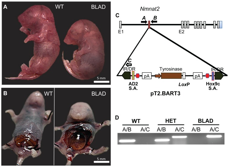
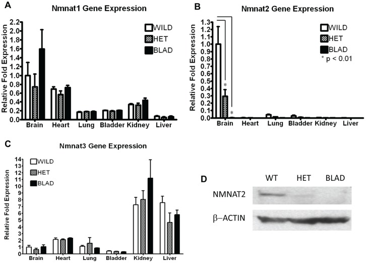
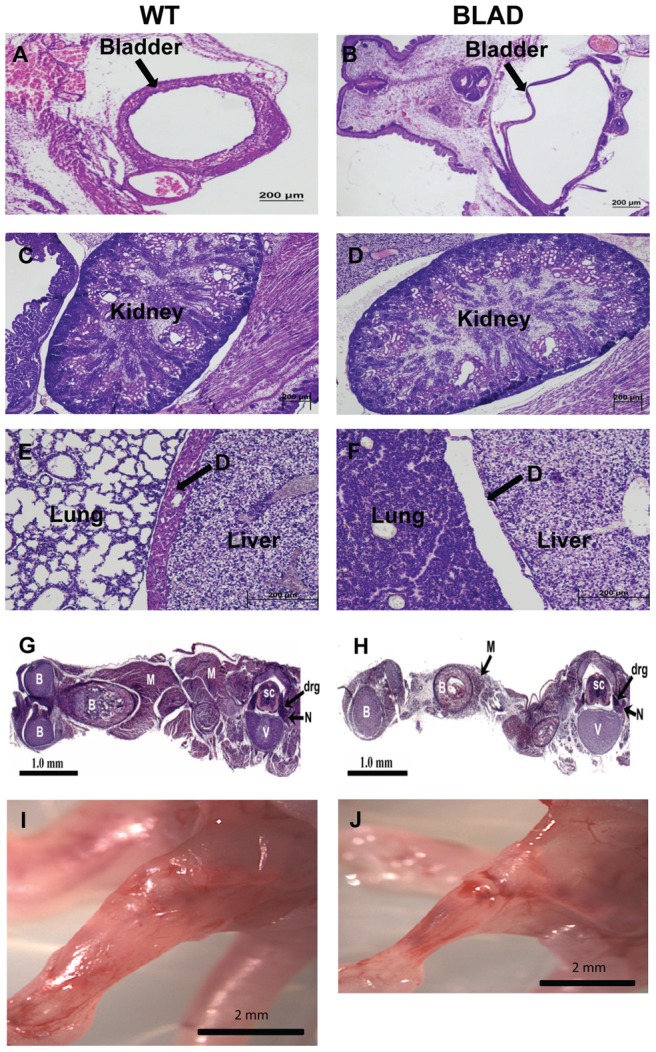
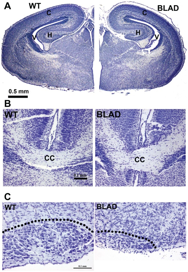
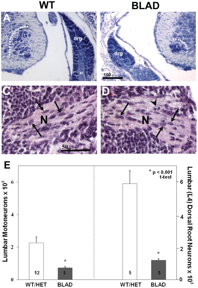
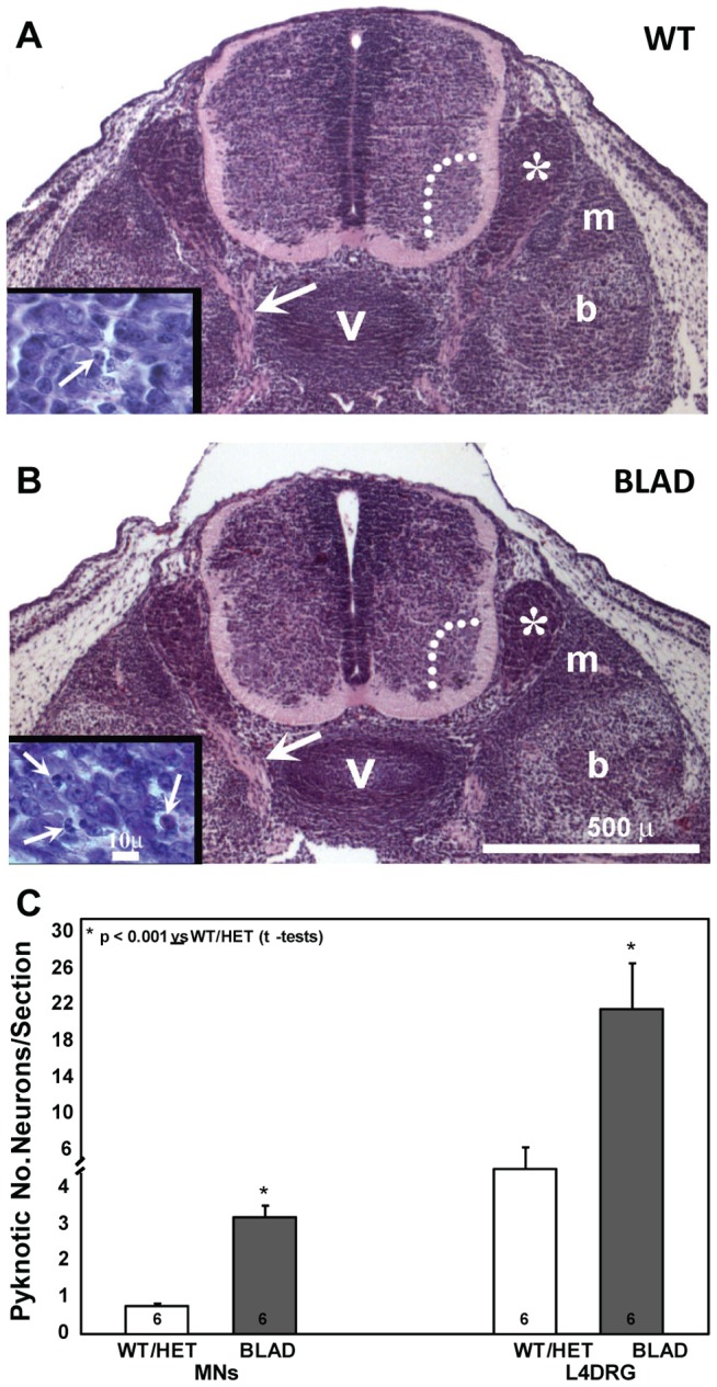
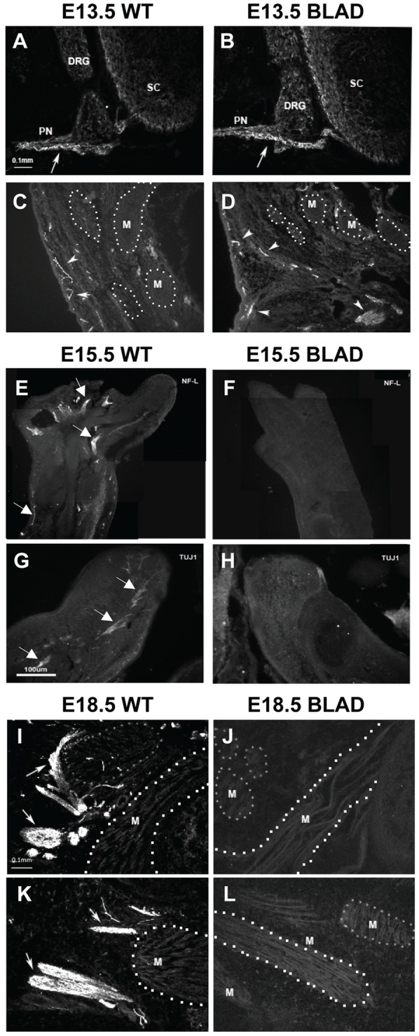
Using transposon-mediated gene-trap mutagenesis, we have generated a novel mouse mutant termed Blad (Bloated Bladder). Homozygous mutant mice die perinatally showing a greatly distended bladder, underdeveloped diaphragm and a reduction in total skeletal muscle mass. Wild type and heterozygote mice appear normal. Using PCR, we identified a transposon insertion site in the first intron of Nmnat2 (Nicotinamide mononucleotide adenyltransferase 2). Nmnat2 is expressed predominantly in the brain and nervous system and has been linked to the survival of axons. Expression of this gene is undetectable in Nmnat2(blad/blad) mutants. Examination of the brains of E18.5 Nmnat2(blad/blad) mutant embryos did not reveal any obvious morphological changes. In contrast, E18.5 Nmnat2(blad/blad) homozygotes showed an approximate 60% reduction of spinal motoneurons in the lumbar region and a more than 80% reduction in the sensory neurons of the dorsal root ganglion (DRG). In addition, facial motoneuron numbers were severely reduced, and there was virtually a complete absence of axons in the hind limb. Our observations suggest that during embryogenesis, Nmnat2 plays an important role in axonal growth or maintenance. It appears that in the absence of Nmnat2, major target organs and tissues (e.g., muscle) are not functionally innervated resulting in perinatal lethality. In addition, neither Nmnat1 nor 3 can compensate for the loss of Nmnat2. Whilst there have been recent suggestions that Nmnat2 may be an endogenous modulator of axon integrity, this work represents the first in vivo study demonstrating that Nmnat2 is involved in axon development or survival in a mammal.


Conflict of interest statement

Competing Interests: The authors have declared that no competing interests exist.

Figures

Figure 1
Figure 1. Identification of Nmnat2 mutants and transposon localization.
Images of E18.5 embryos (A, B). In both images, the wild-type (WT) is on the left and Nmnat2blad/blad (BLAD) on the right. (C) Diagram showing the localization of the pT2. BART3 transposon between exons 1 and 2 in the Nmnat2 gene on chromosome 1. The solid arrows labeled A and B indicate the primer pair used for identification of the native gene, and the solid forward arrow A is combined with the hollow reverse arrow C to identify the presence of the transposon. IR/DR, inverted repeat/direct repeat; S.A., splice acceptor sites (Green/Blue); red hexagon, 3X stop codons; pA, synthetic Poly A signal. (D) PCR results using the primers shown in (C).
Figure 2
Figure 2. Nmnat1, 2, and 3 RNA expression and Nmnat2 protein quantification.
Quantitative RT-PCR for Nmnat1 (A), Nmnat2 (B), and Nmnat3 (C) using total RNA isolated from the brain, heart, lung, bladder, kidney, and liver of E18.5 embryos. Gene expression in wild-type, heterozygote (Nmnat2blad/+) (HET), and homozygous mutant (Nmnat2blad/blad) (BLAD) mice was compared (n = 3 in each group). The data are presented as the mean difference in mRNA fold expression, ± standard deviation, relative to the wild-type brain sample. P-values were calculated using a 2-way ANOVA with a Bonferroni correction. For Nmnat1 and 3, there was no statistical difference in the expression levels between the 3 different genotypes observed in any of the tissues. For Nmnat1, all of the tissues were statistically different except brain/heart and lung/kidney. For Nmnat3, all of the tissues were statistically different except brain/lung and bladder/lung. (D) NMNAT2 and β-ACTIN protein levels from wild-type (WT), heterozygote (HET) (Nmnat2blad/+), and homozygous (BLAD) (Nmnat2blad/blad) E18.5 whole brains.
Figure 3
Figure 3. Gross morphology and histology.
H & E stained E18.5 sections of wild-type (WT) (left) and Nmnat2blad/blad (BLAD) (right) embryos. (A, B) Coronal sections of the bladder. (C, D) Sagittal sections of the kidney. (E, F) Sagittal sections through the diaphragm (D). (G, H) Longitudinal sections of the hind limb and transverse sections of the lumbar region of the spinal cord. (I, J) Fresh E18.5 embryo hind limb with the skin removed. M, muscle; B, bone; N, peripheral nerve; SC, spinal cord; V, vertebra; DRG, dorsal root ganglion.
Figure 4
Figure 4. Brain and facial motor nucleus morphology.
Transverse sections of E18.5 WT (left) and Nmnat2blad/blad (BLAD) (right) forebrains (A), and higher magnification views of the corpus callosum (B). Up is dorsal in (B). (C) Transverse sections of E18.5 WT (left) and BLAD (right) brainstem showing the loss of facial motoneurons (see text for quantitation). Dotted line denotes the boundary of the motor nucleus. H, hippocampus; V, ventricle; C, cortex; CC, corpus callosum.
Figure 5
Figure 5. Spinal cord and dorsal root ganglion
(DRG). (A, B) Transverse sections of the lumbar spinal cord of wild-type (WT) and Nmnat2blad/blad (BLAD) mice at E18.5. Lumbar motoneurons in the spinal cord, sensory neurons in the DRG and the size of DRG are markedly reduced in BLAD mice at E18.5. (C, D) Peripheral nerves (N) from E18.5 WT (C) and BLAD (D) embryos in a lumbar region similar to that indicated by the arrows in Figures 6A–B. The lightly stained regions contain axons and the dark cells in the nerve (arrows) are presumptive Schwann cells. (E) Values (Mean ± SD) for neuron numbers for WT and Nmnat2blad/+ (HET) mice did not differ and have been combined for comparison with BLAD. DRG, dorsal root ganglion; n, nerve; dotted line encloses motoneurons in the ventral horn.
Figure 6
Figure 6. Lumbar spinal cord and pyknotic neurons at E13.5.
A, B: transverse sections of caudal lumbar spinal cord of WT (A) and Nmnat2blad/blad (B) embryos. Large arrows in A and B indicate proximal axons. Insets in A and B depict pyknotic DRG cells (arrows). C: numbers of pyknotic motoneurons (MNs) and DRG sensory neurons. b, bone; m, muscle; v, vertebra; asterisk, DRG; dotted lines enclose motoneurons in the ventral horn.
Figure 7
Figure 7. Peripheral innervation of the hind limb.
Sections of spinal cord (A, B) and thigh (C, D) in E13.5 WT (A, C) and Nmnat2blad/blad (B, D) showing axons (arrows) exiting the spinal cord and innervating thigh muscles (M) in both WT and Nmnat2blad/blad mice. (Sections were stained with a neurofilament antibody and visualized by immunofluorescence). E–H, sections of the distal limb and foot in E15.5 WT (E, G) and Nmnat2blad/blad (F, H) showing NF+ (E, F) and TUJ1+ (G, H) labeled axons (arrows) in peripheral nerves. I–L, longitudinal sections of thigh muscles (M) of E18.5 WT (I, K) and Nmnat2blad/blad (J, L) mice labeled with a neurofilament antibody to visualize intramuscular axons (arrows). DRG, dorsal root ganglia; PN, peripheral nerve; SC, spinal cord; M, the unlabeled background in images of the limbs largely represents muscles many of which are bounded by a dotted line for illustrative clarity.

References

    1. Coleman MP, Freeman MR (2010) Wallerian degeneration, wld(s), and nmnat. Annual review of neuroscience 33: 245–267. - PMC - PubMed
    1. Sorci L, Cimadamore F, Scotti S, Petrelli R, Cappellacci L, et al. (2007) Initial-rate kinetics of human NMN-adenylyltransferases: substrate and metal ion specificity, inhibition by products and multisubstrate analogues, and isozyme contributions to NAD+ biosynthesis. Biochemistry 46: 4912–4922. - PubMed
    1. Berger F, Lau C, Dahlmann M, Ziegler M (2005) Subcellular compartmentation and differential catalytic properties of the three human nicotinamide mononucleotide adenylyltransferase isoforms. The Journal of biological chemistry 280: 36334–36341. - PubMed
    1. Mayer PR, Huang N, Dewey CM, Dries DR, Zhang H, et al. (2010) Expression, localization, and biochemical characterization of nicotinamide mononucleotide adenylyltransferase 2. J Biol Chem 285: 40387–40396. - PMC - PubMed
    1. Raffaelli N, Lorenzi T, Emanuelli M, Amici A, Ruggieri S, et al. (2001) Nicotinamide-mononucleotide adenylyltransferase from Sulfolobus solfataricus. Methods Enzymol 331: 281–292. - PubMed

Publication types

MeSH terms

LinkOut - more resources