

Separation of absorption and scattering properties of turbid media using relative spectrally resolved cw radiance measurements
- PMID: 23082279
- PMCID: PMC3469994
- DOI: 10.1364/BOE.3.002371
Separation of absorption and scattering properties of turbid media using relative spectrally resolved cw radiance measurements
Abstract
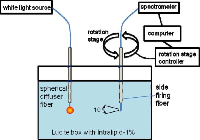
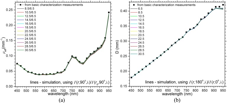
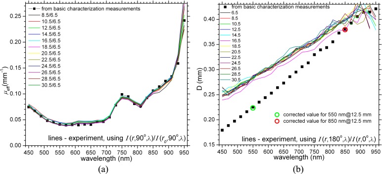
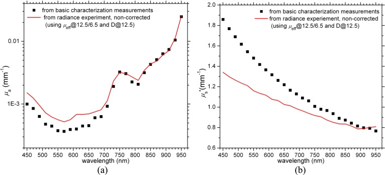
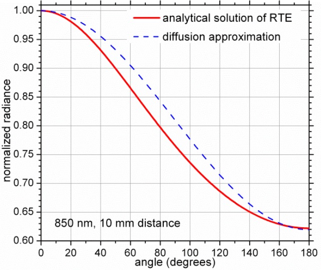
We present a new method for extracting the effective attenuation coefficient and the diffusion coefficient from relative spectrally resolved cw radiance measurements using the diffusion approximation. The method is validated on both simulated and experimental radiance data sets using Intralipid-1% as a test platform. The effective attenuation coefficient is determined from a simple algebraic expression constructed from a ratio of two radiance measurements at two different source-detector separations and the same 90° angle. The diffusion coefficient is determined from another ratio constructed from two radiance measurements at two angles (0° and 180°) and the same source-detector separation. The conditions of the validity of the method as well as possible practical applications are discussed.
Keywords: (170.3660) Light propagation in tissues; (170.6510) Spectroscopy, tissue diagnostics; (170.6935) Tissue characterization; (170.7050) Turbid media; (290.4210) Multiple scattering.
Figures
Similar articles
-
Tagging photons with gold nanoparticles as localized absorbers in optical measurements in turbid media.Biomed Opt Express. 2013 Nov 25;4(12):2989-3006. doi: 10.1364/BOE.4.002989. eCollection 2013. Biomed Opt Express. 2013. PMID: 24409396 Free PMC article.
-
Determination of optical properties of turbid medium from relative interstitial CW radiance measurements using the incomplete P3 approximation.Opt Express. 2017 Oct 16;25(21):25295-25309. doi: 10.1364/OE.25.025295. Opt Express. 2017. PMID: 29041198
-
Optical absorption and scattering properties of bulk porcine muscle phantoms from interstitial radiance measurements in 650-900 nm range.Phys Med Biol. 2014 May 21;59(10):2431-44. doi: 10.1088/0031-9155/59/10/2431. Epub 2014 Apr 17. Phys Med Biol. 2014. PMID: 24743553
-
Simultaneous recovery of a full set of optical properties in turbid media using incomplete P5 approximation to CW radiance.Opt Lett. 2018 Sep 1;43(17):4188-4191. doi: 10.1364/OL.43.004188. Opt Lett. 2018. PMID: 30160748
-
Light dosimetry in vivo.Phys Med Biol. 1997 May;42(5):763-87. doi: 10.1088/0031-9155/42/5/003. Phys Med Biol. 1997. PMID: 9172258 Review.
Cited by
-
In Vivo, Non-Invasive Characterization of Human Bone by Hybrid Broadband (600-1200 nm) Diffuse Optical and Correlation Spectroscopies.PLoS One. 2016 Dec 20;11(12):e0168426. doi: 10.1371/journal.pone.0168426. eCollection 2016. PLoS One. 2016. PMID: 27997565 Free PMC article.
-
Optical characterization of two-layered turbid media for non-invasive, absolute oximetry in cerebral and extracerebral tissue.PLoS One. 2013 May 21;8(5):e64095. doi: 10.1371/journal.pone.0064095. Print 2013. PLoS One. 2013. PMID: 23724023 Free PMC article.
-
Tagging photons with gold nanoparticles as localized absorbers in optical measurements in turbid media.Biomed Opt Express. 2013 Nov 25;4(12):2989-3006. doi: 10.1364/BOE.4.002989. eCollection 2013. Biomed Opt Express. 2013. PMID: 24409396 Free PMC article.
References
-
- Patterson M. S., Wilson B. C., Wyman D. R., “The propagation of optical radiation in tissue II. Optical properties of tissues and resulting fluence distributions,” Lasers Med. Sci. 6(4), 379–390 (1991).10.1007/BF02042460 - DOI
-
- Davidson S. R. H., Weersink R. A., Haider M. A., Gertner M. R., Bogaards A., Giewercer D., Scherz A., Sherar M. D., Elhilali M., Chin J. L., Trachtenberg J., Wilson B. C., “Treatment planning and dose analysis for interstitial photodynamic therapy of prostate cancer,” Phys. Med. Biol. 54(8), 2293–2313 (2009).10.1088/0031-9155/54/8/003 - DOI - PubMed
-
- Pantelides M. L., Whitehurst C., Moore J. V., King T. A., Blacklock N. J., “Photodynamic therapy for localised prostatic cancer: light penetration in the human prostate gland,” J. Urol. 143(2), 398–401 (1990). - PubMed
LinkOut - more resources
Full Text Sources
Other Literature Sources