. 2012 Oct 1;3(10):2500-9.
doi: 10.1364/BOE.3.002500. Epub 2012 Sep 12.

Silica-coated super paramagnetic iron oxide nanoparticles (SPION) as biocompatible contrast agent in biomedical photoacoustics

Affiliations

Silica-coated super paramagnetic iron oxide nanoparticles (SPION) as biocompatible contrast agent in biomedical photoacoustics

Rudolf Alwi et al. Biomed Opt Express. .

Abstract

In this study, we report for the first time the use of silica-coated superparamagnetic iron oxide nanoparticles (SPION) as contrast agents in biomedical photoacoustic imaging. Using frequency-domain photoacoustic correlation (the photoacoustic radar), we investigated the effects of nanoparticle size, concentration and biological media (e.g. serum, sheep blood) on the photoacoustic response in turbid media. Maximum detection depth and the minimum measurable SPION concentration were determined experimentally. The nanoparticle-induced optical contrast ex vivo in dense muscular tissues (avian pectus and murine quadricept) was evaluated and the strong potential of silica-coated SPION as a possible photoacoustic contrast agents was demonstrated.

Keywords: (160.4236) Nanomaterials; (170.3880) Medical and biological imaging; (170.5120) Photoacoustic imaging.


Figures

Fig. 1
Fig. 1
(a) Transmission electron microscopy (TEM) image of silica-coated SPION. (b) SPION-Intralipid PA experimental set-up with a single-element transducer. (c) FTIR-PA spectroscopy results of SiO2 nanoparticles (LUDOX), bare SPION, and silica-coated SPION.
Fig. 2
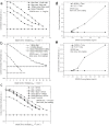
Fig. 2
(a) PA responses of silica-coated SPION of different sizes. (b) Maximum depth detection determination of silica-coated SPION (8 nm core, 3 nm coating). (c) Effect of different solvents on the silica-coated SPION-generated PA signal. Minimum detectable concentration determination of the silica-coated SPION at depth 5 mm (d) and 10 mm (e) inside the Intralipid solution. The experiments were performed using 1-W 1064-nm laser beam incident on the surface of the Intralipid solution and 2 mm beam diameter. One milisecond durations of 1-5 MHz and 0.5-1.5 MHz sinusoidal chirps were employed for the 3.5-MHz focused (a-c) and the 1-MHz unfocused (b,d,e) transducers, respectively. H2O = water; PBS = Phosphate Buffer Saline; BSA = Bovine Serum Albumin.
Fig. 3
Fig. 3
(a) FD-PA array image of a silica coated SPION-filled tube inside chicken breast tissue. (b) Two-dimensional FD-PA image of a rat thigh containing silica-coated SPION obtained using the 3.5-MHz focused transducer.

References

    1. Homan K., Shah J., Gomez S., Gensler H., Karpiouk A., Brannon-Peppas L., Emelianov S., “Silver nanosystems for photoacoustic imaging and image-guided therapy,” J. Biomed. Opt. 15(2), 021316 (2010).10.1117/1.3365937 - DOI - PMC - PubMed
    1. Pramanik M., Song K. H., Swierczewska M., Green D., Sitharaman B., Wang L. V., “In vivo carbon nanotube-enhanced non-invasive photoacoustic mapping of the sentinel lymph node,” Phys. Med. Biol. 54(11), 3291–3301 (2009).10.1088/0031-9155/54/11/001 - DOI - PMC - PubMed
    1. Ungureanu C., Amelink A., Rayavarapu R. G., Sterenborg H. J. C. M., Manohar S., van Leeuwen T. G. C., “Differential pathlength spectroscopy for the quantitation of optical properties of gold nanoparticles,” ACS Nano 4(7), 4081–4089 (2010).10.1021/nn1009165 - DOI - PubMed
    1. Yoon S. J., Mallidi S., Tam J. M., Tam J. O., Murthy A., Johnston K. P., Sokolov K. V., Emelianov S. Y., “Utility of biodegradable plasmonic nanoclusters in photoacoustic imaging,” Opt. Lett. 35(22), 3751–3753 (2010).10.1364/OL.35.003751 - DOI - PMC - PubMed
    1. Yao J., Maslov K., Hu S., Wang L. V., “Evans blue dye-enhanced capillary-resolution photoacoustic microscopy in vivo,” J. Biomed. Opt. 14(5), 054049 (2009).10.1117/1.3251044 - DOI - PMC - PubMed

LinkOut - more resources