

Kaposi Sarcoma Pathogenesis: A Triad of Viral Infection, Oncogenesis and Chronic Inflammation
- PMID: 23082307
- PMCID: PMC3472629
Kaposi Sarcoma Pathogenesis: A Triad of Viral Infection, Oncogenesis and Chronic Inflammation
Abstract
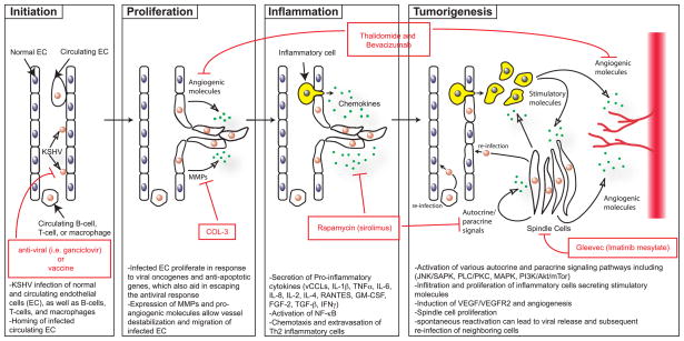
Kaposi sarcoma (KS) is a complex cancer that arises from the initial infection of an appropriate endothelial or progenitor cell by Kaposi Sarcoma Herpesvirus/Human Herpesvirus-8 (KSHV/HHV8). However, the majority of KS cases occur when infected patients also suffer from some coincident form of immune deregulation, providing a favorable microenvironment for tumor development. Cellular hallmarks of KS progression include both the hyper-proliferation of KSHV-infected cells and the infiltration of immune modulatory cells into KS lesions, which together result in chronic inflammation, the induction of angiogenesis and tumor growth. This review describes the current understanding of the interactions between KSHV and host responses that result in this unusual cancer, along with existing treatments and prospects for future therapeutic approaches.
Figures
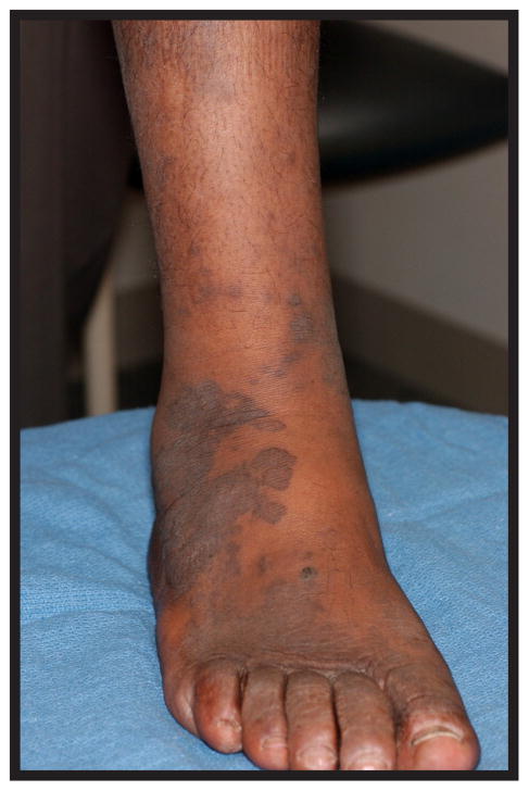
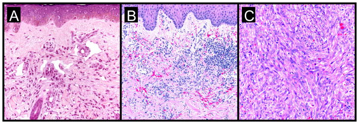
subtle proliferation of irregular vascular channels between normal stromal collagen
the extravasation of erythrocytes and hemosiderin into the stroma
detection of lymphoplasmacytic infiltrate
proliferating spindled cells that form interlacing bundles closely approximated with blood-filled vascular spaces
intracellular hyaline globules within lesional cells (likely representing phagocytosed erythrocytes within lysozomes)
increased inflammatory infiltrate consisting of lymphocytes, plasma cells, macrophages, and dendritic cells
formation of intersecting fascicles and sheets of proliferating spindled cells
References
-
- Martin JN, Ganem DE, Osmond DH, Page-Shafer KA, Macrae D, et al. Sexual transmission and the natural history of human herpesvirus 8 infection. N Engl J Med. 1998;338(14):948–54. - PubMed
-
- Kestens L, Melbye M, Biggar RJ, Stevens WJ, Piot P, et al. Endemic African Kaposi’s sarcoma is not associated with immunodeficiency. Int J Cancer. 1985;36(1):49–54. - PubMed
-
- Rubin AI, Stiller MJ. A listing of skin conditions exhibiting the koebner and pseudo-koebner phenomena with eliciting stimuli. J Cutan Med Surg. 2002;6(1):29–34. - PubMed
-
- Leidner RS, Aboulafia DM. Recrudescent Kaposi’s sarcoma after initiation of HAART: a manifestation of immune reconstitution syndrome. AIDS Patient Care STDS. 2005;19(10):635–44. - PubMed
-
- Ziegler J, Dorfman RF. Overview of kaposi’s sarcoma: History, epidemiology, and biomedical features. In: Ziegler JL, Dorfman RF, editors. Kaposi’s sarcoma: Pathophysiology and clinical management. Marcel Dekker, Inc; New York: 1988. pp. 1–22.
Grants and funding
LinkOut - more resources
Full Text Sources
Other Literature Sources