

The cast imaging of the osteon lacunar-canalicular system and the implications with functional models of intracanalicular flow
- PMID: 23082756
- PMCID: PMC3632224
- DOI: 10.1111/joa.12004
The cast imaging of the osteon lacunar-canalicular system and the implications with functional models of intracanalicular flow
Abstract
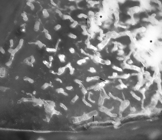
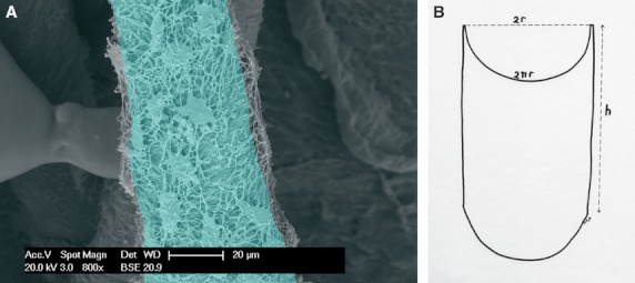
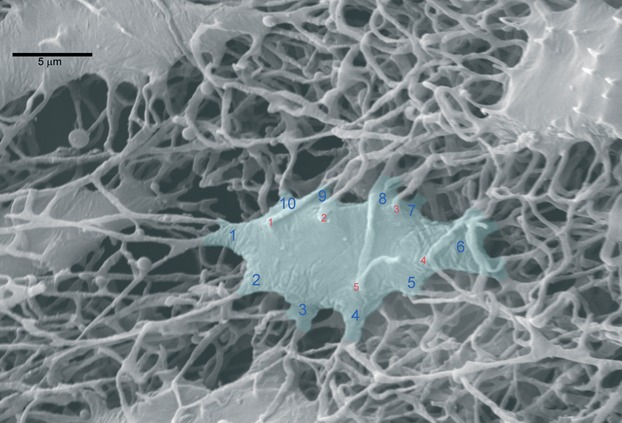
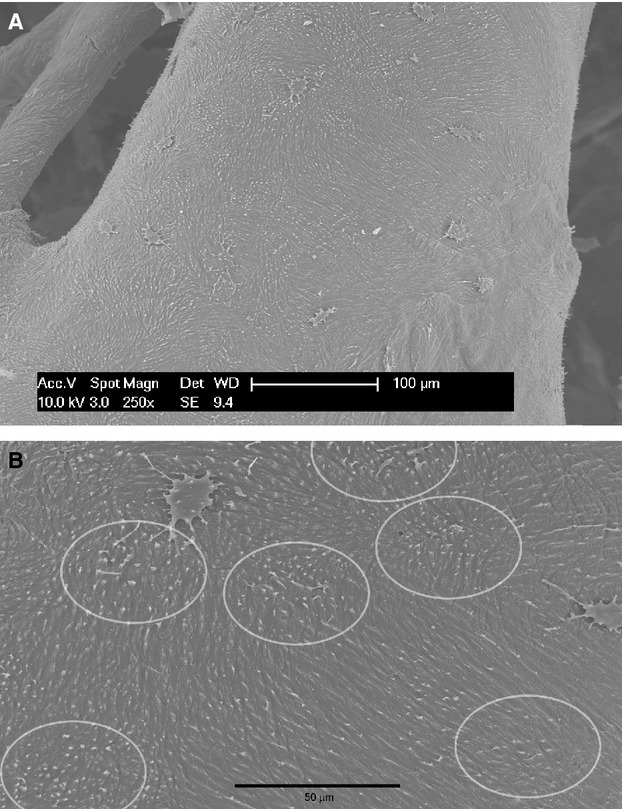
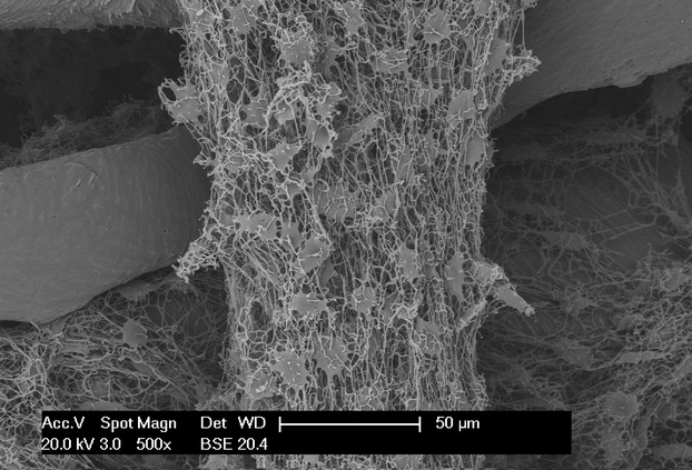
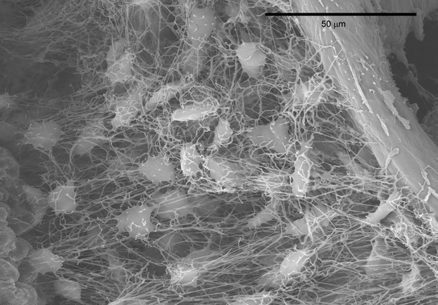
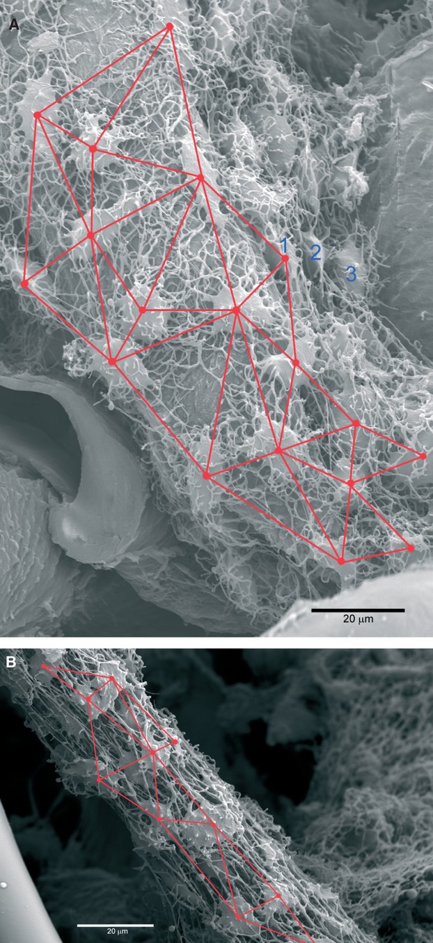
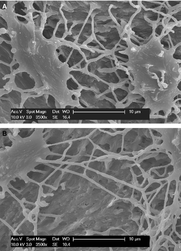
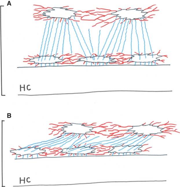
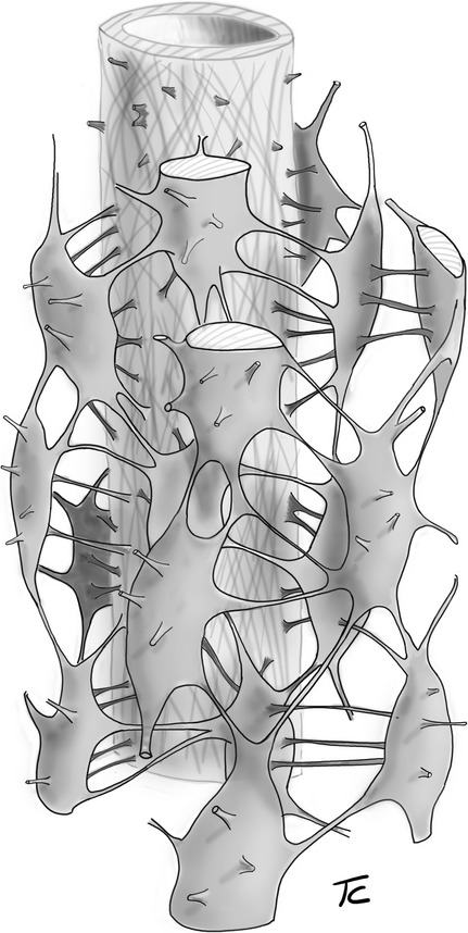
A casting technique with methyl-methacrylate (MMA) was applied to the study of the osteon lacunar-canalicular network of human and rabbit cortical bone. The MMA monomer infiltration inside the vascular canals and from these into the lacunar-canalicular system was driven by capillarity, helped by evaporation and the resulting negative pressure in a system of small pipes. There was uniform, centrifugal penetration of the resin inside some osteons, but this was limited to a depth of four to five layers of lacunae. Moreover, not all of the osteon population was infiltrated. This failure can be the result of one of two factors: the incomplete removal of organic debris from the canal and canalicular systems, and lack of drainage at the osteon external border. These data suggest that each secondary osteon is a closed system with a peripheral barrier (represented by the reversal line). As the resin advances into the osteon, the air contained inside the canalicula is compressed and its pressure increases until infiltration is stopped. The casts gave a reliable visualization of the lacunar shape, position and connections between the lacunae without the need for manipulations such as cutting or sawing. Two systems of canalicula could be distinguished, the equatorial, which connected the lacunae (therefore the osteocytes) lying on the same concentric level, and the radial, which established connections between different levels. The equatorial canalicula radiated from the lacunar border forming ramifications on a planar surface around the lacuna, whereas the radial canalicula had a predominantly straight direction perpendicular to the equatorial plane. The mean length of the radial canalicula was 40.12 ± 10.26 μm in rabbits and 38.4 ± 7.35 μm in human osteons; their mean diameter was 174.4 ± 71.12 nm and 195.7 ± 79.58 nm, respectively. The mean equatorial canalicula diameter was 237 ± 66.04 nm in rabbit and 249.7 ± 73.78 nm in human bones, both significantly larger (P < 0.001) than the radial. There were no significant differences between the two species. The lacunar surface measured on the equatorial plane was higher in rabbit than in man, but the difference was not statistically significant. The cast of the lacunar-canalicular network obtained with the reported technique allows a direct, 3-D representation of the system architecture and illustrates how the connections between osteocytes are organized. The comparison with models derived by the assumption of the role of hydraulic conductance and other mechanistic functions provides descriptive, morphological data to the ongoing discussion on the Haversian system biology.
© 2012 The Authors Journal of Anatomy © 2012 Anatomical Society.
Figures
Similar articles
-
Morphometric analysis of osteonal architecture in bones from healthy young human male subjects using scanning electron microscopy.J Anat. 2013 Sep;223(3):242-54. doi: 10.1111/joa.12079. Epub 2013 Jul 8. J Anat. 2013. PMID: 23834434 Free PMC article.
-
The canalicular system and the osteoblast domain in human secondary osteons.Anat Histol Embryol. 2012 Dec;41(6):410-8. doi: 10.1111/j.1439-0264.2012.01150.x. Epub 2012 Apr 2. Anat Histol Embryol. 2012. PMID: 22469429
-
Network architecture strongly influences the fluid flow pattern through the lacunocanalicular network in human osteons.Biomech Model Mechanobiol. 2020 Jun;19(3):823-840. doi: 10.1007/s10237-019-01250-1. Epub 2019 Nov 28. Biomech Model Mechanobiol. 2020. PMID: 31782029 Free PMC article.
-
Influence of Osteocyte Lacunar-Canalicular Morphology and Network Architecture on Osteocyte Mechanosensitivity.Curr Osteoporos Rep. 2023 Aug;21(4):401-413. doi: 10.1007/s11914-023-00792-9. Epub 2023 Jun 5. Curr Osteoporos Rep. 2023. PMID: 37273086 Review.
-
Osteon: Structure, Turnover, and Regeneration.Tissue Eng Part B Rev. 2022 Apr;28(2):261-278. doi: 10.1089/ten.TEB.2020.0322. Epub 2021 Mar 8. Tissue Eng Part B Rev. 2022. PMID: 33487116 Free PMC article. Review.
Cited by
-
The complex rostral morphology and the endoskeleton ossification process of two adult samples of Xiphias gladius (Xiphiidae).J Fish Biol. 2022 Jul;101(1):42-54. doi: 10.1111/jfb.15069. Epub 2022 May 16. J Fish Biol. 2022. PMID: 35481825 Free PMC article.
-
Contributions of Resin Cast Etching to Visualising the Osteocyte Lacuno-Canalicular Network Architecture in Bone Biology and Tissue Engineering.Calcif Tissue Int. 2023 May;112(5):525-542. doi: 10.1007/s00223-022-01058-9. Epub 2023 Jan 7. Calcif Tissue Int. 2023. PMID: 36611094 Free PMC article. Review.
-
Advancements in composition and structural characterization of bone to inform mechanical outcomes and modelling.Curr Opin Biomed Eng. 2019 Sep;11:76-84. doi: 10.1016/j.cobme.2019.09.011. Epub 2019 Sep 28. Curr Opin Biomed Eng. 2019. PMID: 32864522 Free PMC article.
-
Ptychographic X-ray CT characterization of the osteocyte lacuno-canalicular network in a male rat's glucocorticoid induced osteoporosis model.Bone Rep. 2018 Jul 29;9:122-131. doi: 10.1016/j.bonr.2018.07.005. eCollection 2018 Dec. Bone Rep. 2018. PMID: 30246062 Free PMC article.
-
50 years of scanning electron microscopy of bone-a comprehensive overview of the important discoveries made and insights gained into bone material properties in health, disease, and taphonomy.Bone Res. 2019 May 22;7:15. doi: 10.1038/s41413-019-0053-z. eCollection 2019. Bone Res. 2019. PMID: 31123620 Free PMC article.
References
-
- Cooper RR, Milgram JW, Robinson RA. Morphology of the osteon. An electron microscopy study. J Bone Joint Surg. 1966;48A:1239–1271. - PubMed
-
- Doty SB. Morphological evidence of gap junctions between bone cells. Calcif Tissue Int. 1981;33:509–512. - PubMed
-
- Ejiri S, Ozawa H. Scanning electron microscopic observations of rat tibia using HCl-collagenase method. Arch Histol Jap. 1982;45:399–404. - PubMed
-
- Ennos AR. The aerodynamics and hydrodynamics of plants. J Exp Biol. 1999;202:3281–3284. - PubMed
Publication types
MeSH terms
Substances
LinkOut - more resources
Full Text Sources
Research Materials
Miscellaneous
