. 2012 Nov;336(2):139-47.
doi: 10.1111/j.1574-6968.12000.x. Epub 2012 Sep 24.

The Yersinia pseudotuberculosis degradosome is required for oxidative stress, while its PNPase subunit plays a degradosome-independent role in cold growth

Affiliations

The Yersinia pseudotuberculosis degradosome is required for oxidative stress, while its PNPase subunit plays a degradosome-independent role in cold growth

Amanda Henry et al. FEMS Microbiol Lett. 2012 Nov.

Abstract

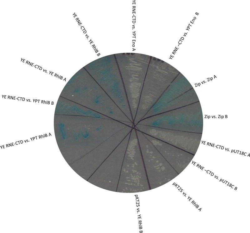
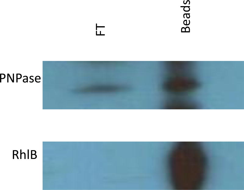
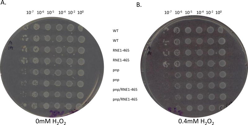
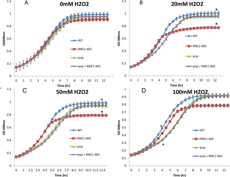
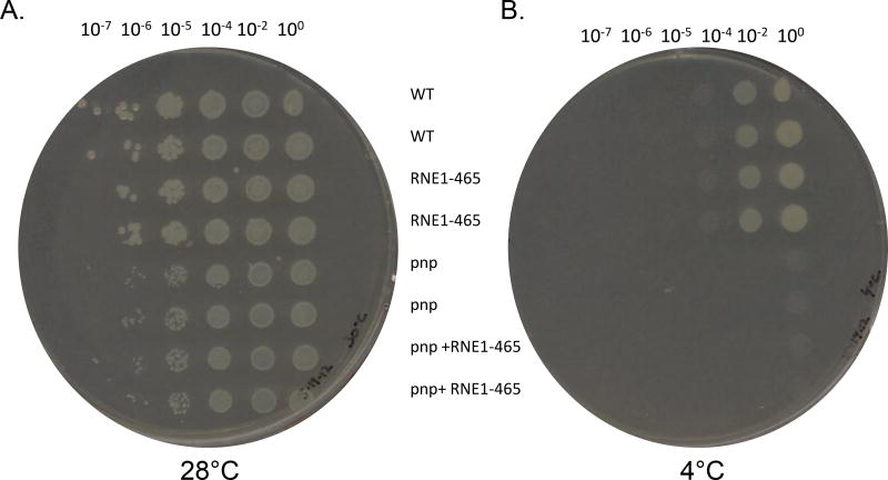
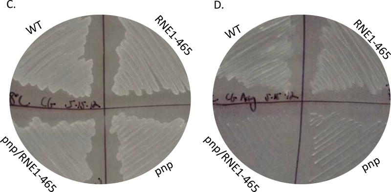
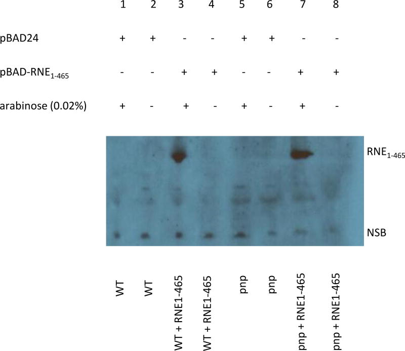
Yersinia polynucleotide phosphorylase (PNPase), a 3'-5' exoribonuclease, has been shown to affect growth during several stress responses. In Escherichia coli, PNPase is one of the subunits of a multiprotein complex known as the degradosome, but also has degradosome-independent functions. The carboxy-terminus of E. coli ribonuclease E (RNase E) serves as the scaffold upon which PNPase, enolase (a glycolytic enzyme), and RhlB helicase all have been shown to bind. In the yersiniae, only PNPase has thus far been shown to physically interact with RNase E. We show by bacterial two-hybrid and co-immunoprecipitation assays that RhlB and enolase also interact with RNase E. Interestingly, although PNPase is required for normal growth at cold temperatures, assembly of the yersiniae degradosome was not required. However, degradosome assembly was required for growth in the presence of reactive oxygen species. These data suggest that while the Yersinia pseudotuberculosis PNPase plays a role in the oxidative stress response through a degradosome-dependent mechanism, PNPase's role during cold stress is degradosome-independent.


Figures

Figure 1
Figure 1
Representative B2H experiment testing f or interactions between the Y. pseudotuberculosis RNase E CTD scaffolding region and full length RhlB, enolase, or PNPase. Individual bacterial colony forming units were evaluated, and the positive control was pKT25-Zip vs. pUT18C-Zip. The negative control was pKT25-RNE1–465 vs. pUT18Cempty.
Figure 2
Figure 2
Representative B2H experiment testing for interactions between the Y. enterocolitica RNase E CTD scaffolding region and full length Y. pseudotuberculosis and Y. enterocolitica RhlB. Single colony bacterial isolates were evaluated, and the positive control was pKT25-Zip vs. pUT18C-Zip. The negative controls were pKT25 vs. pUT18C-Y. enterocolitica RhlB and pKT25- Y. enterocolitica RNE1–465 vs. pUT18C.
Figure 3
Figure 3
Co-immunoprecipitations using anti-RNase E antibody fused Protein G sepharose beads. Depicted is an immunoblot probing the co-precipitated protein complex (bead) and probing the flow through (FT) using either rabbit polyclonal anti-PNPase or -RhlB antibodies.
Figure 4
Figure 4
Representative H2O2 plate experiment. Y. pseudotuberculosis + empty vector pBAD24 (WT), Y. pseudotuberculosis Δpnp + empty vector pBAD24 (pnp), Y. pseudotuberculosis Δpnp + pBAD-RNE1–465 (pnp/RNE), and Y. pseudotuberculosis + pBAD-RNE1–465 (RNE) strains were grown on 0mM H2O2 (A) or 0.4mM H2O2 (B) at 30°C for 16 hours at which point the plates were scanned. All strains were spotted in duplicate for internal dilution and spotting controls.
Figure 5
Figure 5
Representative H2O2 liquid growth experiment. Subcultures of Y. pseudotuberculosis + empty vector pBAD24 (WT), Y. pseudotuberculosis Δpnp + empty vector pBAD24 (pnp), Y. pseudotuberculosis Δpnp + pBAD-RNE1–465 (pnp/RNE), and Y. pseudotuberculosis + pBAD-RNE1–465 (RNE) were grown (in triplicate) in 100µl volumes using a 96 well plate. Following 1 hour of static growth, 0mM (A), 20mM (B), 50mM (C), or 100mM (D) H2O2 was a added to the appropriate wells, and plates were constantly agitated while grown at 30°C for 12 hours. Samples were read at optical density 600nm every 30 minutes. Asterisk between the WT and pnp samples in panel D at 4 hours and between WT and RNE in panel B and C at 12 hours denote statistical significance (p< 0.5). All statistical tests employed the Student’s T-test.
Figure 6
Figure 6
Representative cold-growth experiments. A. Various dilutions of saturated Y. pseudotuberculosis + empty vector pBAD24 (WT), Y. pseudotuberculosis Δpnp + empty vector pBAD24 (pnp), Y. pseudotuberculosis Δpnp + pBAD-RNE1–465 (pnp/RNE), and Y. pseudotuberculosis + pBAD-RNE1–465 (RNE) cultures were spotted (using a pronger) on LB agar plates and grown at 30°C for 16 hours (A) or at 4°C for 11 days (B) at which point the plates were scanned. The same aforementioned strains were streaked on plates and images were acquired following 16 hours of growth at 30°C (C) or following 11 days of growth at 4°C (D).
Figure 6
Figure 6
Representative cold-growth experiments. A. Various dilutions of saturated Y. pseudotuberculosis + empty vector pBAD24 (WT), Y. pseudotuberculosis Δpnp + empty vector pBAD24 (pnp), Y. pseudotuberculosis Δpnp + pBAD-RNE1–465 (pnp/RNE), and Y. pseudotuberculosis + pBAD-RNE1–465 (RNE) cultures were spotted (using a pronger) on LB agar plates and grown at 30°C for 16 hours (A) or at 4°C for 11 days (B) at which point the plates were scanned. The same aforementioned strains were streaked on plates and images were acquired following 16 hours of growth at 30°C (C) or following 11 days of growth at 4°C (D).
Figure 7
Figure 7
Arabinose induction of the truncated RNE1–465. Y. pseudotuberculosis + empty vector pBAD24 (WT), Y. pseudotuberculosis Δpnp + empty vector pBAD24 (pnp), Y. pseudotuberculosis Δpnp + pBAD-RNE1–465 (pnp + RNE), and Y. pseudotuberculosis + pBAD-RNE1–465 (WT + RNE) cultures were grown and induced for 1.5 hours with 0.02% arabinose. An immunoblot was performed employing polyclonal anti-RNase E antibody. CTD-deficient RNE1–465 was expressed in strains that contained the appropriate plasmid and that were induced with 0.02% arabinose. NSB= non-specific band.

Similar articles

Cited by

References

    1. Achtman M, Zurth K, Morelli G, Torrea G, Guiyoule A, Carniel E. Yersina pestis, the cause of plague, is a recently emerged clone of Yersinia pseudotuberculosis. Proceedings of the National Academy of Science of the United States of America. 1999;96:14043–14048. - PMC - PubMed
    1. Briegel KJ, Baker A, Jain C. Identification and analysis of Escherichia coli ribonuclease E dominant-negative mutants. Genetics. 2006;172(1):7–15. - PMC - PubMed
    1. Bölin I, Wolf-Watz H. Molecular cloning of the temperature-inducible outer membrane proteins of yersiniae. Infect Immun. 1984;43:72–78. - PMC - PubMed
    1. Cairrão F, Cruz A, Mori H, Arraiano CM. Cold shock induction of RNase R and its role in the maturation of the quality control mediator SsrA/tmRNA. Mol Microbiol. 2003;50(4):1349–60. - PubMed
    1. Carpousis AJ. The Escherichia coli RNA degradosome: structure, function and relationship in other ribonucleolytic multienzyme complexes. Biochem Soc Trans. 2002;30(2):150–5. - PubMed

Publication types

MeSH terms